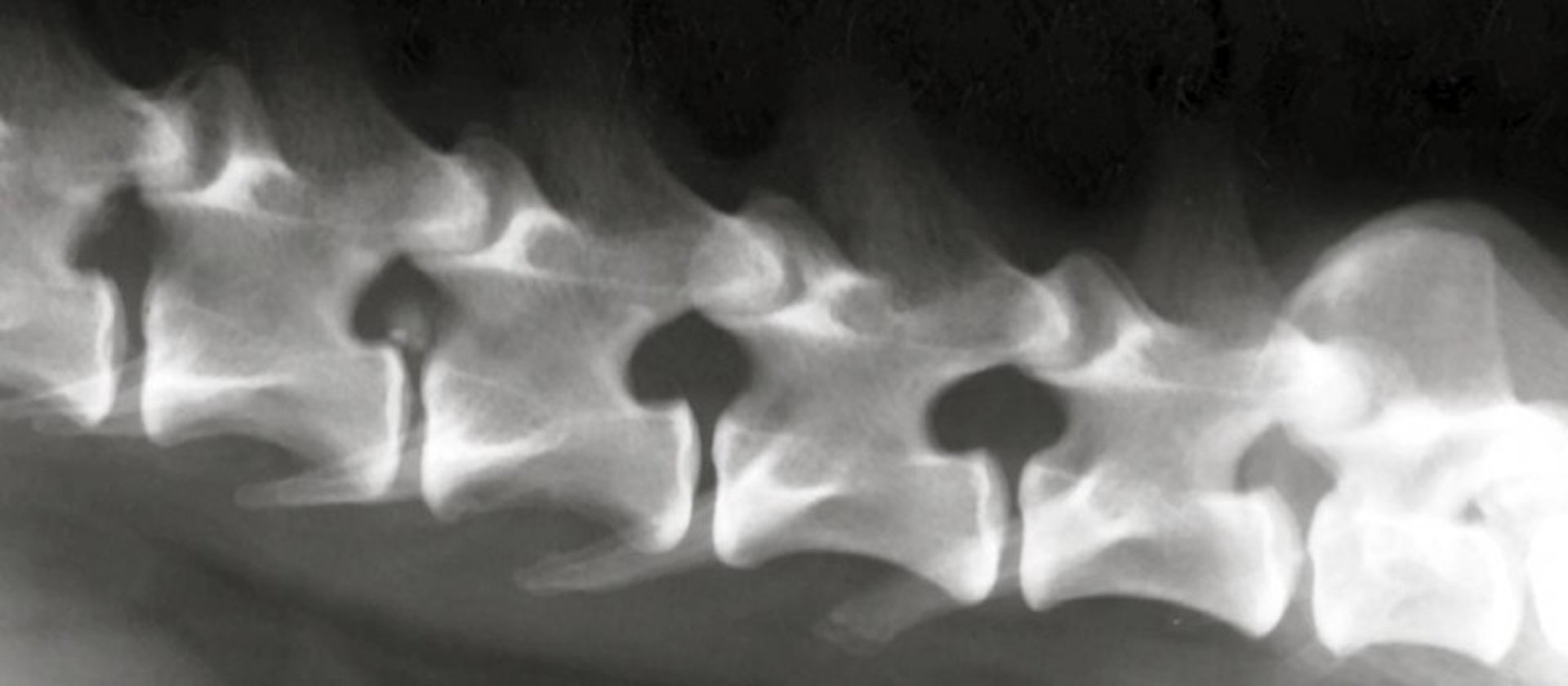
Intervertebral disk extrusion, dog, MRI
Transverse plane T2-weighted MRI image of a dog with intervertebral disk extrusion, showing compression of the left lateral aspect of the spinal cord.
Courtesy of Dr. William Thomas.
Transverse plane T2-weighted MRI image of a dog with intervertebral disk extrusion, showing compression of the left lateral aspect of the spinal cord.
Courtesy of Dr. William Thomas.