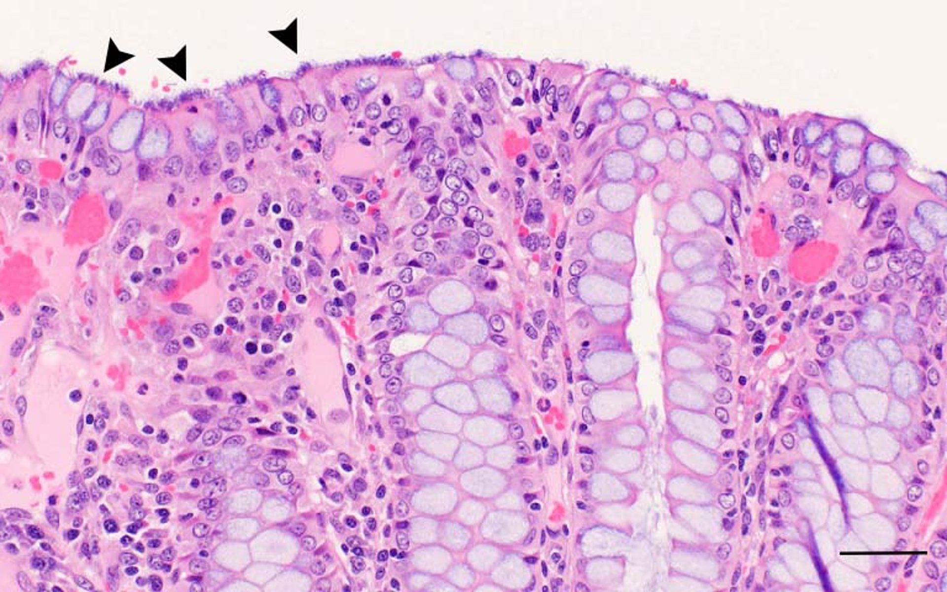
Intestinal spirochetosis, pig, histological image
Photomicrograph showing intestinal spirochetosis in a pig. Note the numerous end-on attached spirochetes (arrowheads) lining the luminal surface of the colonic mucosa. H&E stain; scale bar = 50 mcm.
Courtesy of Dr. Eric R. Burrough.
In these topics