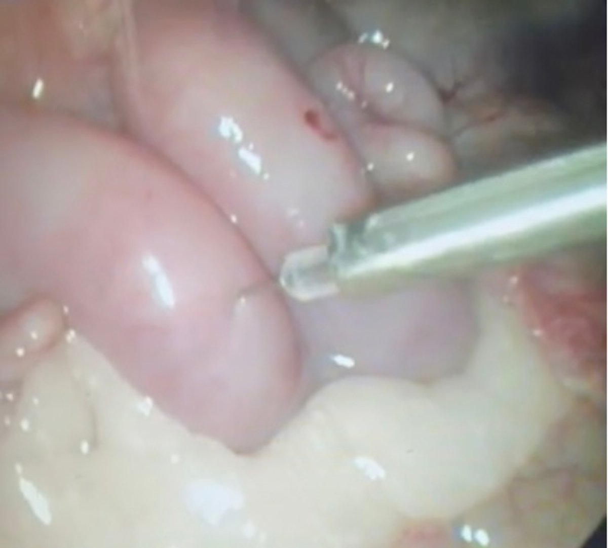
Intrauterine laparoscopic insemination, ewe
Laparoscopic view of an needle penetrating the uterine horn for deposition of semen during intrauterine laparoscopic artificial insemination (LAI) in a ewe. During intrauterine LAI in the ewe, the uterus is visualized through the laparoscope inserted via one of two abdominal ports placed caudal to the umbilicus and lateral to midline. Once the abdominal cavity is insufflated and the reproductive tract is exposed, the uterine horns appear as smooth pale pink tubular structures extending cranially from the uterine body toward their respective ovaries. An insemination needle, passed through the auxiliary port, is introduced under direct visualization. The needle is long and slender, with a needle tip designed to penetrate the uterine wall with minimal trauma. The small amount of blood in the other uterine horn shows the location of the first insemination.
Courtesy of Dr. Clare Scully.