


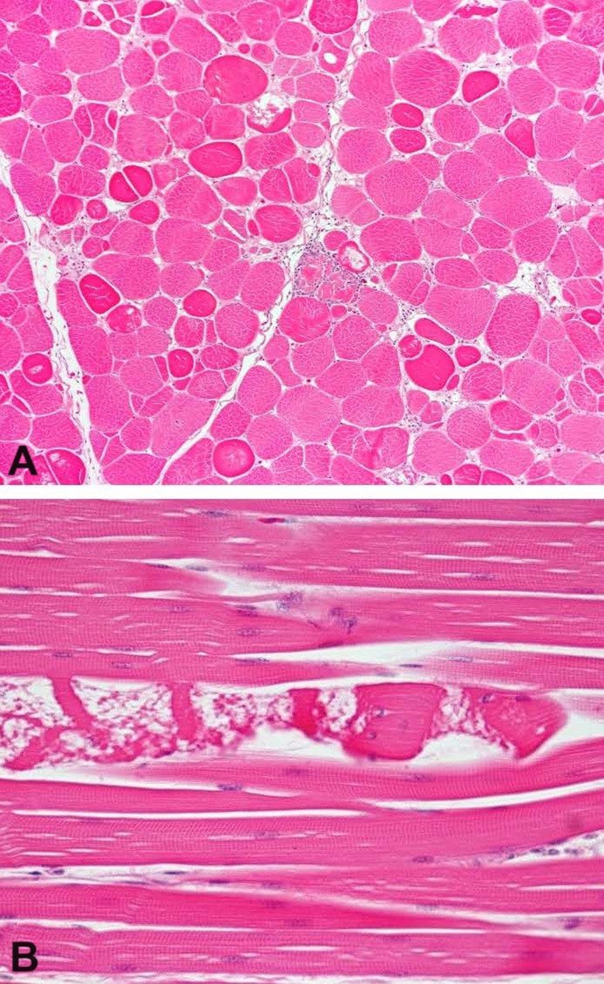
Minimal myopathy, photomicrographs, broiler
Photomicrographs of transverse (A) and longitudinal (B) sections of the pectoral muscle from a broiler chicken with minimal myopathy. (A) Note the focal area showing variation in myofiber size, swollen rounded hyalinized myofibers, and myofiber degeneration with fragmentation, with and without inflammation. H&E stain; original magnification 200X. (B) Note the myofiber degeneration and necrosis. H&E stain; original magnification 400X.
Images courtesy of Dr. H. J. Barnes.
In these topics