


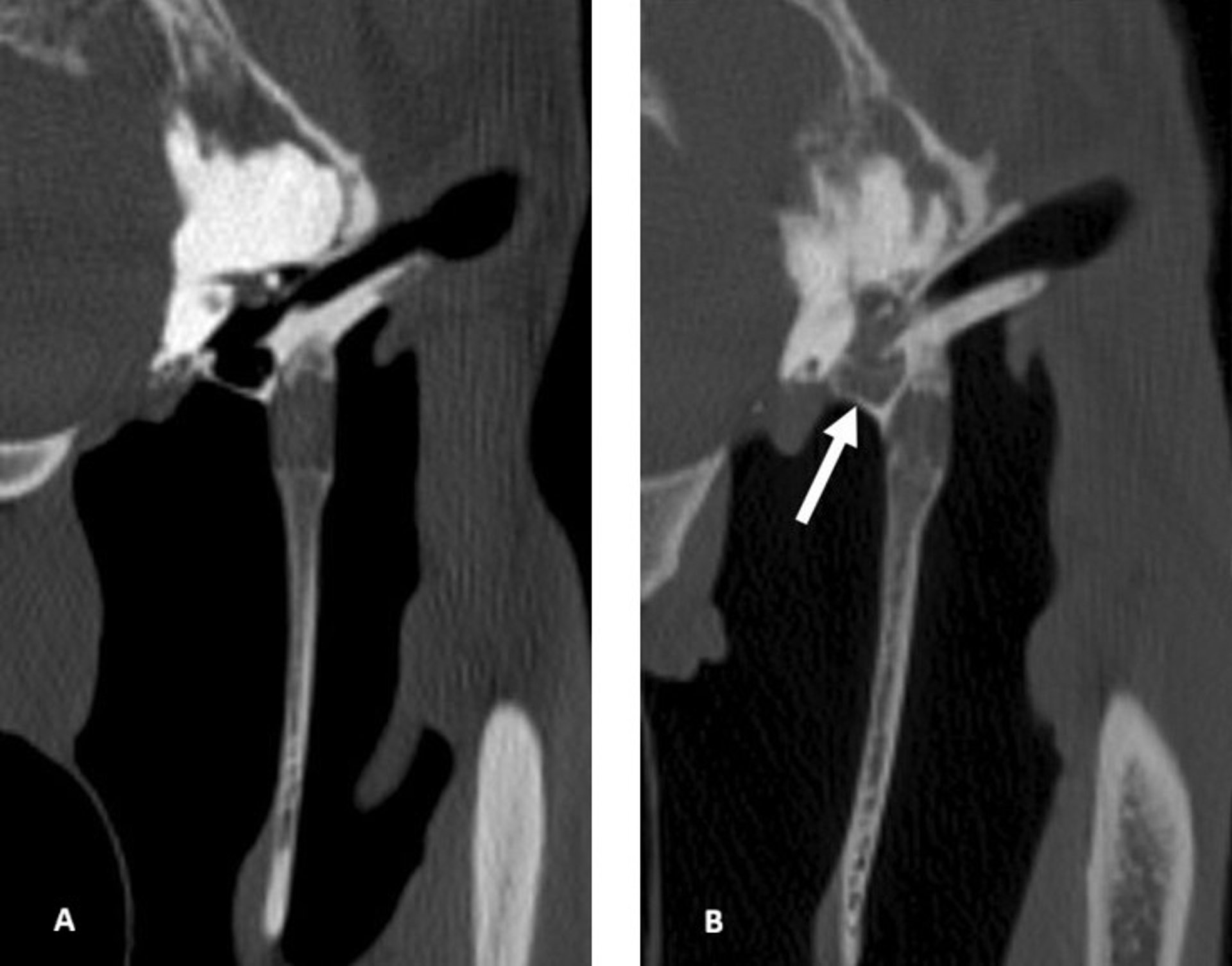
Otitis media, CT scan, horse
Transverse CT scans of the temporohyoid joint, tympanic bulla, horizontal ear canal, stylohyoid bone, and guttural pouch of a normal horse (A) and a horse with otitis media (B). A. Horse without otitis media: The horizontal ear canal, tympanic bulla, and guttural pouch are air-filled, and the stylohyoid bone and temporohyoid joint are of normal contour. B. Horse with otitis media: The tympanic bulla is filled with fluid (arrow). Helical acquisition, 120 kV and 400 mA, 512 × 512 matrix, 0.625 mm slice thickness, 0.3 mm slice interval, WW: 2800, WL: 800, bone kernel reconstruction.
Images from Dash RF, Perkins JD, Chang Y-M, Morgan RE. Computed tomography of the equine temporohyoid joint: association between imaging changes and potential risk factors. Equine Vet J. 2025. doi:10.1111/evj.14495. © 2025 The Author(s). Equine Veterinary Journal published by John Wiley & Sons Ltd on behalf of EVJ Ltd. Used under CC BY 4.0. Images extracted from Figure 1 (part A) and Figure 3 (part A).