


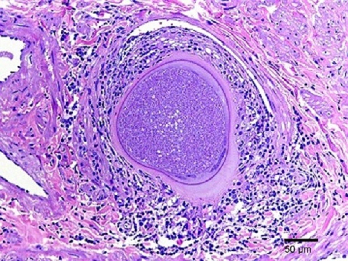
Pampiniform plexus tissue cysts, bull
Photomicrograph of a cross section of a tissue cyst sampled from a blood vessel, surrounded by an inflammatory infiltrate, in the pampiniform plexus of a bull with besnoitiosis. H&E stain; scale marker = 50 mcm.
Courtesy of Dr. Gema Álvarez García.
In these topics