

Atresia and normal distal nasolacrimal duct, foal

Atresia and normal distal nasolacrimal duct, foal
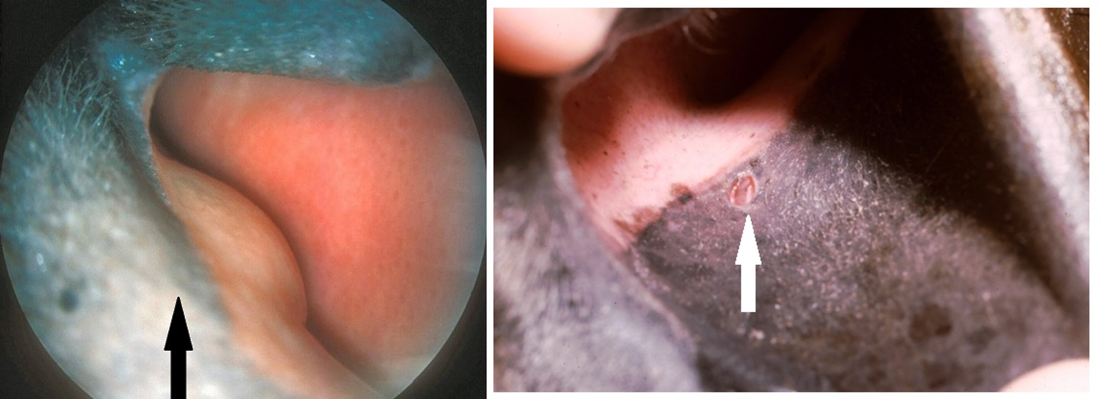
Left: Atresia of the distal nasolacrimal duct in a foal's eye. The 3- to 4-mm oval orifice that is typically found on the ventral floor of the nasal vestibule just outside the mucocutaneous junction (arrow) on the pigmented skin is missing. Right: Normal equine nasolacrimal duct (white arrow pointing to opening).
Left image courtesy of K. Gelatt. Right image courtesy of Dr. Bret Moore.
In these topics