

Transcutaneous cloacopexy, iguana

Transcutaneous cloacopexy, iguana
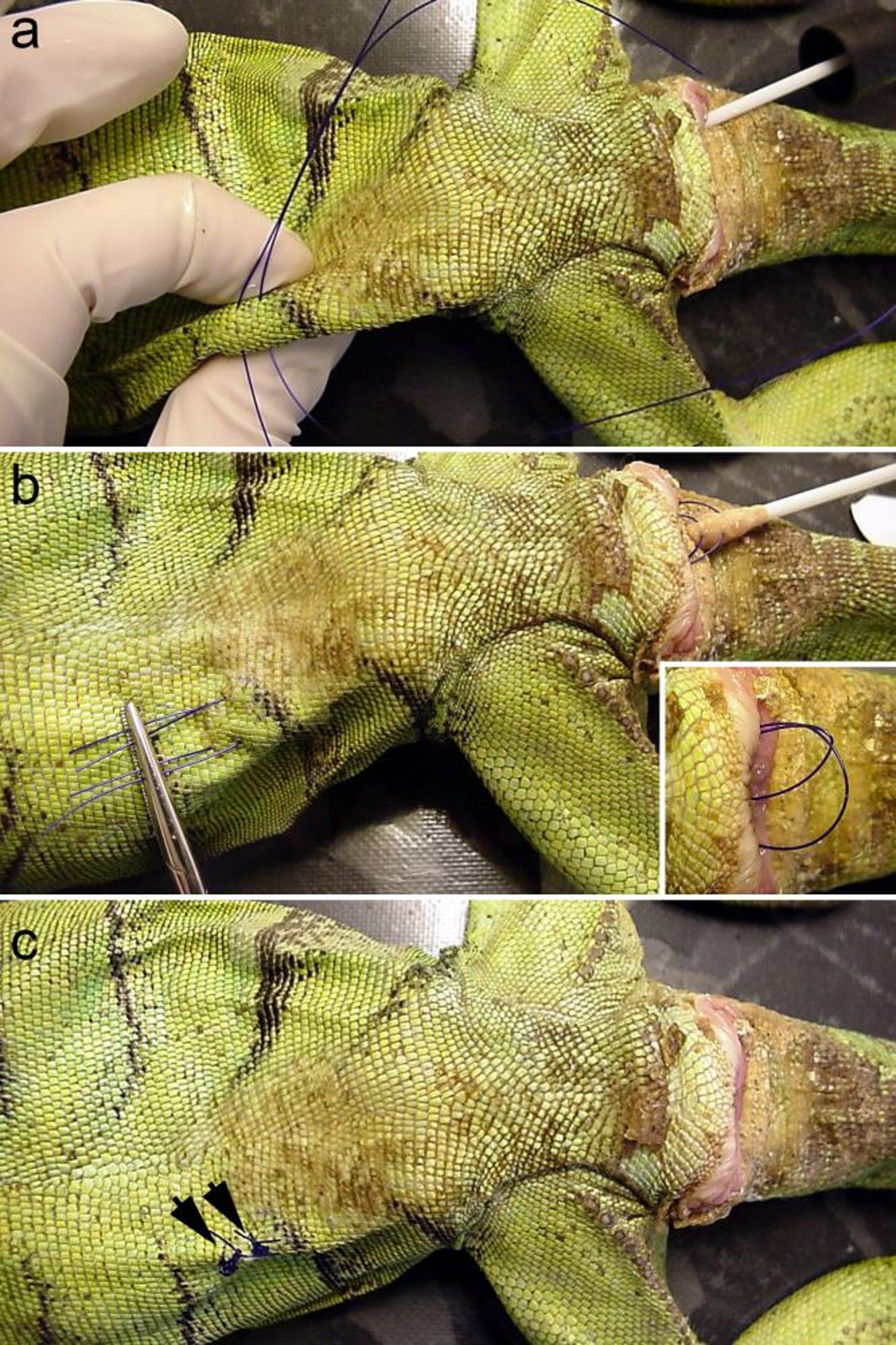
Transcutaneous cloacopexy in an iguana. a) After replacement of the viable prolapsed tissue, a large, lubricated, cotton-tipped applicator is inserted into the cloaca and tented against the ventrolateral body wall. Two or three simple interrupted polydioxanone sutures are placed through the skin and cotton tip. b) The suture ends are left long and held by hemostats. The cotton-tipped applicator, carrying the sutures, is removed from the cloaca. The cotton is teased away from the sutures (inset). c) After the sutures are pulled back into the cloaca with the hemostats, they are tied. The cloacocolonic wall is now sutured to the body wall, and a fibrous adhesion generally develops over the next 6–8 weeks.
Courtesy of Dr. Stephen Divers.
In these topics