

Canine distemper virus, mink

Canine distemper virus, mink
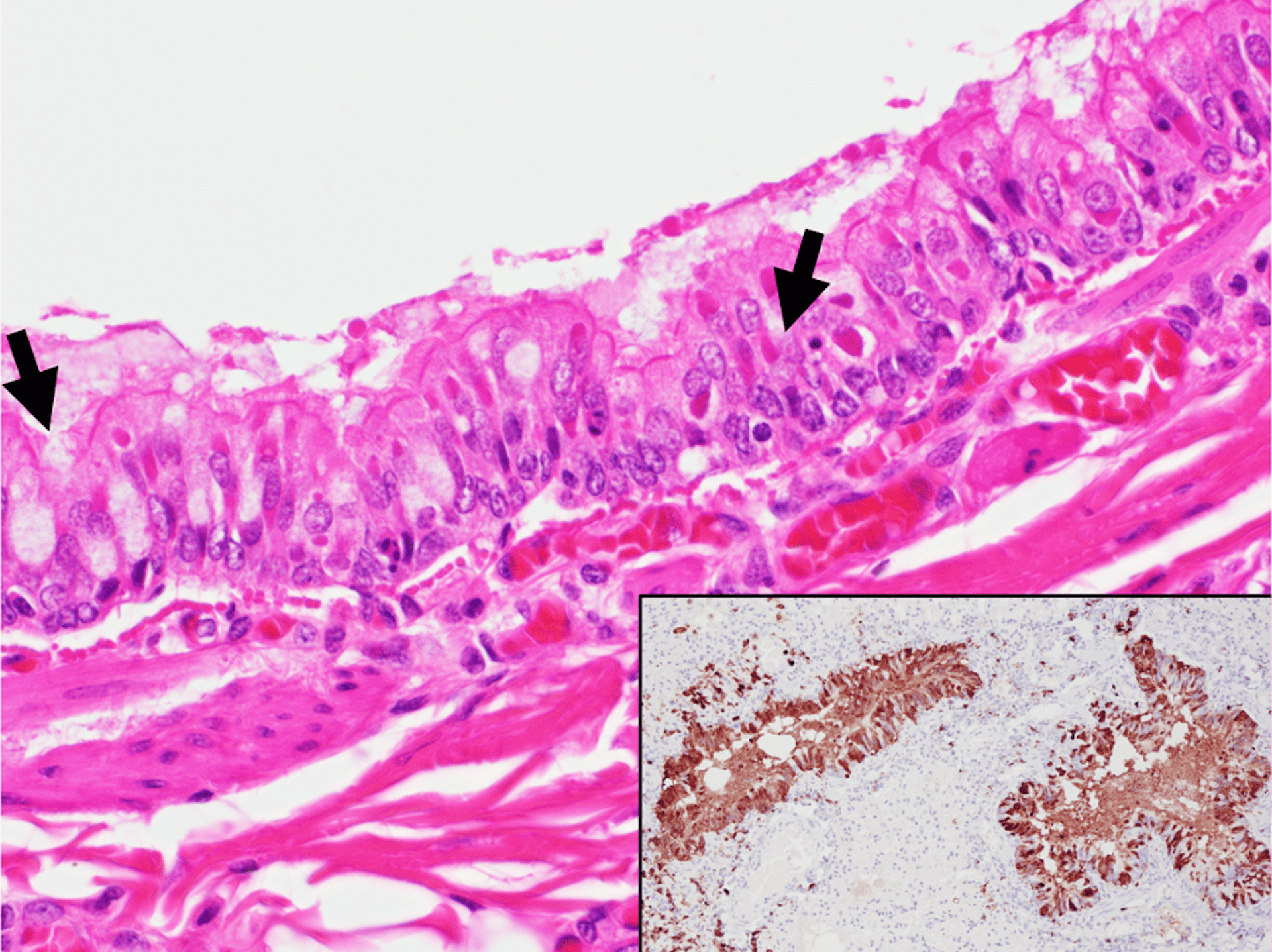
Photomicrograph of a histologic section of lung tissue from a mink infected with canine distemper virus. Eosinophilic intracytoplasmic inclusion bodies can be seen in the apical cytoplasm of some bronchial epithelial cells (black arrows). Inset: Section of lung tissue from an infected animal immunohistochemically labelled for canine distemper virus antigen, counterstained with Mayer’s hematoxylin. Note strong positive nuclear and cytoplasmic immunolabeling (brown staining) of airway epithelial cells.
Courtesy of Dr. Emily Brouwer and Dr. Marina Brash.
In these topics