


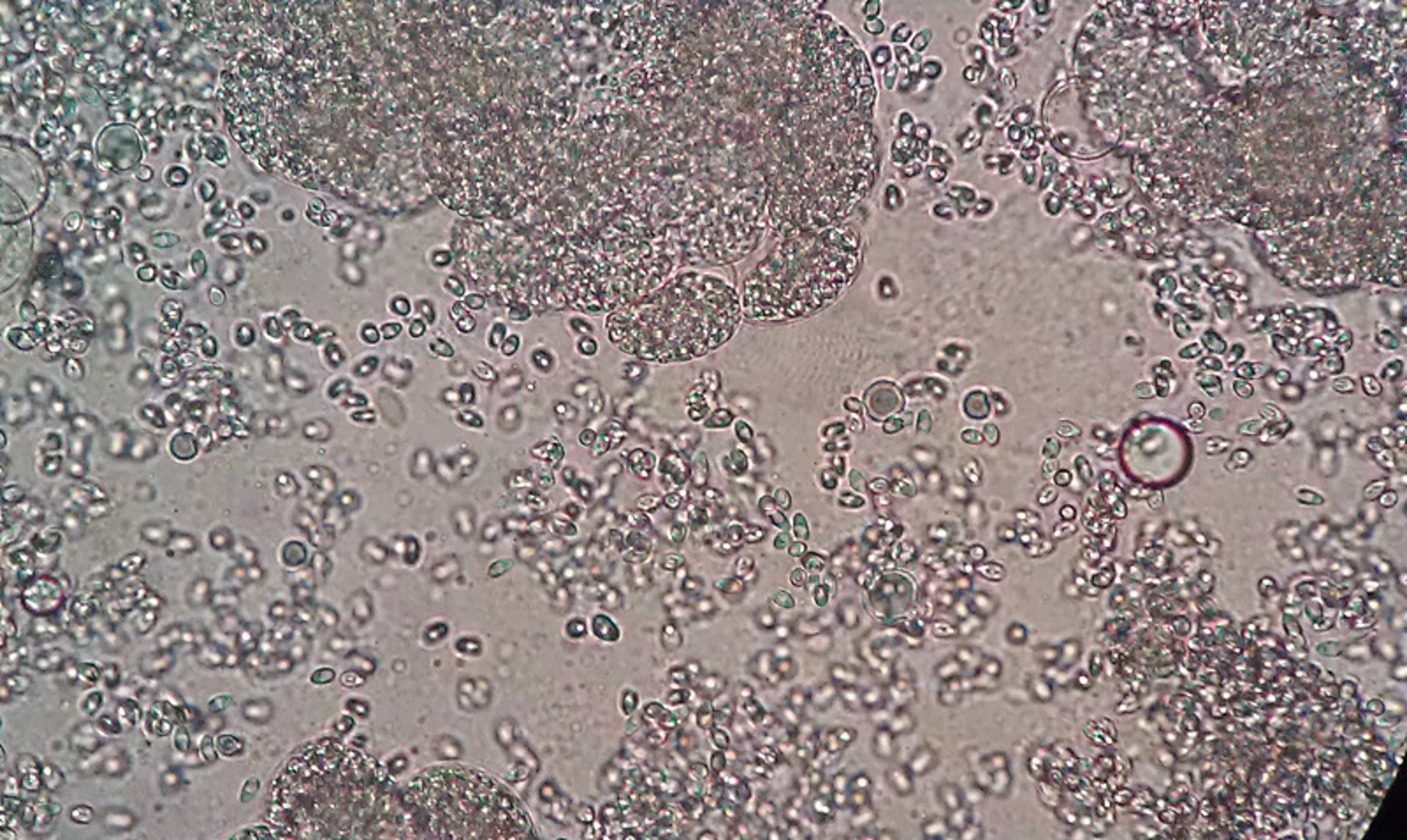
Wet mount of microsporidia, zebrafish
Photomicrograph of a section of skeletal muscles of a zebrafish squashed for an unstained wet mount. Pale areas in the dorsal skeletal muscles were observed at necropsy. The large clumps are xenomas filled with spores, and individual spores can be seen scattered in between the xenomas. Unstained wet mount; original magnification, 400×.
Courtesy of Dr. Denise Petty.
In these topics