

Infectious Hoof Lesions
Digital Dermatitis in Cattle

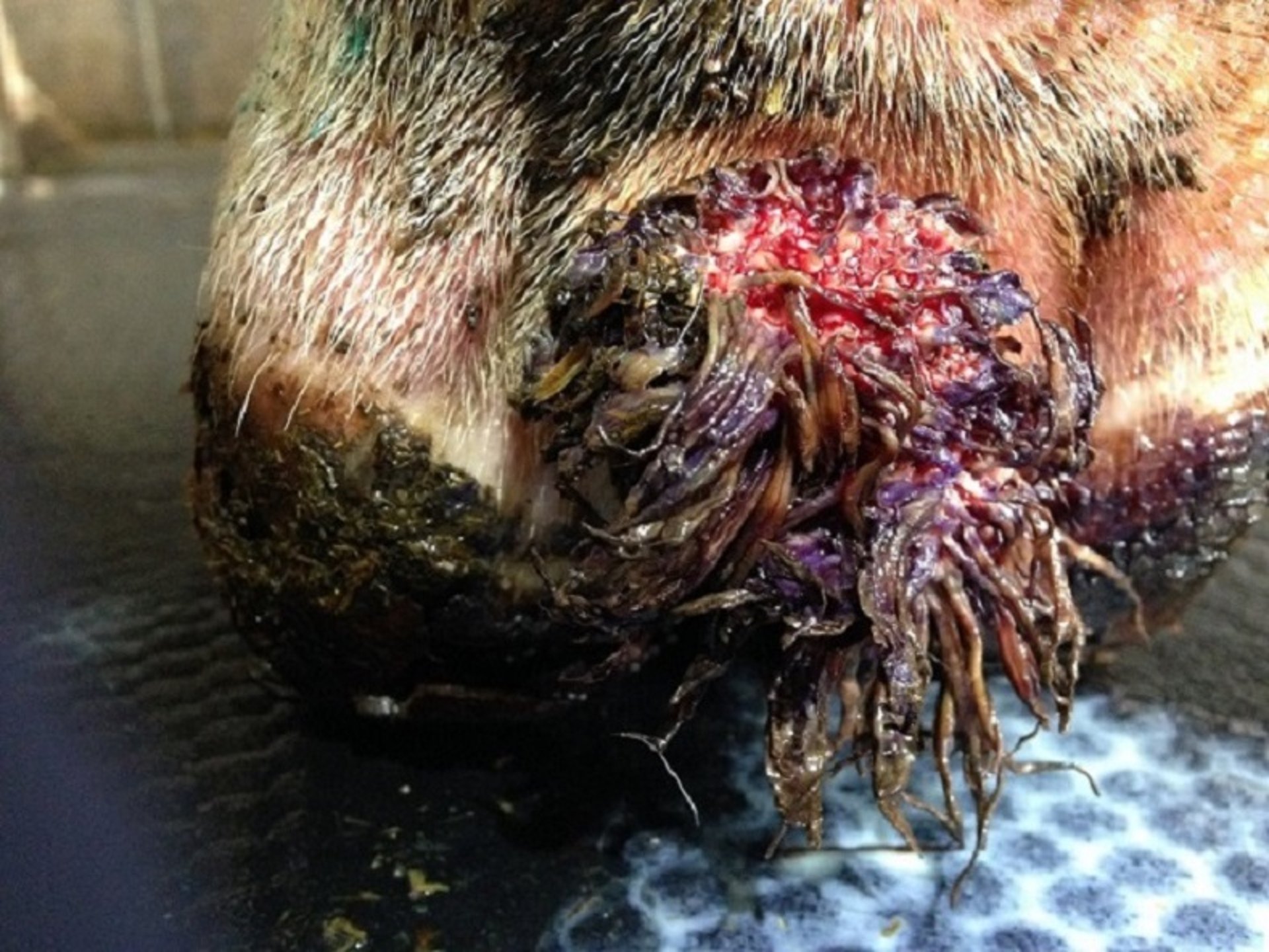
This photograph shows a chronically recurring, proliferative, painful digital dermatitis lesion in the interdigital cleft of the plantar surface of a cow’s foot.
Courtesy of Dr. Laura Solano.
Digital dermatitisis an infectious condition of the skin that commonly occurs in the interdigital cleft of the foot.
Pathogenesis
When the skin of a cow's foot are damaged by mechanical irritation and maceration by water and chemicals, a synergistic group of bacteria can invade the compromised skin barrier and create acute inflammation and infection of the dermis and epidermis. These bacteria are common in the environment and normally live in the rumen. The fact that digital dermatitis is not present on all farms suggests that more virulent strains of these bacteria are present on some farms. Treponema spp are a necessary component of this group of bacteria for generating disease. Treponema spp are gram-negative spirochetes that are microaerophilic and can encyst to protect themselves. As the bacteria invade the epidermis and damage the different layers, the body responds with a local inflammatory process that can result in hyperkeratosis and proliferative lesions.
Diagnosis
Digital dermatitis presents in a variety of stages ranging from painful, bright red, ulcerated skin lesions to less painful, gray-black, circular, granulomatous skin lesions. Edges can have a white margin and/or “hairs” protruding from them. Lesions are clearly demarcated and are typically located in the interdigital cleft; they can also occur at other locations, such as in the interdigital space or at the front of the foot. Severe lesions can become either proliferative with filamentous projections or hyperkeratotic. For practical purposes, it is useful to classify lesions as active (painful and ulcerative lesions > 2 cm) or chronic (gray-black hyperkeratotic lesions without painful ulcerative lesions > 2 cm).
Prevention
The main focus to prevent digital dermatitis is hygiene. Providing a clean environment without wet and/or abrasive walking surfaces decreases the chances of weakening the skin barrier. Foot baths are a preventive measure that should be used regularly, at whatever frequency is necessary to minimize the occurrence of active painful lesions. Foot baths are typically filled with disinfectant solutions such as copper sulfate or formalin and should be at least 10 feet (3 m) long to achieve a minimum of two immersions per rear foot. Other preventive measures include not allowing infected animals to enter the herd and ensuring that replacement animals are managed to prevent new infections.
Treatment
The number of licensed products that exist to treat digital dermatitis is sparse and varies from place to place. Treatment typically consists of applying topical tetracycline-based antimicrobials to active lesions by using a wrap or a paste. The microaerophilic nature of the bacteria makes wraps unnecessary; if they are used, however, they should be removed within 24 hours of application. Other topical agents that contain heavy metals such as copper are also often used in the field. The role of topical treatment is to treat active lesions, hastening their transition to a chronic stage, and to decrease pain. Once the lesion is in a chronic stage, it is the role of the foot bath to prevent recurrence.
Foot Rot in Cattle

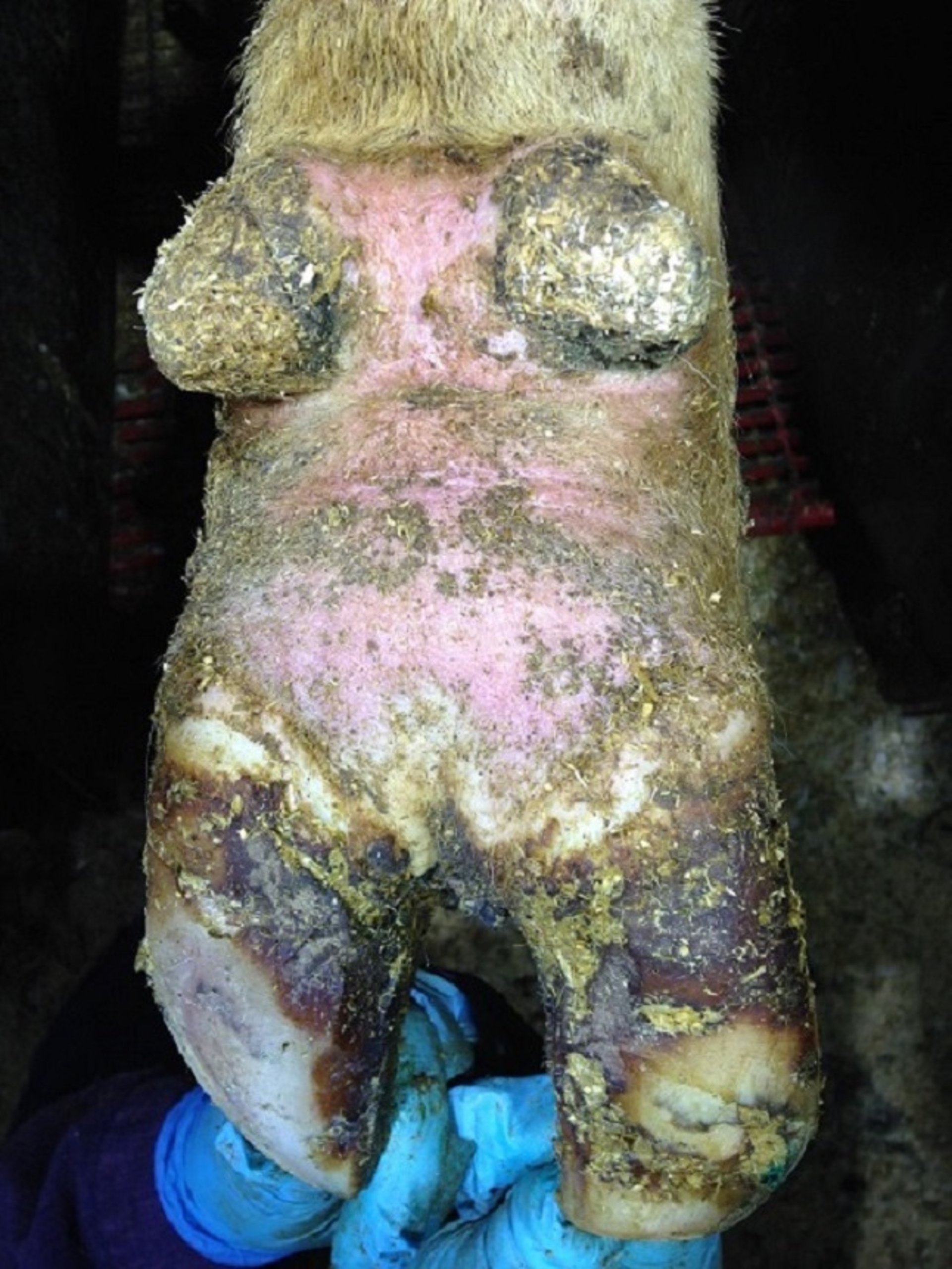
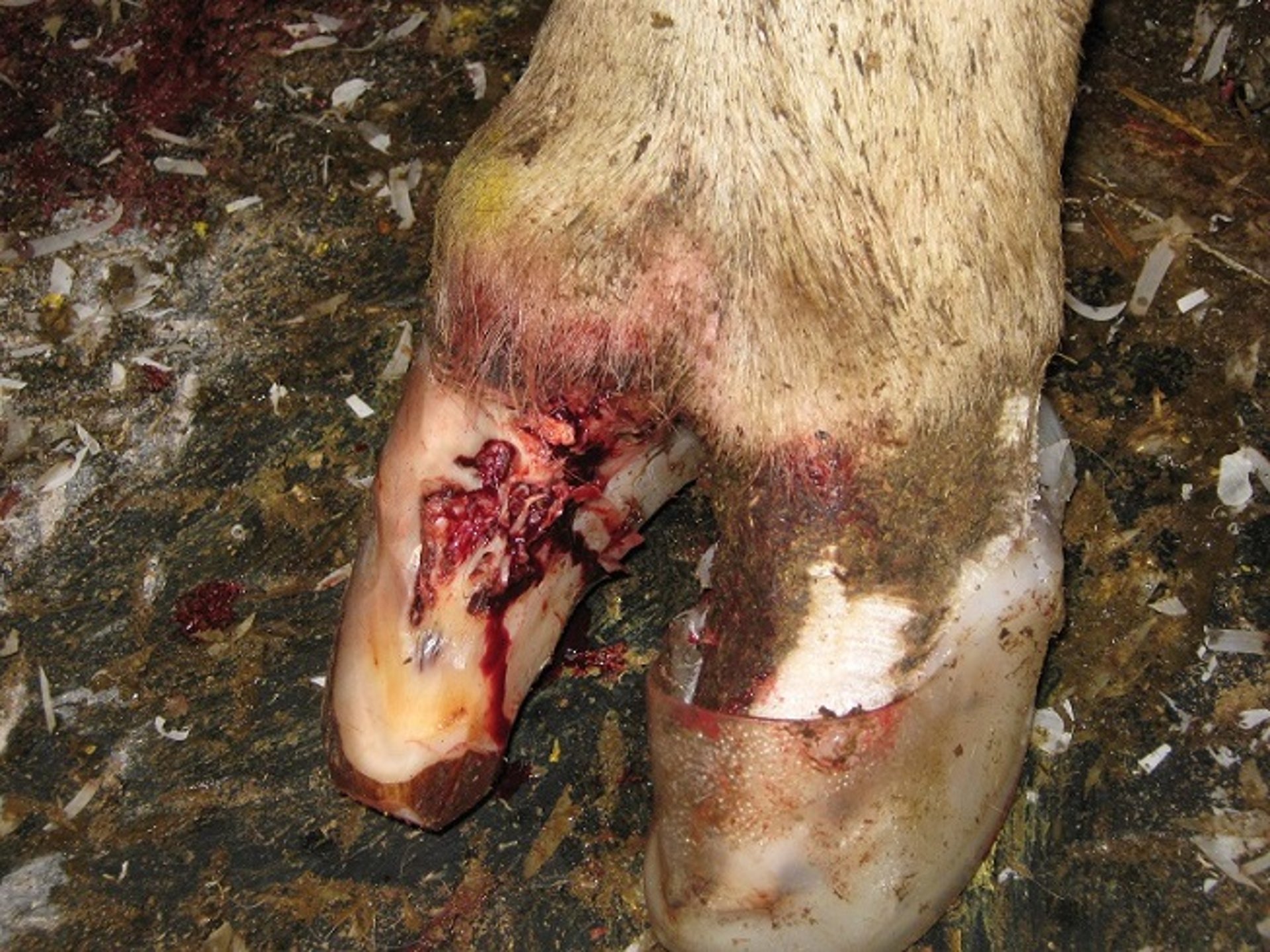
Extensive swelling above the hoof, a characteristic of foot rot, is evident in this photograph of a cow's foot.
Courtesy of Dr. Laura Solano.

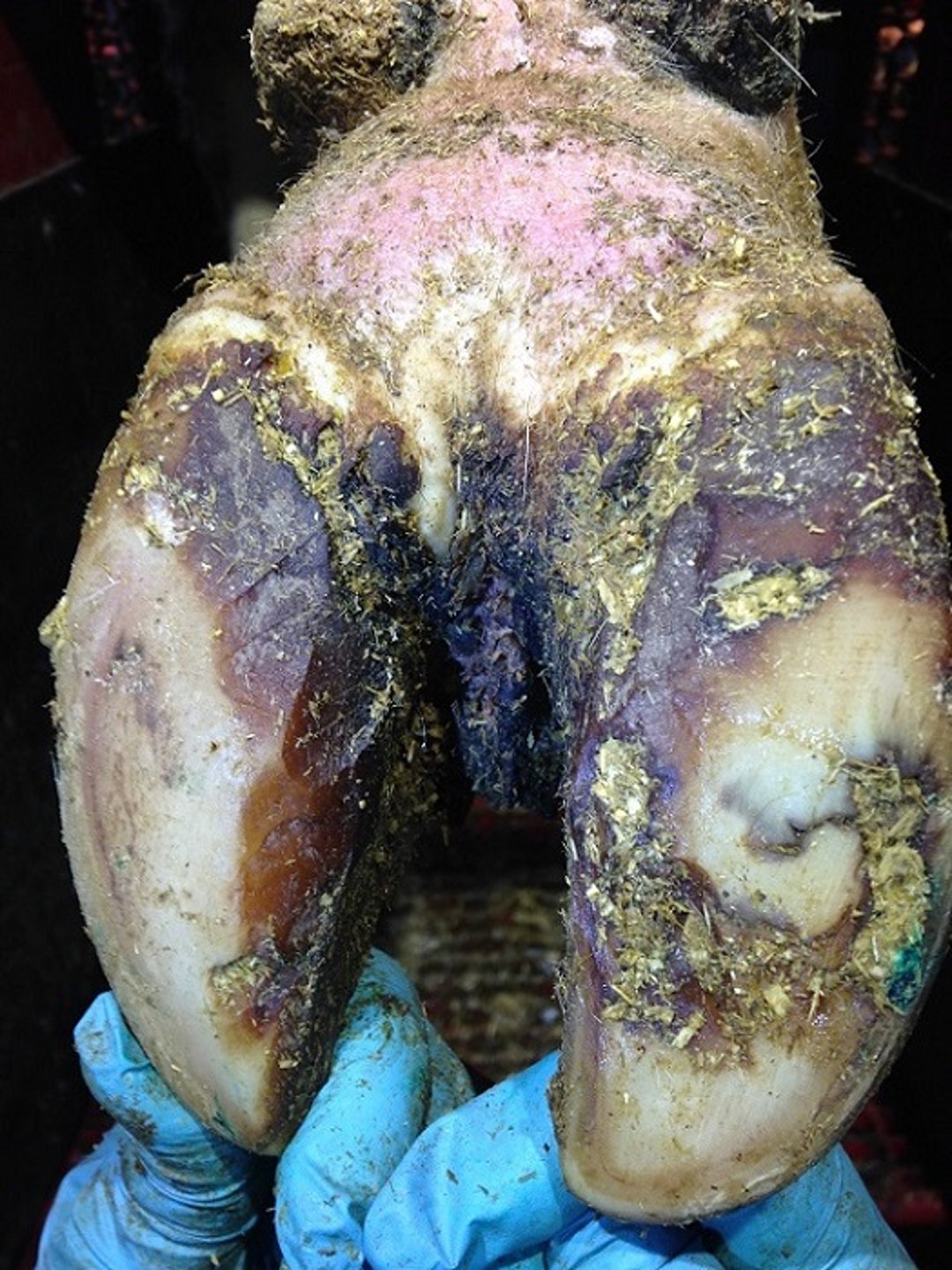
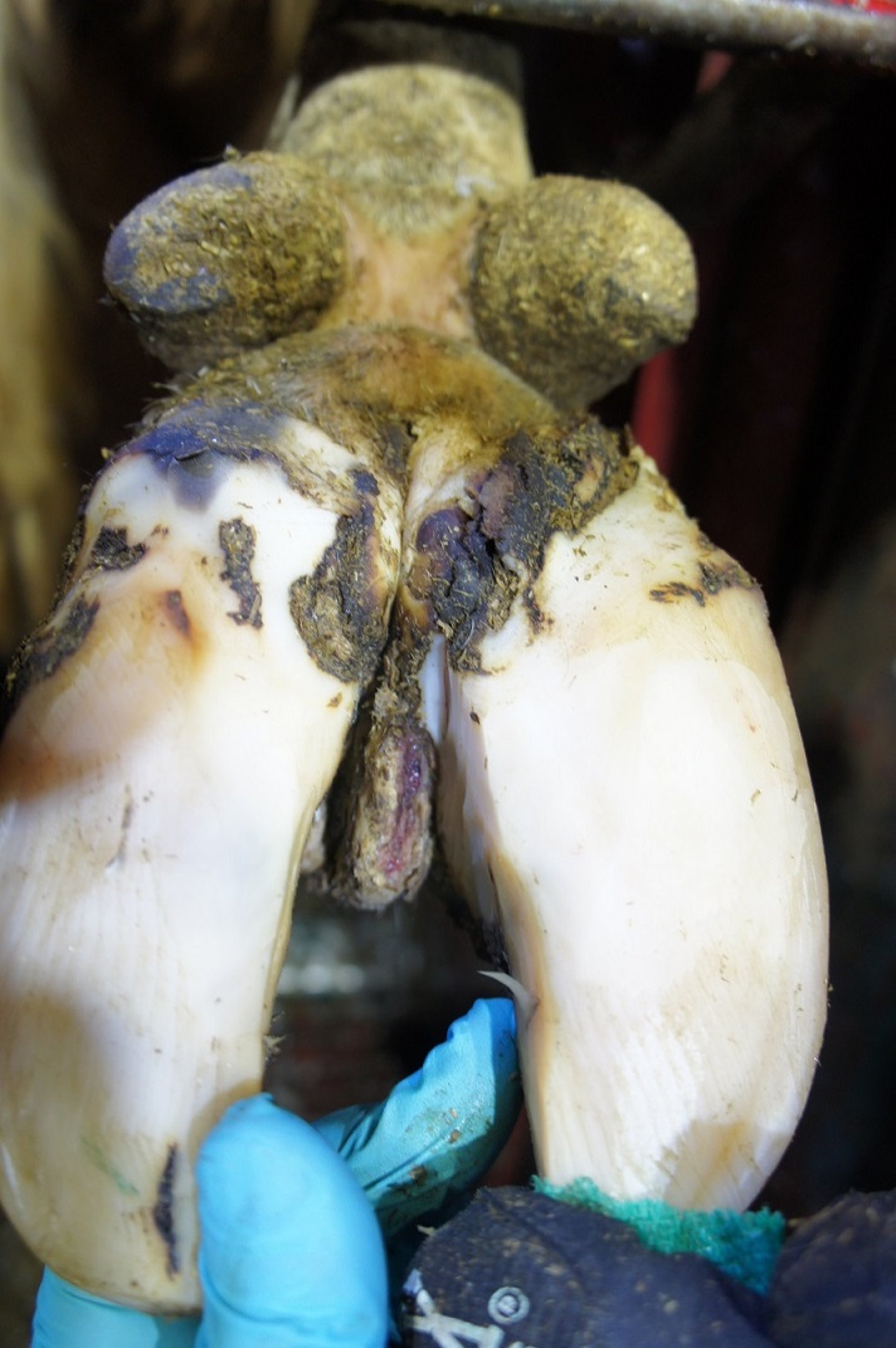
This photograph shows necrosis in the interdigital space of a cow with foot rot.
Courtesy of Dr. Laura Solano.

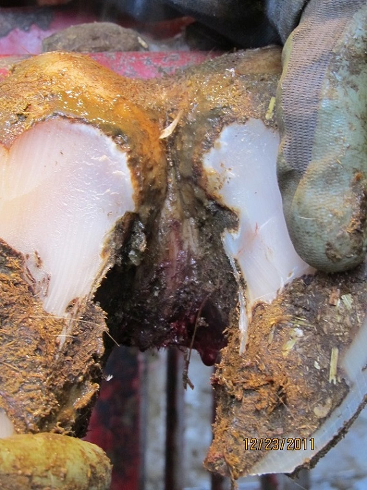
This photograph shows necrosis in the interdigital space of a cow with acute foot rot.
Courtesy of Dr. Gerard Cramer.
Foot rot is a sporadic infection of the soft tissues of the foot in dairy and beef cattle, creating a sudden onset of mild to severe lameness.
Pathogenesis
The most common bacteria associated with foot rot are Fusobacterium necrophorum, Dichelobacter nodosus, Trueperella pyogenes, Porphyromonas levii, and Prevotella intermedia. They are all gram-negative anaerobes that are present in the GI system of cattle and, as a result, are also found in the animal’s environment. These bacteria invade through a breach in the interdigital skin and then work synergistically to cause inflammation and necrosis of the soft tissues in the lower leg.
Diagnosis
Foot rot is recognized by the sudden onset of lameness, accompanied by symmetrical swelling of the lower leg above the hoof. It can present in an outbreak at herd level as a result of the sudden exposure to a risk factor for the disease. Depending on the stage of the disease, the interdigital skin splits open, and a foul-smelling discharge is noticeable. In more severe cases, loose pieces of necrotic tissue can be easily removed from the interdigital space.
Prevention
The key focus to prevent foot rot is to prevent skin damage, thereby removing the opportunity for bacterial infection. Skin damage, typically of mechanical origin, results from contact with rocks, sharp edges, cables, and other hazardous objects or protrusions in the animal's housing environment. Skin damage can also result from chronic wetting of the foot in muddy or wet and dirty environments. On dairy farms, the use of foot baths with a range of disinfectants is an aid to clean and disinfect the interdigital skin. Currently, no pharmaceutical products are labeled with a claim to prevent foot rot.
Treatment
Foot rot should be treated with systemic antimicrobials according to label directions. There is typically no need to remove necrotic tissue or apply bandages. Treated animals should visually improve within 2–3 days. If animals do not respond, the diagnosis should be reevaluated. In severe cases, the infection can extend to tendons and joints, resulting in very severe lameness that is unresponsive to regular systemic antimicrobial treatment.
Heel Horn Erosion and Interdigital Dermatitis in Cattle

This photograph of a cow's foot shows the typical V-shaped cracks that occur with heel horn erosion.
Courtesy of Dr. Gerard Cramer.
Interdigital dermatitis is an infectious and contagious bacterial infection that results in erosion of the heel bulbs or a mild dermatitis (different from digital dermatitis) that typically does not cause lameness. Heel horn erosion is part of the interdigital dermatitis disease complex.
Pathogenesis
The most common bacterium associated with heel horn erosion and interdigital dermatitis is Dichelobacter nodosus, a gram-negative, anaerobic, commensal, and opportunistic bacterium. Typically, the bacteria invade skin and horn that have been damaged by wet and dirty environments. The bacteria then create a cycle of further damage that is due to their virulence factors and the body’s response of excessive horn growth.
Diagnosis
Heel horn erosion and interdigital dermatitis are common findings at routine hoof trimmings. Heel horn erosion typically presents as superficial damage to the non-weight-bearing heel horn that causes undermining and grooves in a V-like pattern on both heels. Interdigital dermatitis presents as pale and damaged skin in the interdigital space. Typically, lameness results only if the erosion extends to the level of the corium.
Prevention
Prevention of heel horn erosion should focus on avoiding prolonged exposure to wet and dirty environments. As with other infectious hoof lesions, using foot baths regularly and appropriately to clean and disinfect hooves is a preventive measure in housed dairy cows.
Treatment
Heel horn erosion and interdigital dermatitis are treated during the trimming process mainly by the removal of loose horn from the non-weight-bearing surface of the heel. Anaerobic pockets in the grooves of the heel horn are removed at the same time, eliminating the environment that supports the growth of the causative bacteria.
Noninfectious Hoof Lesions
Sole Hemorrhages in Cattle
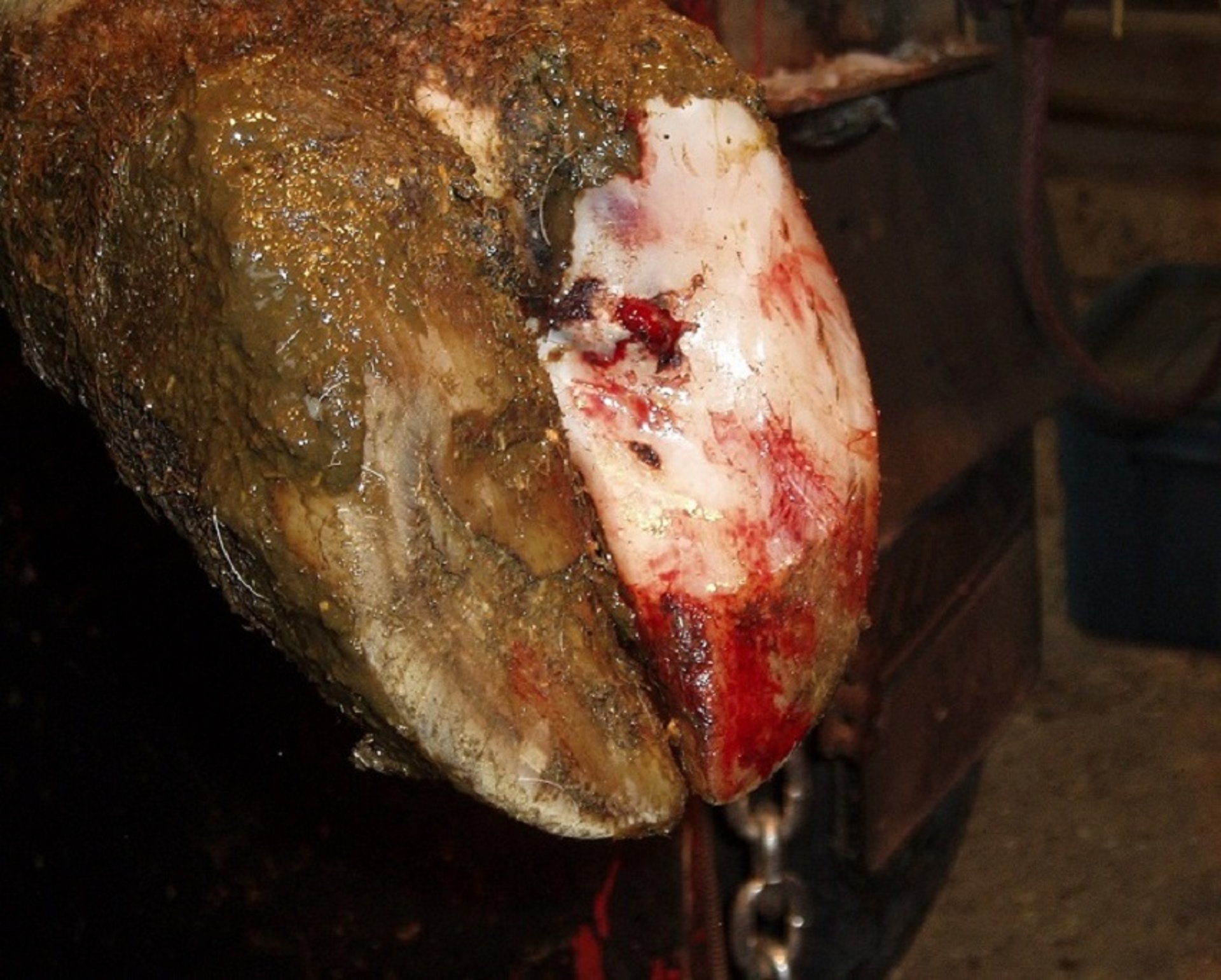
Sole hemorrhages typically occur beneath the flexor tuberosity of the third phalanx (P3) or along the white line; they can also occur on any part of the weight-bearing surface of the hoof. The lameness associated with a sole hemorrhage varies in relation to the time since onset and the size of the hemorrhage. Sole hemorrhages should be thought of as precursors to more severe hoof lesions, such as sole ulcers (below).
Pathogenesis
Sole hemorrhages result from the pressure of P3 on the corium. This pressure is a consequence of changes in the suspending and supporting structures of that are due to mechanical and or metabolic processes. As a result of the pressure, the corium leaks blood into keratinocytes at the dermal-epidermal interface. The timing and amount of pressure determine the amount of hemorrhage visible on the hoof. Extended periods of pressure lead to more severe hoof lesions, such as sole ulcers and white line disease.
Diagnosis

This photograph shows a recent, ongoing, painful sole hemorrhage in a cow.
Courtesy of Dr. Gerard Cramer.

This photograph shows an experimentally induced chronic sole hemorrhage in a cow, that was nonresponsive to hoof testers.
Courtesy of Dr. Gerard Cramer.
Sole hemorrhages present as red, yellow, blue, and/or purple discoloration of the hoof horn. Cows with a sole hemorrhage may or may not show clinical signs of lameness at the time of diagnosis. This inconsistency in lameness is due to the inherent lag between the development of a sole hemorrhage and the visibility of this hemorrhage on the sole surface. Sole hemorrhages can be an incidental finding at the time of hoof trimming. In addition, cows with a history of lameness may present with sole hemorrhage at the site of their previous lesions.
Prevention
Sole hemorrhage prevention focuses on minimizing the pressure on the corium by decreasing standing time, minimizing negative energy balance, and maintaining appropriate and strategic hoof trimming. More details are described under Sole Ulcers below.
Treatment
The treatment of sole hemorrhages depends on the size of the hemorrhage and the amount of pain associated with it. Small, nonpainful hemorrhages can be treated simply by decreasing pressure on the affected hoof by lowering the heel or placement of a thin hoof block. A large sole hemorrhage that persists throughout the hoof-trimming process and produces a withdrawal response with hoof testers, indicating pain, should be treated like a sole ulcer; ie, with a hoof block and NSAIDS.
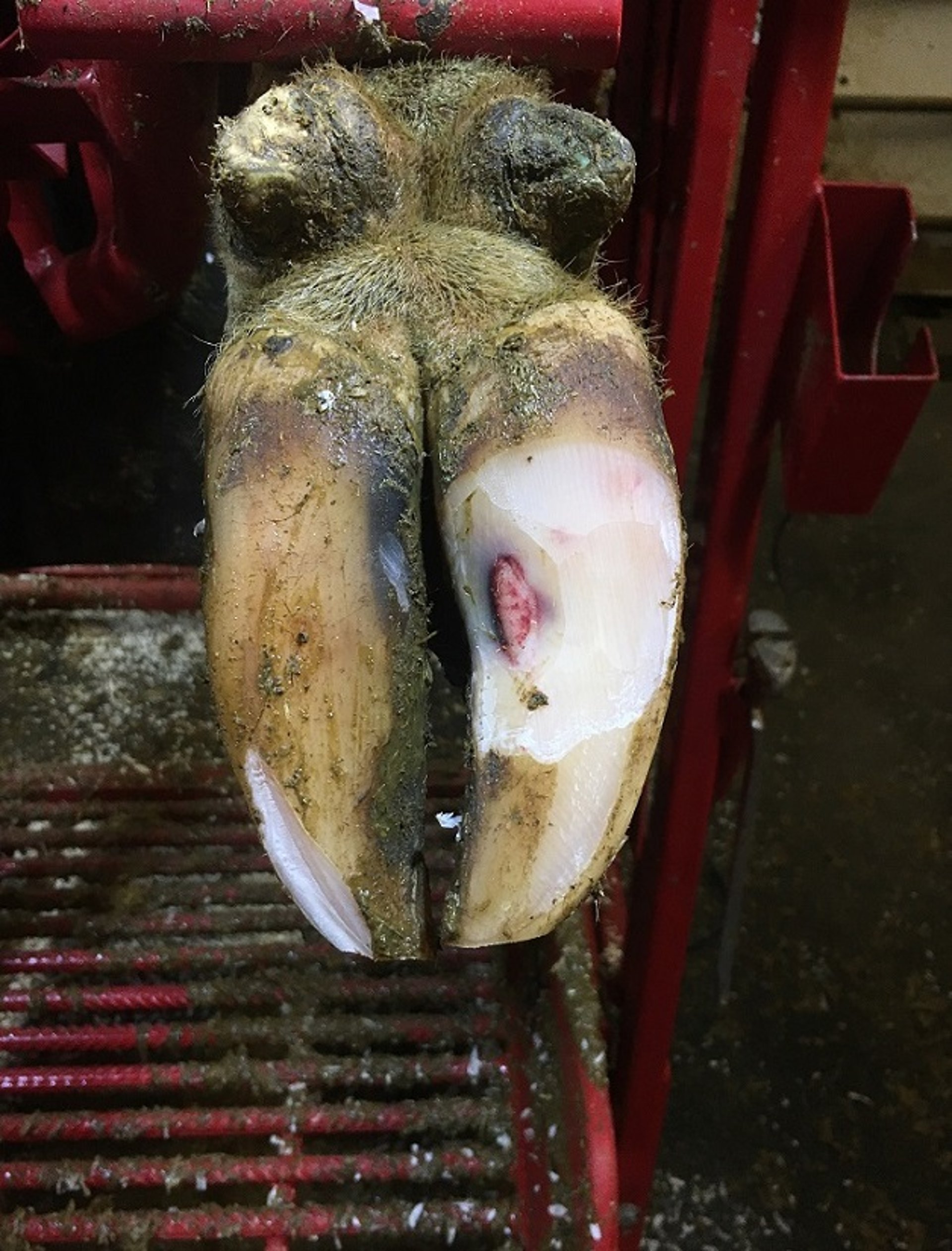
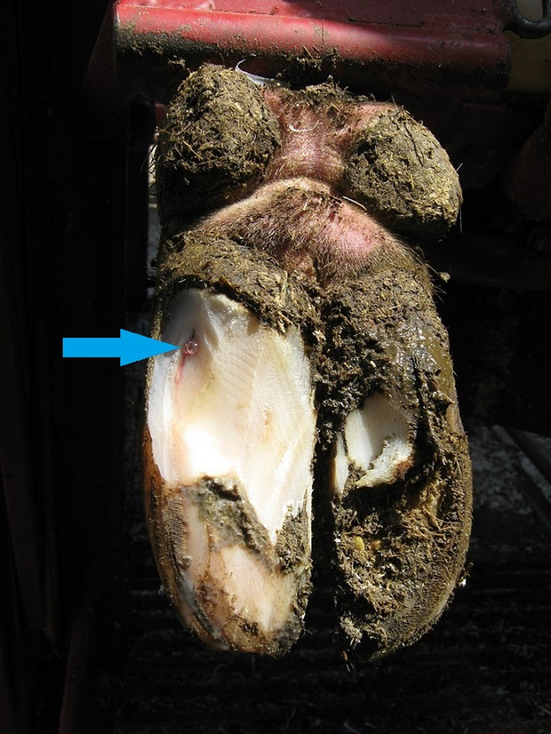
Sole Ulcers in Cattle
Sole ulcers are damaged or penetrated areas of the sole horn due to increased pressure and repeated compression of the corium. They commonly occur beneath the flexor tuberosity of P3. They are associated with varying amounts of pain and resulting changes in weight bearing.
Pathogenesis
In their early stages, sole ulcers and sole hemorrhages share a common pathogenesis. Sole ulcers develop as a result of continuous pressure by the flexor tuberosity of P3 on the corium. This pressure, triggered by movement and sinking of P3, leads to changes and weakening of the suspending and supporting structures of P3 because of mechanical, hormonal, and/or metabolic processes. As a result of this pressure, the corium initially leaks blood into keratinocytes at the dermal-epidermal interface (resulting in the hemorrhage). Over time, the pressure from P3 leads to the destruction of keratinocytes and the interruption of horn growth, causing the corium to protrude through the horn defect. The pressure on the corium also initiates an inflammatory pathway, resulting in longterm structural changes to P3 and the corium.
Diagnosis

All loose horn has been removed from the sole ulcer pictured in this photograph.
Courtesy of Dr. Gerard Cramer.
Typically, sole ulcers are recognized by protrusion of the corium at the ulcer site. The exposed corium can range from a fresh/red to a brown/necrotic appearance. Sole ulcers typically occur in the lateral hoof of the rear legs because it bears more weight compared to the medial hoof.
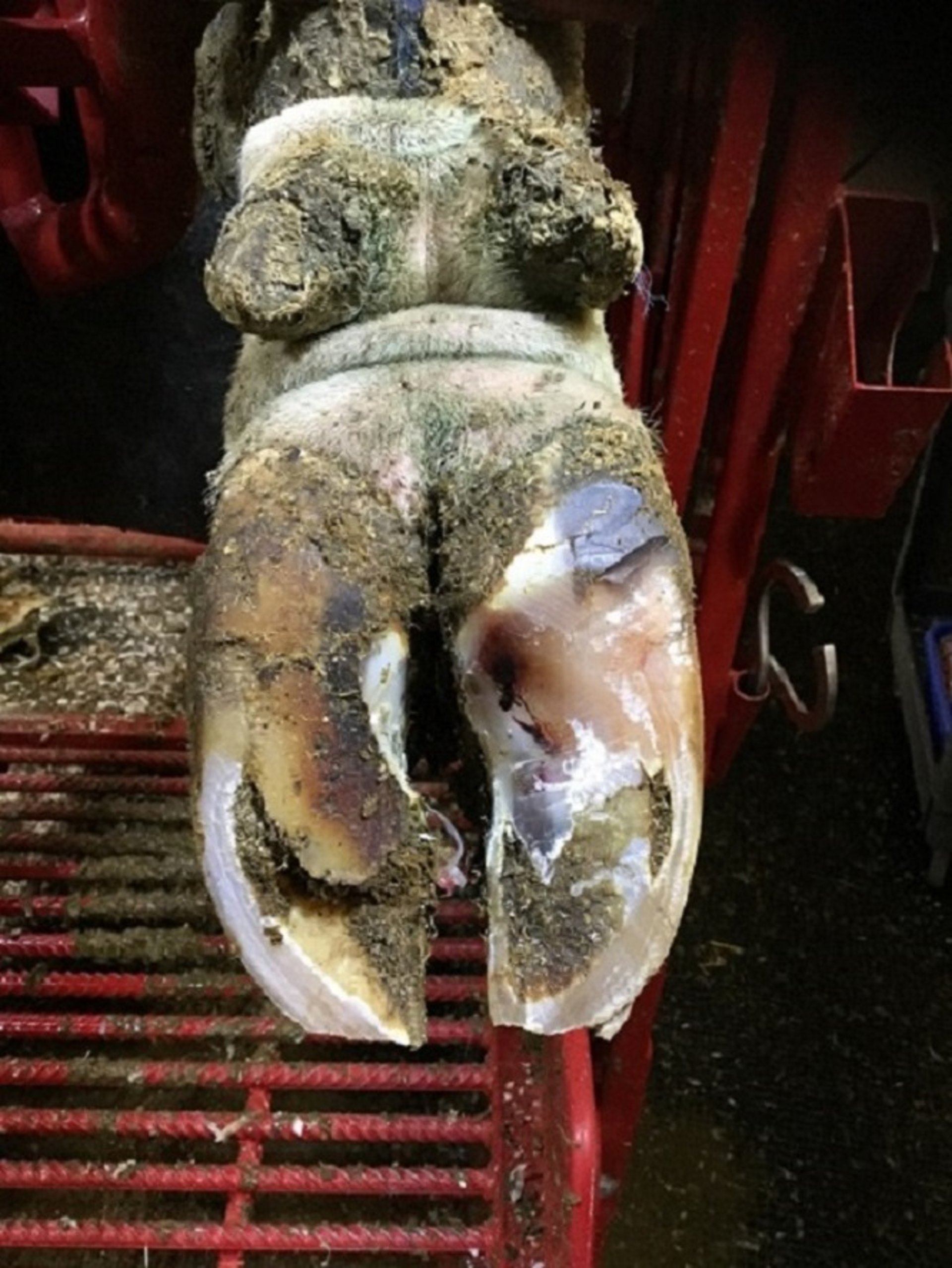
Prevention

This photograph of the left front foot of a cow shows how the hoof can be trimmed with a deep model to prevent sole ulcers.
Courtesy of Dr. Gerard Cramer.
As with sole hemorrhages, the focus in preventing sole ulcers is to minimize pressure on the corium by decreasing the amount of time the animal stands on hard surfaces and to minimize negative energy balance and associated inflammatory processes. To ensure an adequate lying time of 12–14 hours per day, cows should not be away from their resting area for more than 3–4 hours per day. The comfort and design of the lying surface, stocking density, and heat stress management, along with strategies to minimize problems during the transition period, all have an important role in decreasing the amount of time standing. Another strategy to prevent prolonged standing time in animals at risk is to ensure that first-lactation cows are allowed to become accustomed to adult cow housing at least 60 days before calving. Finally, appropriately timed and correctly performed hoof trimming to balance weight distribution between hooves and decrease pressure on the common sole ulcer site is a key component of a sole ulcer and sole hemorrhage prevention program.
Treatment

Photograph of a polyurethane hoof block applied to the contralateral hoof of a cow with a sole ulcer to reduce pressure on the affected hoof.
Courtesy of Dr. Gerard Cramer.
Sole ulceration results in chronic changes and is a painful condition. Appropriate, early treatment is critical to the successful resolution of clinical signs and to minimizing the impact of longterm changes. The treatment of sole ulcers is to remove all loose horn around the affected corium. This removal should be handled delicately, with great care taken to minimize further damage to the corium. Once the loose horn has been removed around the lesion, pressure on the ulcer site should be decreased to maximize the speed of horn growth. The pressure is reduced by removal of the horn around the lesion and by application of a properly sized hoof block to transfer weight to the sound hoof. Cows with sole ulcers should be rechecked in 3–6 weeks to assess healing and to either remove or reposition the block, if necessary. The administration of an NSAID in early sole ulcer cases should be considered to counteract the inflammatory changes and to lower the risk of new bone development in P3 that could cause irreversible anatomical damage.
White Line Disease in Cattle
The term white line disease encompasses a range of lesions (hemorrhages, fissures, separations, abscesses) that occur in the white line region.
Pathogenesis
The exact cause of white line lesions is unclear. The white line is made up of three different types of horn, all of which are weaker than the wall and sole horn. Currently, the pathogenesis of white line disease is thought to be similar to that described for sole ulcers, in which P3’s suspensory mechanisms are compromised, resulting in damage to the keratinocytes that grow white line horn. Internal and external traumatic forces generate a shearing action at the white line that can cause hoof wall separation. These gaps can allow the entry of bacteria and foreign bodies, resulting in damage to the corium and inflammatory changes to P3.
Diagnosis

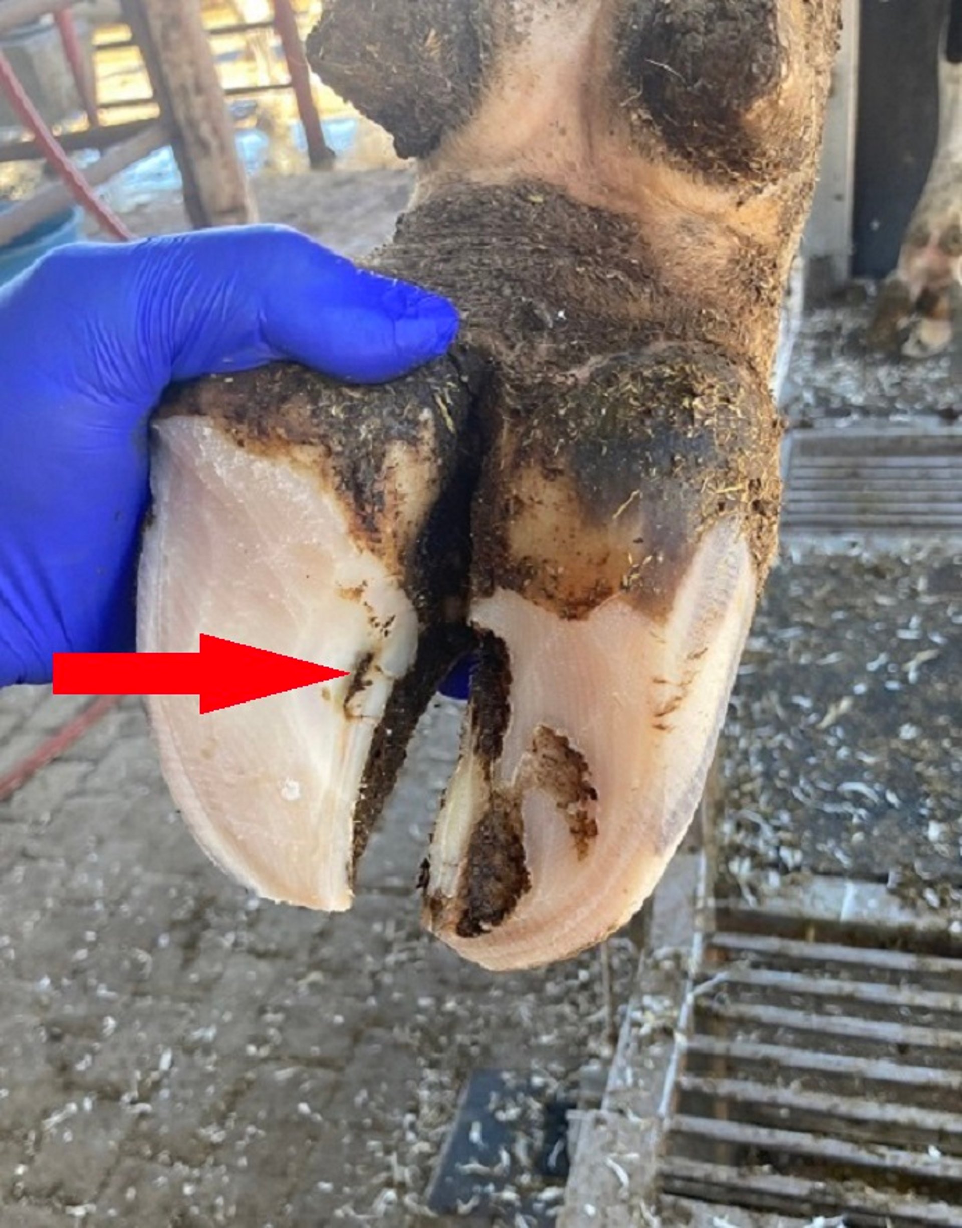
This photograph, taken before the removal of loose horn, shows the swelling and opening of the drainage tract (arrow) associated with white line disease in the lateral left hind hoof of a cow.
Courtesy of Dr. Gerard Cramer.
White line lesions can present as a hemorrhage, a separation, or an abscess. Not all white line lesions are painful when tested with hoof testers. Painful lesions can present as minor white line separation at the sole level; however, this separation can lead to an abscess or a draining tract that extends up to the coronary band or bulb of the heel. The presence of an abscess depends on the timing of the foot evaluation relative to the course of disease. The typical location of white line lesions is the abaxial white line of the heel area on the lateral hooves of the rear feet; however, similar types of lesions can be found in different white line regions of the hooves and should be treated similarly.
Prevention
White line disease can be prevented by minimizing forces exerted on the white line that result in its separation or damage. The forces can be minimized by decreasing the risk of trauma to the white line with proper flooring and proper cattle handling. Thus, prevention starts with providing optimal walking surfaces, ensuring that floors have good traction, are not abrasive, and are not slippery. Proper walking surfaces vary depending on the housing system and bedding material used. Key preventive practices include the strategic use of rubber flooring, proper sizing and spacing of floor grooves, and good maintenance of pasture tracks. Optimal flooring should be combined with proper cattle handling, allowing cows to walk at their own pace. An additional prevention strategy is to incorporate supplemental minerals (copper or zinc) and biotin in the animals' feed to increase horn strength. Finally, appropriately timed and correctly performed hoof trimming should be a key component of a prevention program.
Treatment
White line disease can appear as an acute and painful condition; however, the severity and extent of separation of the white line varies. Appropriate and early treatment is critical to a successful recovery. The treatment of painful white line lesions is to remove all loose horn around the lesion, including the wall. As with sole ulcers, this horn removal should be managed delicately, with great care taken to minimize further damage to the corium. Once the loose horn has been removed from around the lesion, pressure on the lesion should be decreased by thinning the lesion margins and lowering the heel to maximize the speed of horn growth. Pressure on the lesion is lowered further by the application of a properly sized hoof block to transfer weight to the sound hoof. Cows with white line lesions should be rechecked in 3–6 weeks to assess healing and to either remove or reposition the block, if necessary. As with sole ulcers, the administration of NSAIDs in early white line cases should be considered to counteract the inflammatory changes and associated bony proliferations in P3.
Thin Soles in Cattle
Thin sole lesions occur when the sole horn has worn away enough that the sole flexes with digital pressure but the corium is not exposed.
Pathogenesis
Thin soles can result from overtrimming or from excessive wear as a result of walking on an abrasive surface. Abrasive walking surfaces or bedding materials are typically necessary factors in thin sole development. These factors, combined with long walking distances or aggressive or poor handling practices, lead to slipping and increased wear of the sole. Another predisposing factor to increased wear of the sole is abrupt changes in the animal's walking surfaces, such as going from pasture or dirt lots to concrete flooring.
Diagnosis

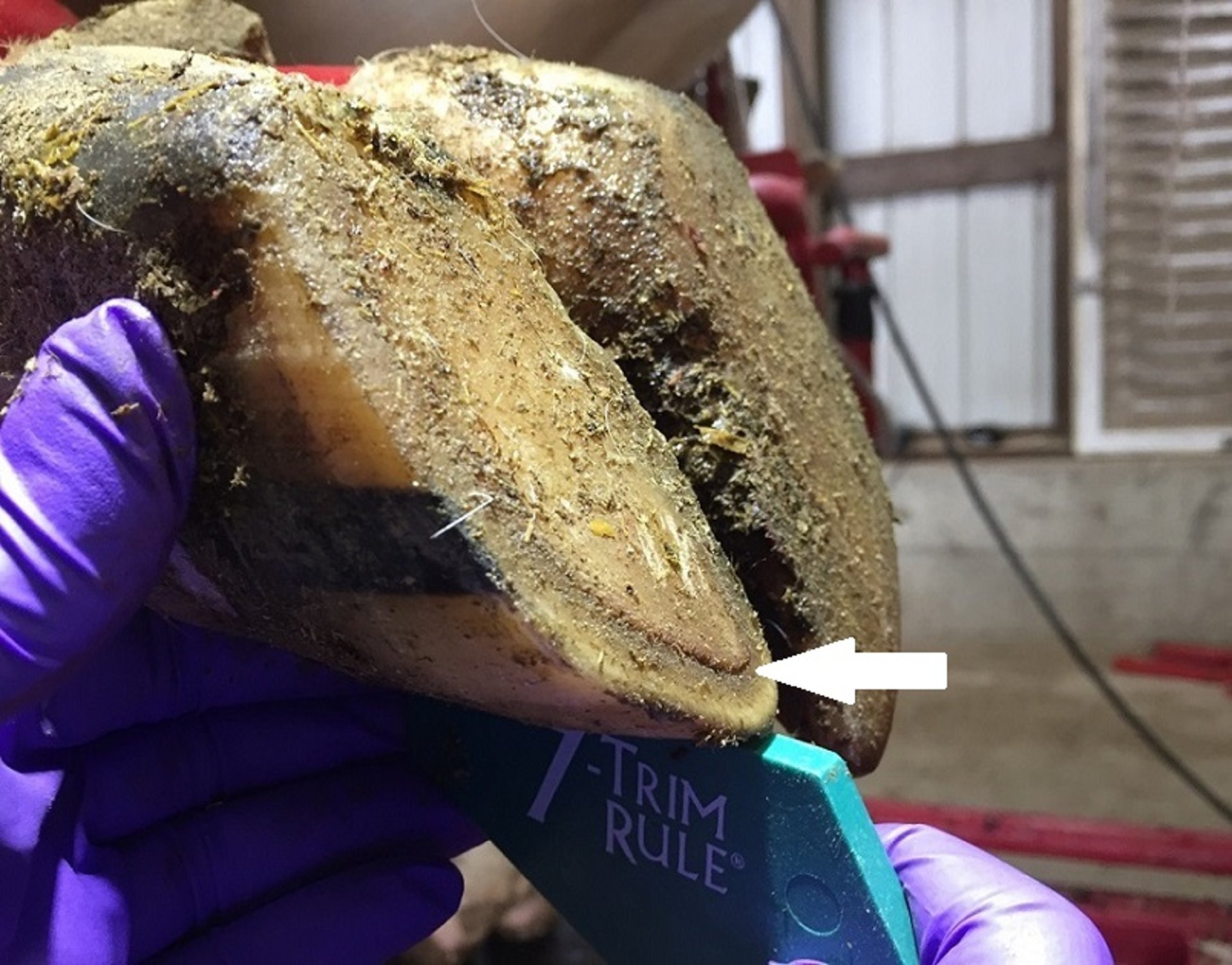
In this photograph of the left front foot of a cow, a thin sole is evident on the lateral hoof, as illustrated by its being shorter than a 7.5-cm gauge and showing bulging (arrow) of the sole compared to the wall at the toe.
Courtesy of Dr. Gerard Cramer.

In adult animals, a thin sole should be suspected when the dorsal wall length is less than 7.5 cm (3 inches). Upon pressure with a finger or hoof tester/knife, the sole will feel flexible and soft. In more severe cases, the hoof tester will evoke a withdrawal response. Typically, the white line areas will appear the thinnest, and the corium will appear to bulge at this location. In cows with thin soles, multiple hooves may be affected, and the animal may walk in a characteristically tender manner.
Prevention
To prevent thin soles in cows, it is important to balance growth and wear. Horn growth will increase in response to excessive wear; however, this process takes time. In environments that produce excessive wear, changes in cattle handling can be the quickest way to decrease thin soles. The use of rubber flooring in strategic areas such as transfer and return lanes, as well as loading and unloading areas, will also decrease wear. In high-wear environments, the use of appropriate and less frequent strategic hoof trimming should be considered. An additional prevention strategy is to incorporate supplemental minerals (copper or zinc) and biotin in the animals' feed to increase horn strength.
Treatment
The goal of treating thin soles is to prevent further wear of the sole and further exposure of the corium. Toward that goal, a thin hoof block is placed on the affected hoof. This thin block prevents further abrasion and enables the affected hoof to grow more horn. Additional treatment strategies include decreasing the walking distance of affected cows and providing a deeply bedded lying area for recovery. Cows with thin soles should be rechecked in 4–6 weeks to remove the block.
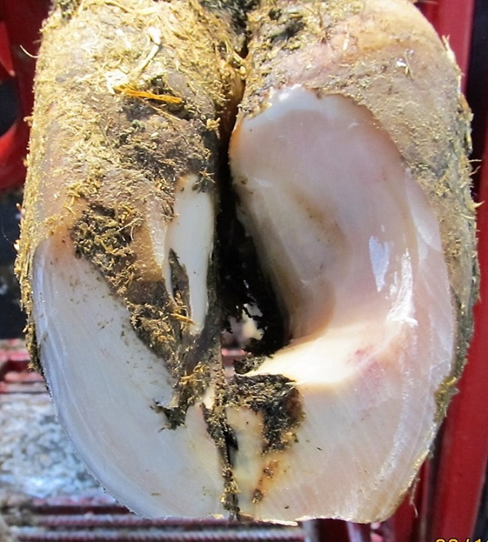
Toe Ulcers and Toe Necrosis in Cattle
Toe ulcers and toe necrosis are sequelae of thin soles that cause severe lameness.
Pathogenesis
The factors that result in toe ulcers and toe necrosis are similar to those that cause thin soles. Together, these toe lesions should be thought of as a continuum. Toe ulcers and toe necrosis develop because thin soles go undiagnosed or because the abrasion is so excessive that the thin-sole stage is very short. Once the corium is exposed, bacteria have an entry point and can proliferate. Furthermore, an inflammatory process develops as a result of bacterial invasion or the original traumatic process. Both the bacterial and inflammatory processes result in widespread damage to the corium that can eventually extend to parts of P3.
Diagnosis

The sagittal section of a cow hoof pictured in this photograph shows toe necrosis—in particular, extensive damage to the third phalanx and to the surrounding corium.
Courtesy of Dr. Gerard Cramer.
Cattle with toe ulcers or toe necrosis present with a characteristic lameness in which they attempt to shift weight toward the heel. In hoof evaluation of cows with toe ulcers, the corium is exposed at the toe. If the lesion has progressed to toe necrosis, the corium will show extensive damage, with darkened and necrotic tissue surrounding P3. If left untreated, this process will extend to P3 as well.
Prevention
As with thin soles, prevention of toe ulcers and toe necrosis focuses primarily on minimizing wear and trauma to the sole. Early identification and treatment of thin soles can also prevent the progression to toe ulcers and toe necrosis.
Treatment
Treatment principles for toe ulcers and toe necrosis are similar to those for sole ulcers and white line lesions. For toe ulcers, the primary focus should be to remove all loose horn surrounding the lesion and to apply a hoof block. In most cases, adding two blocks to produce more lift at the toe will facilitate healing. The treatment of toe necrosis is more involved because it requires, in addition to application of the hoof block, removal of the infected bone of P3 and soft tissue. A partial resection of P3 is typically warranted. Toe ulcers and necrosis should be treated by using intravenous regional anesthesia, administering follow-up NSAIDs, and providing a deeply bedded lying area for recovery. The patient should be rechecked after 2–4 weeks to ensure proper healing and block positioning.
Corkscrew Claw in Cattle
A corkscrew claw is a structural abnormality of the phalanges that occurs in both beef and dairy cattle. Historically, it occurred on the lateral hind hooves of older animals. but is now reported in medial hooves of both the front and back feet of younger animals as well.
Pathogenesis

This photograph of the right hind leg of a cow shows a corkscrew of the medial hoof. Note the excessive curvature of the dorsal wall away from the midline.
Courtesy of Dr. Gerard Cramer.

This photograph of a transverse section of both hooves of the right hind leg of a cow shows a corkscrew on the medial hoof, illustrating the rotation and changes in the third phalanx.
Courtesy of Dr. Gerard Cramer.
The exact cause of corkscrew claws is unknown. Historically, the presentation in older animals was thought to have both genetic and environmental components. The newer development of corkscrew syndrome in younger animals seems to have more of an environmental component, because it appears to occur in specific situations. The anatomical changes present in corkscrew claws—such as rotation, bone remodeling, and narrowing of P3—suggest that specific factors experienced during an animal’s development lead to these changes. It is currently unclear whether the changes in P3 are the cause of external changes or the external changes occur first, followed by changes in P3. Breed and growth rate have been associated with the development of corkscrew claws. Similarly, raising young animals in an environment that has lots of traction (eg, sand bedding), as well as competition or infrequent feed delivery in the feed bunk area, appears to increase the prevalence of corkscrew claw. All of these risk factors suggest that the pathogenesis is due to excessive rotational forces on P3 during periods of increased laxity in ligaments and tendons.
Diagnosis
Lameness associated with corkscrew claw is variable and depends on the size of the animal, extent of the hoof rotation, and abrasiveness of the walking surfaces. Upon evaluation, the affected hoof will appear longer and narrower, with an inward and upward spiral rotation creating a corkscrew appearance. The sole of a corkscrew claw typically shows excessive growth under the flexor tuberosity and curling over of the abaxial wall if the animal is not walking in an abrasive environment. If the animal is housed in an abrasive environment, the affected hoof will likely be larger, with hemorrhaging at the white line near the toe, and the opposite hoof is likely to have a thin sole.
Prevention
The best way to decrease the incidence of corkscrew claw is to modify management practices, including ensuring that rapidly growing animals have access to resources without excessive competition and without exposure to abrasive flooring surfaces. Additional factors include proper bunk management to ensure that all animals can access feed at once or that feed is always available. Because corkscrew claw may have a genetic component, it is important to select replacement animals from sires that lack this trait.
Treatment

This photograph shows excessive modeling and maintenance of the axial wall in the toe region to provide stability to a cow with a corkscrew on the lateral hoof of the right hind leg.
Courtesy of Dr. Gerard Cramer.
Treatment of corkscrew claw focuses on managing external changes on the hoof because the internal changes are permanent. To prevent lameness, animals with corkscrew claws should have their hooves trimmed every 3 months to remove the excess growth under the flexor tuberosity to avoid excessive force on the hoof. Key components of treatment include excessive modeling of the sole to compensate for the excessive growth under the flexor tuberosity and avoiding excessive decrease in the sole thickness of the affected hoof. Excessive horn growth on the dorsal wall can be removed; excess horn on the axial wall, however, should be left in place to ensure the stability of a narrower P3.
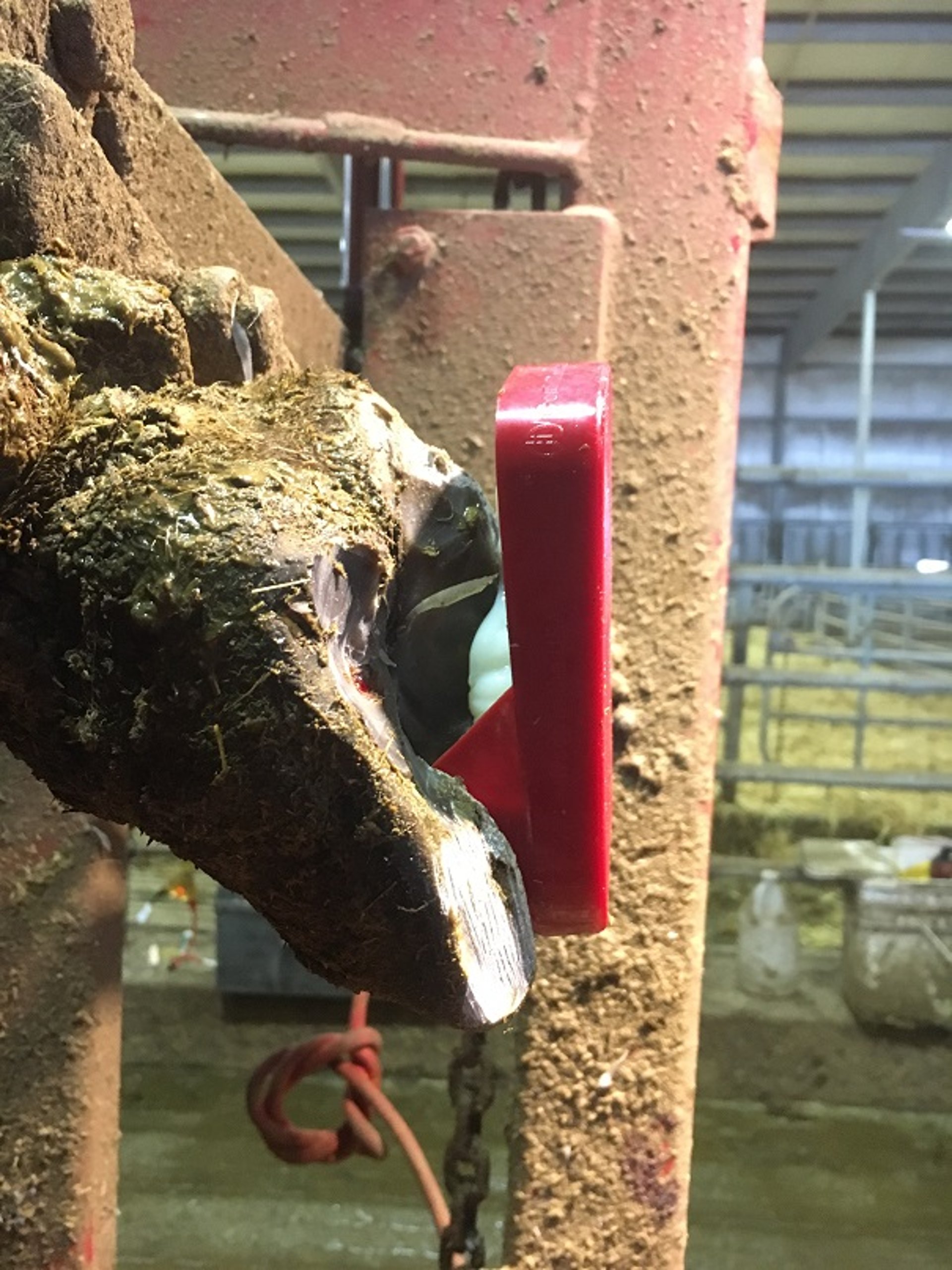
Fissures in Cattle
Fissures form in the walls of hooves in both dairy and beef cattle and may cause lameness if they extend to the corium. Fissures are uncommon in most herds. By direction and location, they can be classified into horizontal, vertical, and axial wall fissures.
Pathogenesis
The pathogenesis of axial and vertical fissures is not well understood. Vertical wall fissures are more common in beef cattle housed in hot, dry, and sandy climates. Vitamin and mineral deficiencies are thought to be contributing factors because of their role in hoof horn development and integrity. Axial wall fissures are even less researched, although some association with digital dermatitis has been reported anecdotally. Horizontal fissures occur when a variety of severe metabolic or physiological events disrupt horn production. Because of the multiple segments that make up the wall, the grooves do not typically extend to the corium.
Diagnosis

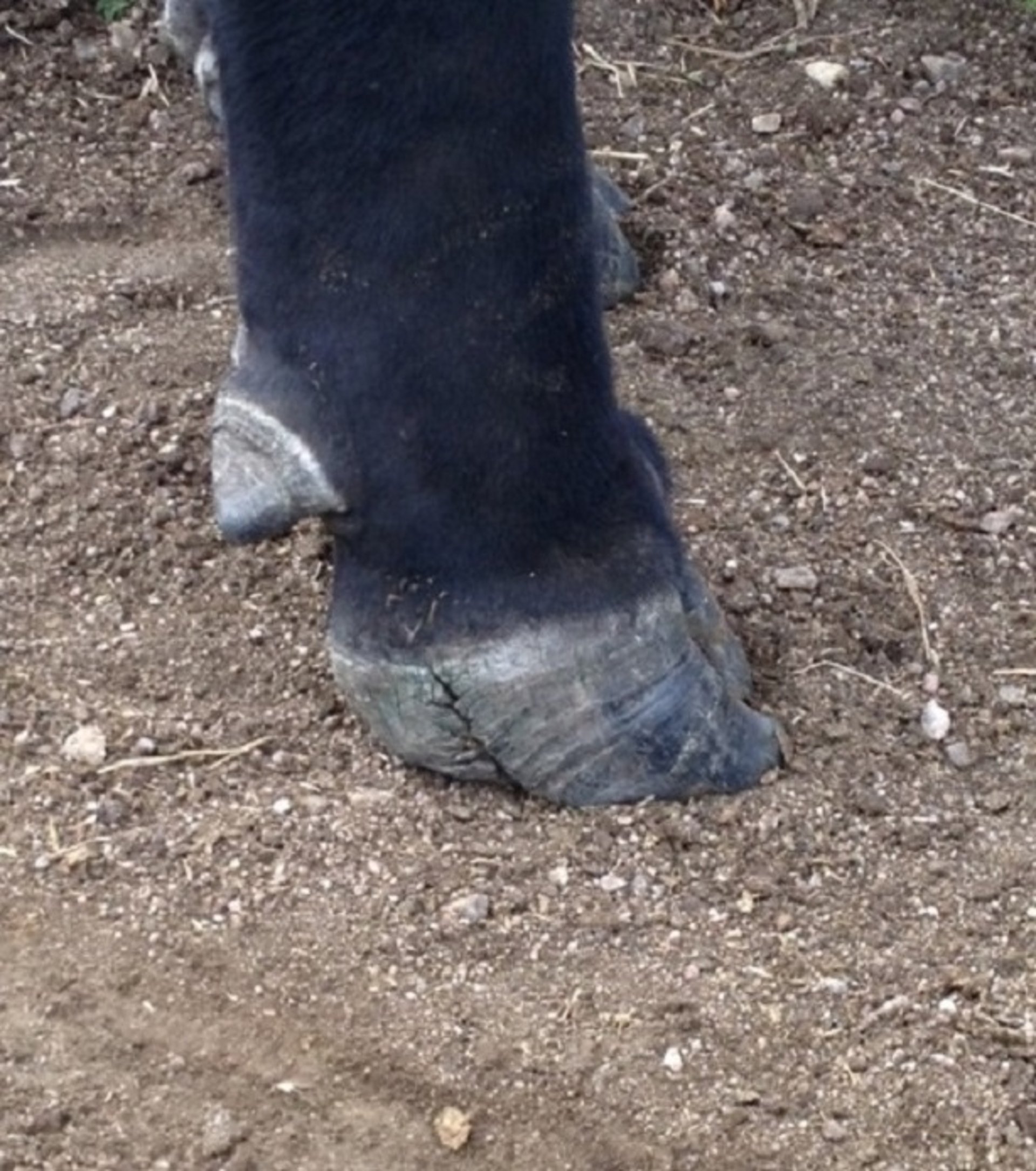
This photograph shows an axial wall fissure (arrow) on the lateral hoof of the left hind leg of a cow.
Courtesy of Dr. Gerard Cramer.

This photograph shows a vertical wall fissure on the right hind lateral hoof in a large beef bull.
Courtesy of Dr. Richard Touret.
Horizontal and vertical fissures occur on the dorsal wall and can be identified without picking up the cow's foot. Vertical wall fissures are more common in the front feet of beef cattle on pasture and can originate from various locations, including the coronary band, toe, or middle of the wall. Horizontal wall cracks typically occur on multiple hooves on the same animal at approximately the same location. When the horizontal wall crack approaches the toe area, it is likely to become separated from the underlying wall. Axial wall cracks extend from the sole to the coronary band on the axial wall.
Prevention
Because there is no clear pathogenesis for fissures, there are also no clear preventive strategies, other than ensuring optimal vitamin and mineral supplementation. This is especially true in regions with known mineral deficiencies in the soil. Prevention of horizontal wall cracks should focus on preventing severe metabolic events such as toxic mastitis or other off-feed events.
Treatment

This photograph of a cow with a severe axial fissure in its hoof shows that all loose horn has been removed and a block has been applied to treat the fissure.
Courtesy of Dr. Gerard Cramer.
Most fissures do not cause lameness or warrant treatment. Treatment is warranted only if the lesion extends to the corium or the coronary band. Treatment of any lameness-causing fissure should focus on removing the loose horn and thinning the margins around the fissure. The use of hoof blocks for vertical and axial wall fissures will improve healing by decreasing the forces on the wall as it regrows. Once a horizontal wall fissure reaches the toe area, the distal horn can become a pinch point when the cow pushes off in her stride; therefore, removal of loose horn at this point is warranted to prevent damaging the underlying horn and corium.
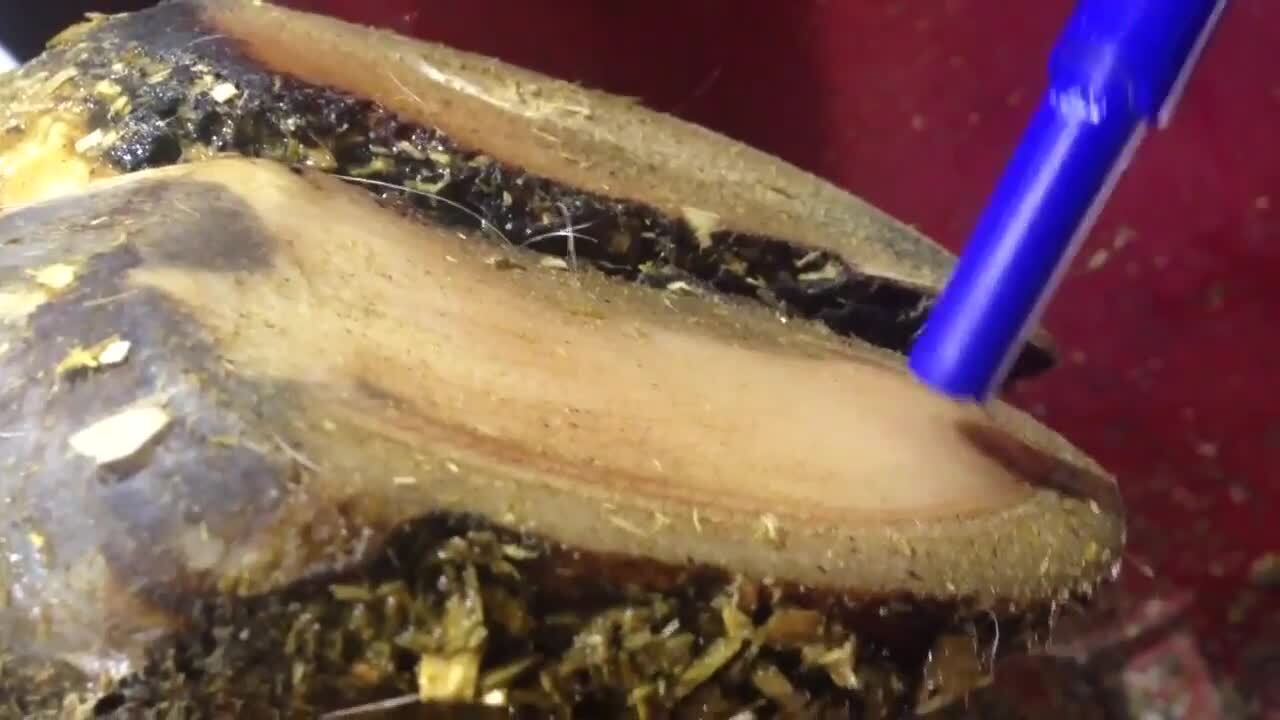
Interdigital Hyperplasia in Cattle
Interdigital hyperplasia is the growth of fibrous tissue in the interdigital space of beef and dairy cattle.
Pathogenesis
The pathogenesis of interdigital hyperplasia is thought to be multifactorial, but it is not well understood. Chronic interdigital skin irritation is the primary factor; factors that contribute to this irritation are thought to include poor hygiene, hindered walking surfaces, and other infectious hoof lesions. In dairy herds, hyperplasia appears to be related to other infectious hoof lesions, such as foot rot and digital dermatitis. The irritation and bacteria that cause infectious hoof lesions probably also can cause hyperplasia of the skin cells. Another relevant factor is the strain exerted on interdigital ligaments by inappropriate hoof-trimming practices, abnormal conformation, or unstable walking surfaces; this strain is believed to predispose to interdigital hyperplasia.
Diagnosis

This photograph of the left hind foot of a cow shows interdigital hyperplasia that is affected by digital dermatitis.
Courtesy of Dr. Laura Solano.
The growth may be evident in various locations in the interdigital space initially, but as the mass grows, it will protrude dorsally. Texture of the hyperplasia will vary depending on the extent of irritation and the foot-bathing practices used. Irritated growths can become affected with digital dermatitis.
Prevention
Preventive practices for interdigital hyperplasia are similar to those for infectious hoof lesions; they include foot bathing and ensuring hygienic environmental conditions. To minimize strain on the interdigital ligaments, appropriate hoof-trimming practices and environments with stable footing are important.
Treatment
Treatment of interdigital hyperplasia varies depending on the severity. Small lesions may not result in lameness nor require treatment, as long as the axial wall is not rubbing on the lesion. All interdigital hyperplasia lesions should be evaluated for concurrent digital dermatitis lesions and treated appropriately if present. Larger lesions that cause lameness and are unresponsive to conservative treatment may require surgical removal; however, care should be taken to dissect the fat pad in the interdigital space and avoid damage to the interdigital ligaments.
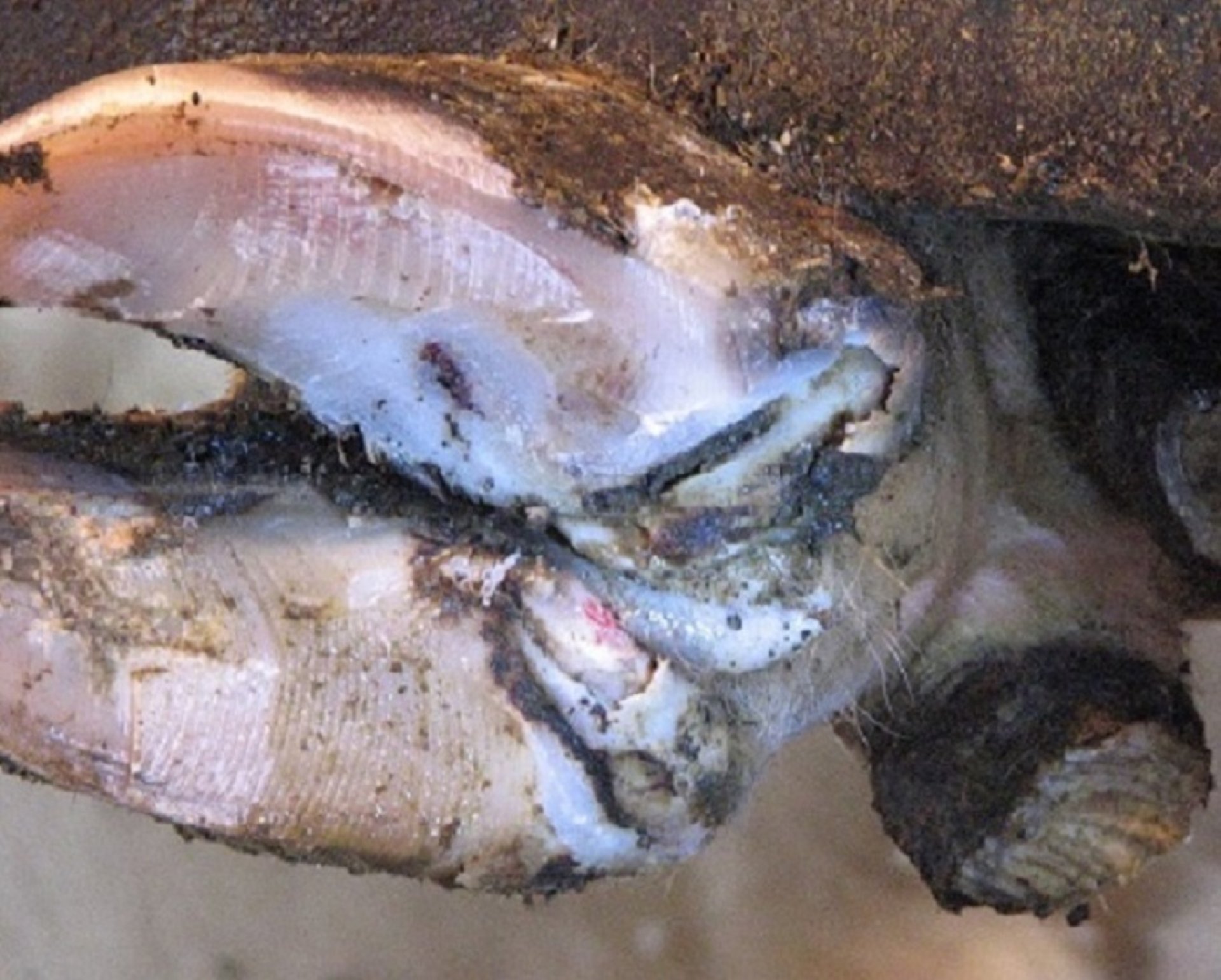
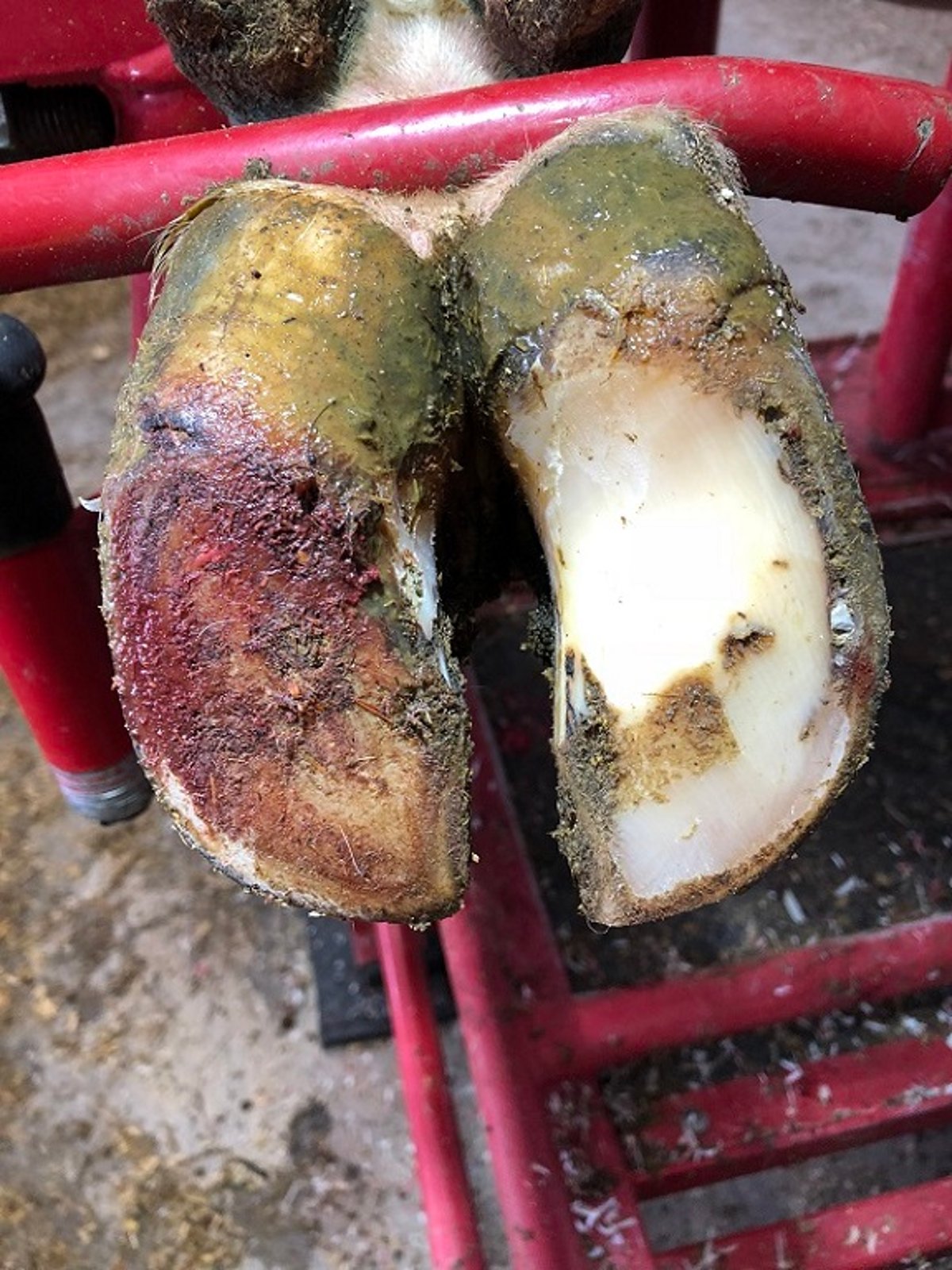
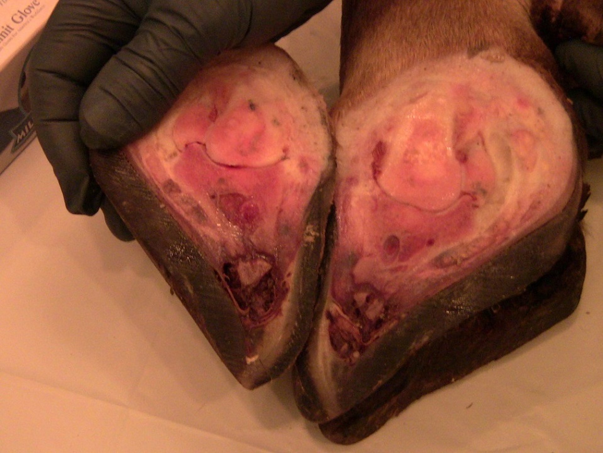
Deep Digital Sepsis in Cattle
Deep digital sepsis is an infection of the foot that involves the deeper structures, typically including the distal interphalangeal joint and flexor tendons. Deep sepsis can be an infrequent sequela to lesions such as foot rot, sole ulcers, and white line disease.
Pathogenesis

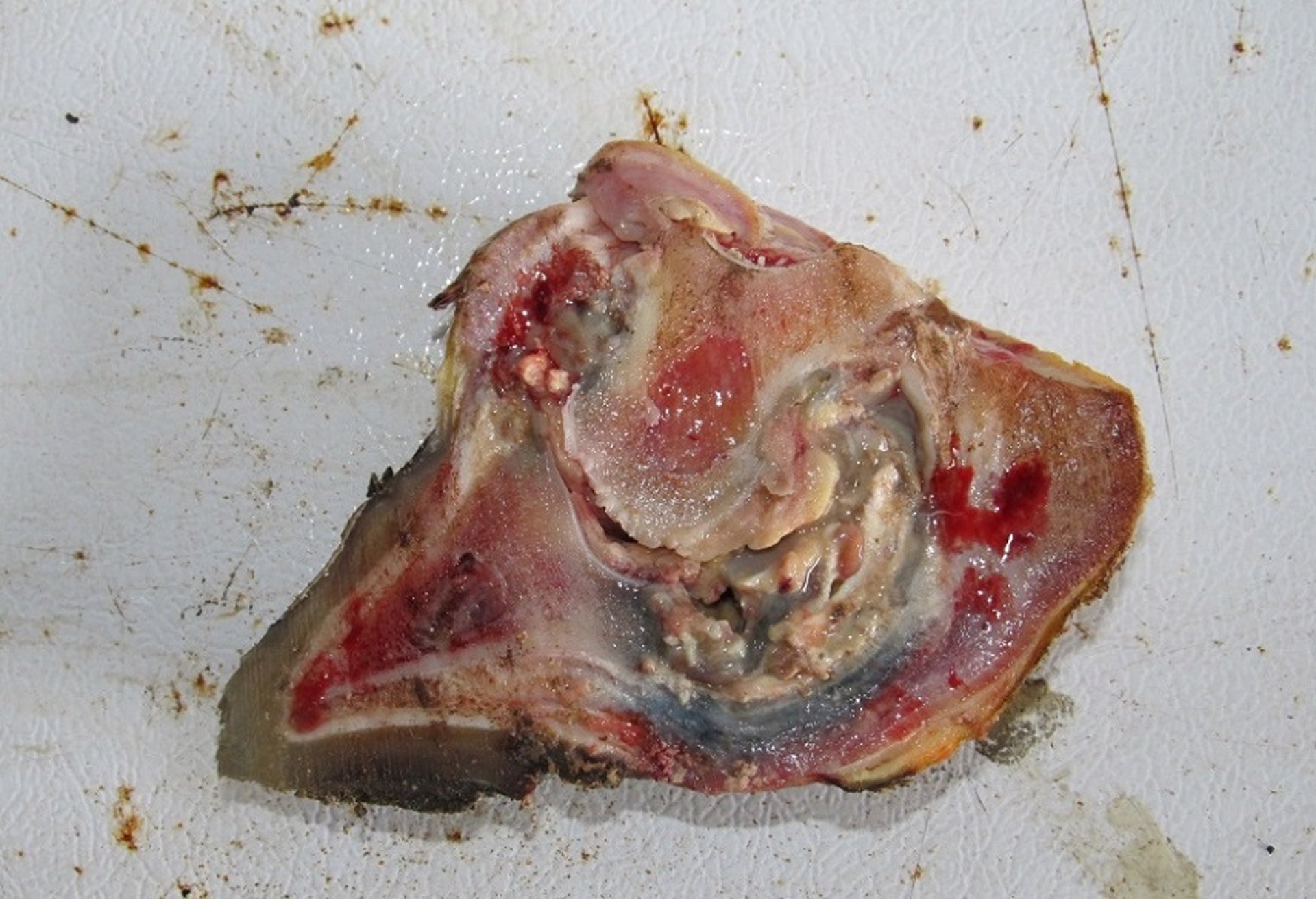
The sagittal section of the hoof of a cow affected with deep sepsis shows extensive necrosis and damage to the third phalanx, the deep digital flexor, the navicular bone, and the P2-P3 joint space.
Courtesy of Dr. Gerard Cramer.
Deep digital sepsis is a sequela of other hoof lesions, such as foot rot, sole ulcers, white line disease, and more rarely, puncture wounds. Delayed or inappropriate treatment of these lesions can increase the occurrence of deep sepsis. Deep digital sepsis occurs after the inflammation, bacterial contamination, and necrosis associated with these original hoof lesions have reached the distal interphalangeal joint, the deep flexor tendon, or other deeper structures, such as the navicular bursa. At this stage, normal treatments for the original lesions are infective, and the animal responds with a localized inflammatory response.
Diagnosis
Deep digital sepsis is recognized by the presence of asymmetrical swelling of the distal limb and foot, with most of the swelling located on the abaxial side of the affected hoof, and non-weightbearing lameness. If the deep flexor tendon and tendon sheath are involved, the swelling can extend to or above the dewclaws.
Prevention
Prevention of deep digital sepsis involves preventing, detecting, and appropriately treating the primary hoof lesion that precedes it. The primary hoof lesions should be rechecked in 4–6 weeks to monitor progress and reevaluated if there is no improvement or if swelling increases.
Treatment
Deep digital sepsis is an extremely painful condition and requires immediate intervention to address the pain. Conservative treatment using solely antimicrobials, hoof blocks, and therapeutic trimming is not sufficient or appropriate in these cases. Treatment options for deep digital sepsis include surgery (arthrodesis or amputation), and in some cases euthanasia may be the most humane course of action. Sending animals with deep digital sepsis to slaughter is not appropriate, because of the high risk that these animals will become downer animals. Various surgical approaches exist to treat these lesions, and the choice of technique should include animal- and herd-level factors such as age, value, productivity, stage of gestation, footing, and ability to provide postoperative care. Generally, amputation results in a shorter productive life but has a quicker recovery period than other treatment options.


