

A systematic investigation of a lame horse may be time consuming when the cause is not obvious. The examination benefits from standardized facilities such as a level, firm, nonslip surface to walk and trot the horse and a soft support area to lunge and ride the horse. The examiner must be knowledgeable in equine anatomy, normal conformation and gaits, regional anesthesia, and imaging techniques and be able to recognize forelimb and hindlimb lameness.
The examination begins with a comprehensive medical history. Factors that may give important clues to the type of lameness include the following:
type and age of horse
training regimen
time since onset of lameness
interim management since lameness onset
whether the lameness improves with either rest or exercise
The interval since the last shoeing should also be noted. Response to anti-inflammatory or analgesic medications may provide useful information. Results of hematologic and biochemical analyses may occasionally shed light on infections, metabolic disturbances, electrolyte imbalances, myopathies, and other problems that influence overall performance.
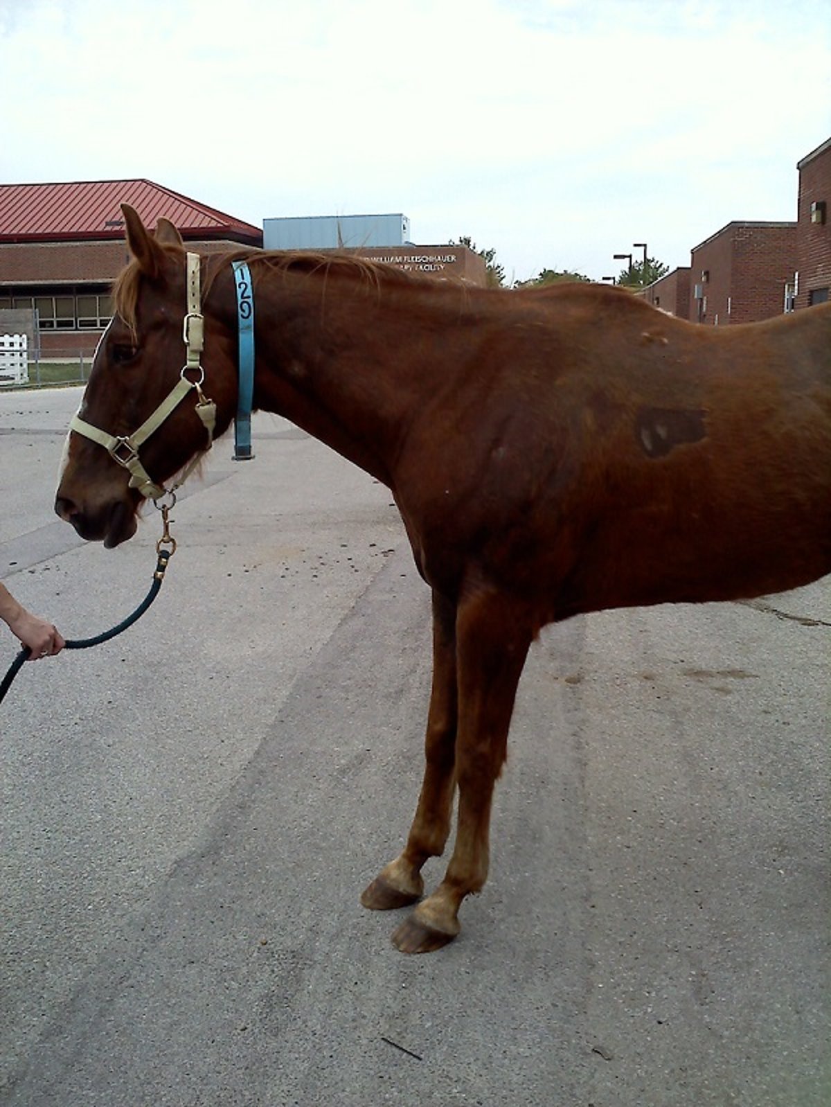
A thorough visual evaluation and manual palpation of the limbs in weightbearing and non-weightbearing positions is critical at the beginning of the lameness examination. Conformation should be evaluated and the horse visually checked for symmetry, swellings, muscle loss, abnormal stance, and obvious injuries. The trunk and limbs should be palpated for heat, pain, swellings, and joint effusion. There is variation between horses, and comparison with the contralateral limb can provide a useful control. The reaction of the horse to palpation and the range of flexion and extension of all joints should be noted.

Courtesy of Dr. Stephen Adams.
The feet should be thoroughly examined, including compression of the walls and sole with hoof testers. Wear patterns of shoes and feet should be noted. A number of abnormalities such as an abnormal hoof/pastern axis; mismatched hoof angles; under-run, contracted, and sheared heels; and disproportionate hoof size are seen more frequently in lame than in sound horses. Shoes should be left on during the initial stages of the examination, because removing them might make the horse footsore and preclude further examination. Shoes should be removed and the sole and frog cleaned and pared for complete and thorough examination of the foot only when the lameness has been localized to the foot and any exercise needed for diagnosis has been completed.

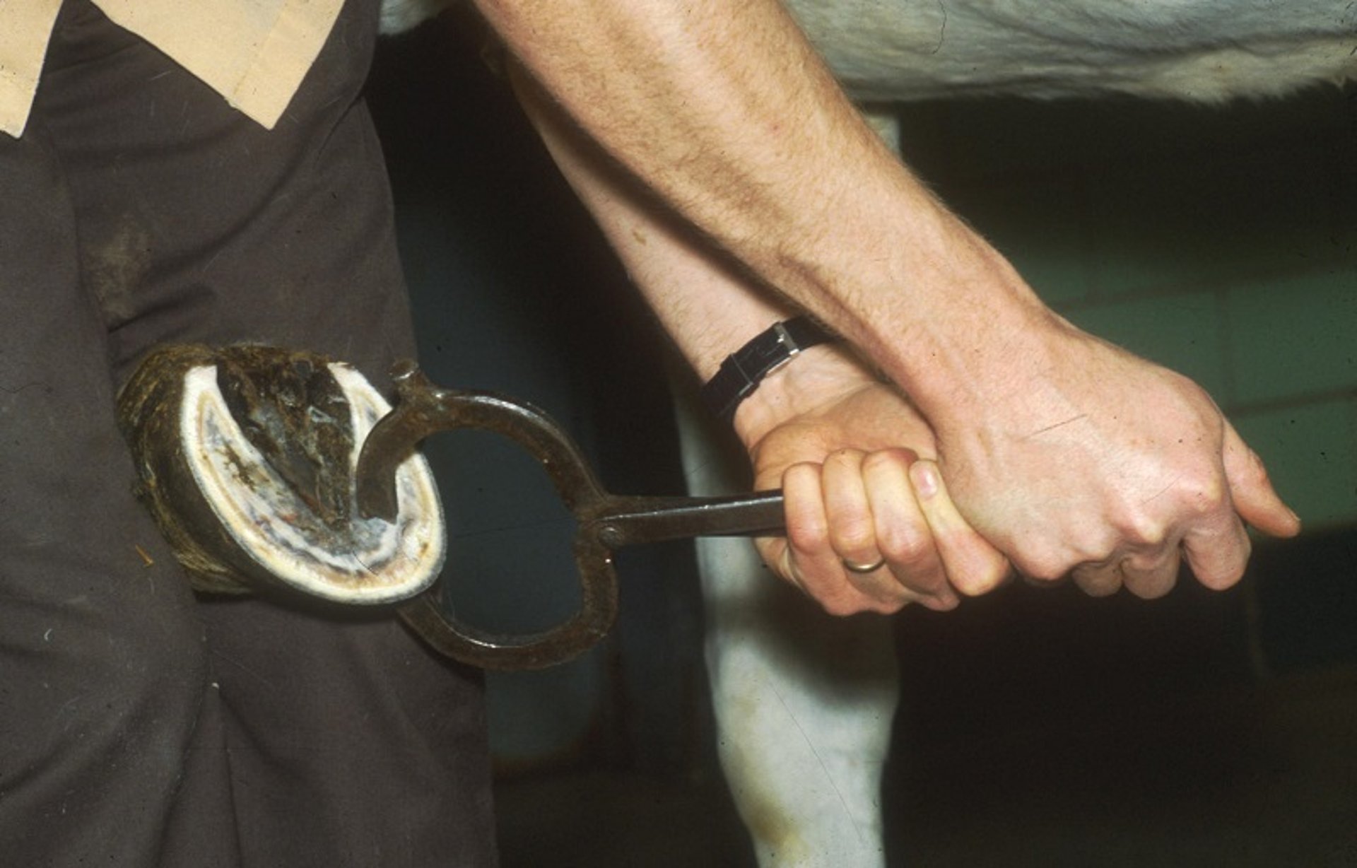
Hoof testers are used to compress structures inside the hoof capsule to detect pain. Note the sole has been pared clean for thorough inspection of the sole, frog, sulci, and white line.
Courtesy of Dr. Stephen Adams.
The back and neck should be thoroughly examined with the horse restrained and standing square on a level surface. Mobility and pain of the back and trunk can be difficult to detect, so the examiner should practice a consistent technique and series of palpations and assessments in these regions, in order to develop their observational skills.
Examination during exercise is often required to localize the lameness to a specific limb or site and to evaluate the response to diagnostic regional anesthesia. If lameness has an acute onset, is severe, and a fracture is suspected, exercise should not be undertaken or a catastrophic breakdown may result. Similarly, diagnostic regional anesthesia should not be performed when a fracture is suspected. It is important to determine whether the horse may have been given analgesic medication before the lameness examination.
Recognition of lameness is a key skill to successful diagnosis. The most consistent sign of a unilateral forelimb lameness is the head nod. The head and neck of the horse rise when the lame forelimb strikes the ground and is weightbearing, and they fall when the sound limb strikes the ground. The sacral rise, also called a pelvic rise or hip hike, is the most consistent and easily observed sign of hindlimb lameness. The entire pelvis and sacrum rise when the lame limb strikes the ground and is weight bearing, and fall when the sound limb strikes the ground. Both head nod and sacral rise serve to reduce concussion on the lame limb. Gait analysis systems are also available to augment lameness detection; these systems usually employ video capture of reflective markers or inertial sensors on the head, withers and tubera sacrale.

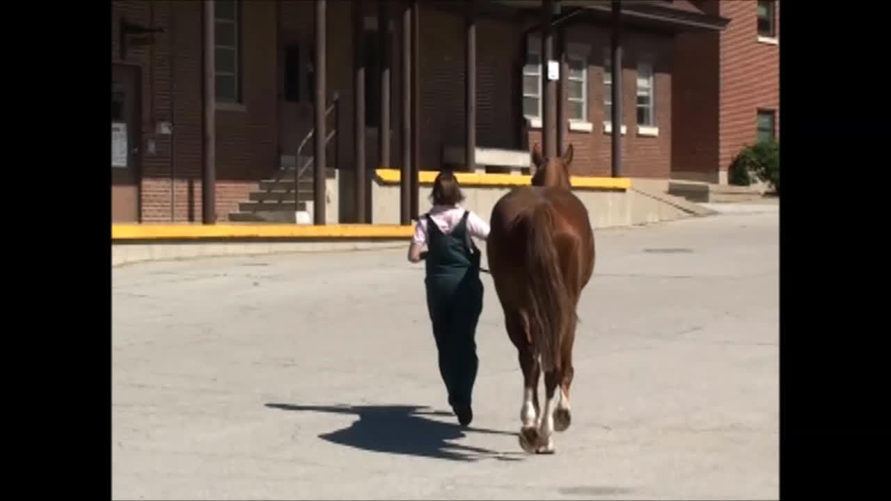
The horse should initially be examined by walking and jogging in hand with a loose lead line so that the movement of the horse is not restricted. A firm, nonslippery surface (eg, hardpack fine gravel) is ideal for trotting on a straight line and lunging on a firm surface. It also provides an opportunity to listen to the footfall and consider this information along with the visual appraisal.
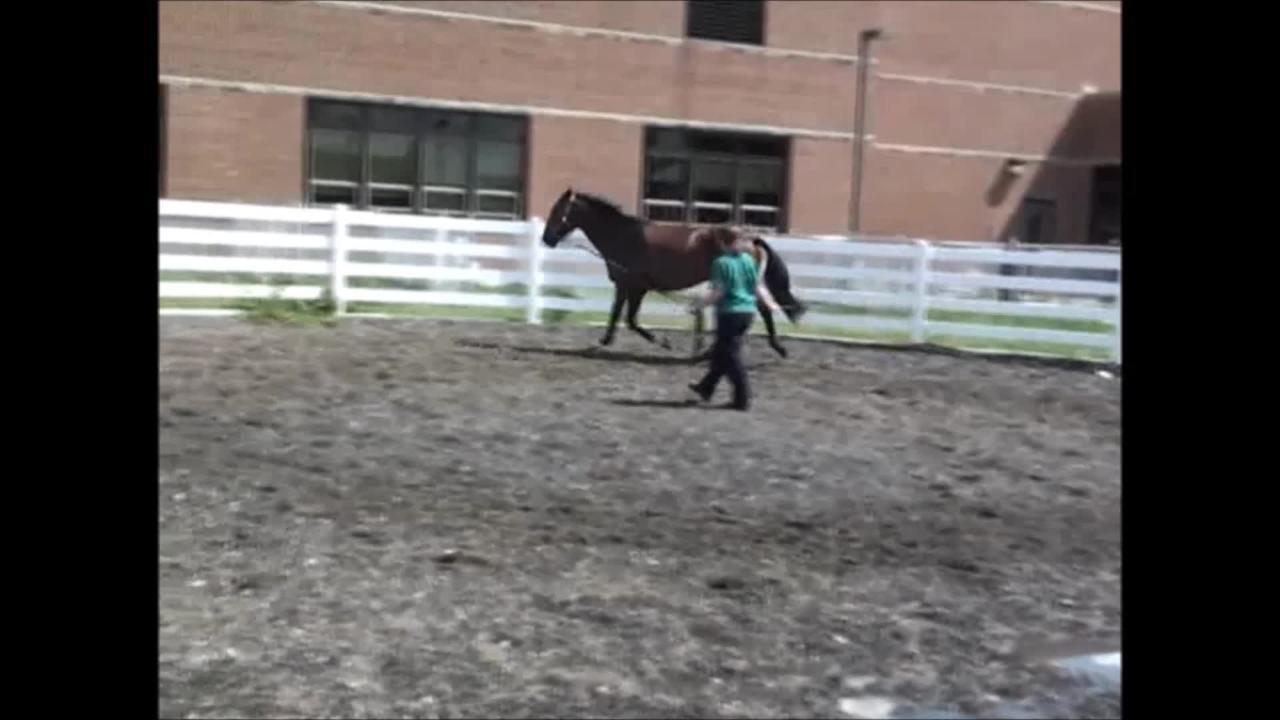
Frequently, lameness is more pronounced when the horse is worked in a circle. Circling can be done on a lunge line, by free exercise in a large round pen, in hand, or under saddle. Lunging on asphalt or concrete predisposes the horse to slipping and injury but may be done in selected cases to accentuate a very subtle hoof or lower limb lameness. Lunging is better performed on the soft footing of an arena or round pen. Both forelimb and hindlimb lameness may become worse when the horse is circled.
Flexion tests are performed during the evaluation on the straight line. After a baseline trot-up is established, a series of flexion tests are performed one at a time and the horse's response is assessed by trotting off immediately afterward, comparing to the baseline appearance. In addition, the range of movement and response to passive flexion should be observed. The distal phalanges in both forelimbs and hindlimbs should be flexed independently of the carpus and hock to obtain maximal information. Bending pressure should be firm but not excessive, which can create false-positive responses. All tests should be done on both sound and lame limbs for comparison. Consistency should always be applied, and individual experience used. A single positive flexion test without associated lameness may not be of significance.

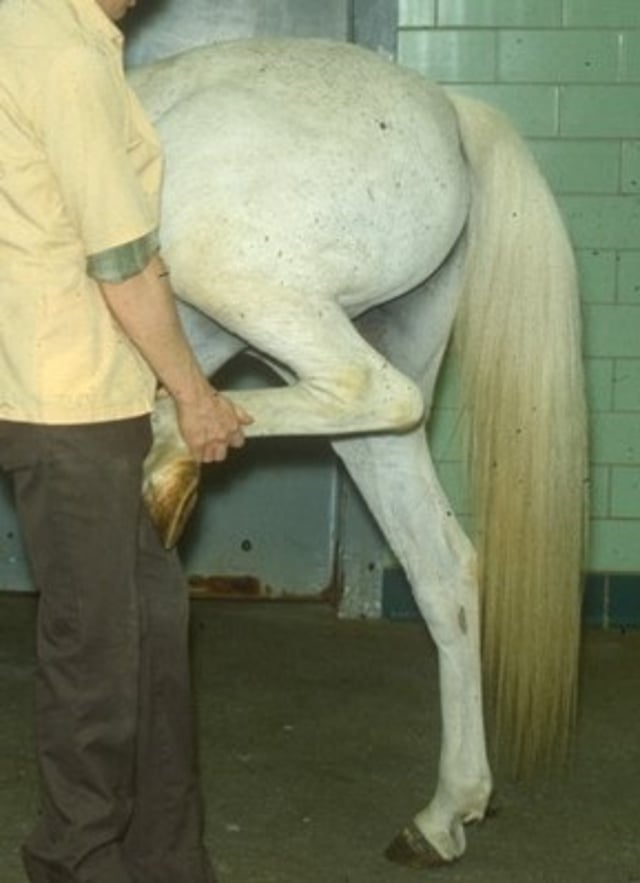
This full limb flexion test is frequently called the spavin test because the hock is a common source of pain when this test is positive. This test can also accentuate problems in the stifle and hip joints and some ligaments and tendons. The limb is flexed for 30–60 seconds and the horse is trotted away from the examiner to determine whether lameness is created or accentuated.
Courtesy of Dr. Stephen Adams.
To establish consistency, the entire examination should involve the same handler, the same bridle and tack when the horse is under saddle, and the same surfaces under foot. The horse should be controlled so that it is trotting at a useful, repeatable pace to evaluate the lameness. Slowing down the pace at the trot often illustrates a subtle lameness better, because the horse loses its momentum and struggles with suspension in the affected limb(s).
A ridden assessment of the horse may be necessary, particularly with a subtle lameness that can only be observed under saddle. A multiple-limb lameness without an obvious single-limb lameness may also be detected. The clinical signs may be minor (eg, the horse refusing certain movements or activities, slight head tilts, or tail swishing). However, a good rider can, often inadvertently, hide a problem by his or her inherent expertise and ability to “correct” deficiencies in the horse's gait.
Occasionally, a horse appears to be sound when lunged and ridden, but the rider feels that the performance is impaired. In such cases, it may be worth working the horse on concomitant analgesic or anti-inflammatory medication at therapeutic levels for an adequate period (eg, phenylbutazone 2–3 g/day, PO, for 7–14 days for an adult horse) to assess whether improvement occurs. Some clinical signs purported to be caused by lameness are training problems. If improvement on medication occurs, the medication should be withdrawn and diagnostic anesthesia used beginning in an arbitrary limb, most often a forelimb. In this way, multiple-limb lamenesses (as many as four) can be investigated.
Diagnostic regional anesthesia should be used to determine the area of pain whenever the lameness can be localized to a specific limb but not to a specific site on the limb (ie, occasionally the origin of pain is identified on palpation). A consistently observable lameness must be present for the clinician to evaluate response to anesthesia.
Because lameness may be caused by neuromuscular disorders, a complete neurologic examination should be part of the lameness examination whenever an obvious painful or mechanical cause of lameness has not been found. The examination should include evaluation of cranial nerve and upper and lower motor neuron function.
Observing the horse execute movements such as stopping short, backing, negotiating a curb, turning in tight circles, and walking uphill and downhill should be done. These tests help determine whether reduced proprioception, weakness, or spasticity may be the cause of the gait abnormality.
For More Information
Also see pet health content regarding the lameness examination in horses.


