


Courtesy of Dr. Stephen Adams.
Tendons act as bridging and attachment structures for the muscles to bone; some tendons are long and therefore prone to injury, especially because they are often loaded to the extreme and are only minimally capable of elastic elongation. A prime example is the superficial flexor tendon of horses, which is frequently injured by partial tearing that leads to tendinitis. Another acquired injury of tendons involves traumatic disruptions by internal forces or by external trauma. Healing of tendons and ligaments is prolonged, and the resultant repair tissue is usually of inferior mechanical strength compared to the original, healthy tendon. Newer treatment modalities (eg, stem cell injection, platelet-rich plasma, extracorporeal shock wave therapy) combined with specific rehabilitation programs have improved the prognosis for recovery from tendon and ligament disorders and return of animals to soundness for their intended use.
Tenosynovitis
Tenosynovitis, an inflammation of the synovial membrane and usually of the fibrous layer of the tendon sheath, is characterized by distention of the tendon sheath due to synovial effusion. Tenosynovitis may be idiopathic, acute, chronic, or septic (infectious). Idiopathic synovitis is the synovial distention of tendon sheaths in young animals, in which the cause is uncertain. The acute and chronic types of tenosynovitis are due to trauma. Tenosynovitis is often caused by lesions in the tendons within the affected tendon sheath. Septic tenosynovitis may be associated with penetrating wounds, local extension of infection, or a hematogenous infection.
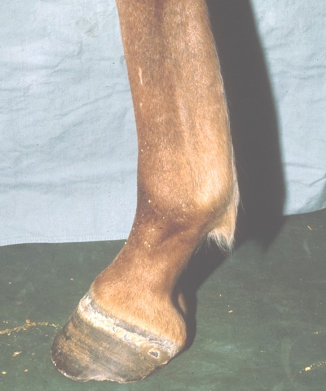
Clinical Findings and Diagnosis of Tenosynovitis in Large Animals

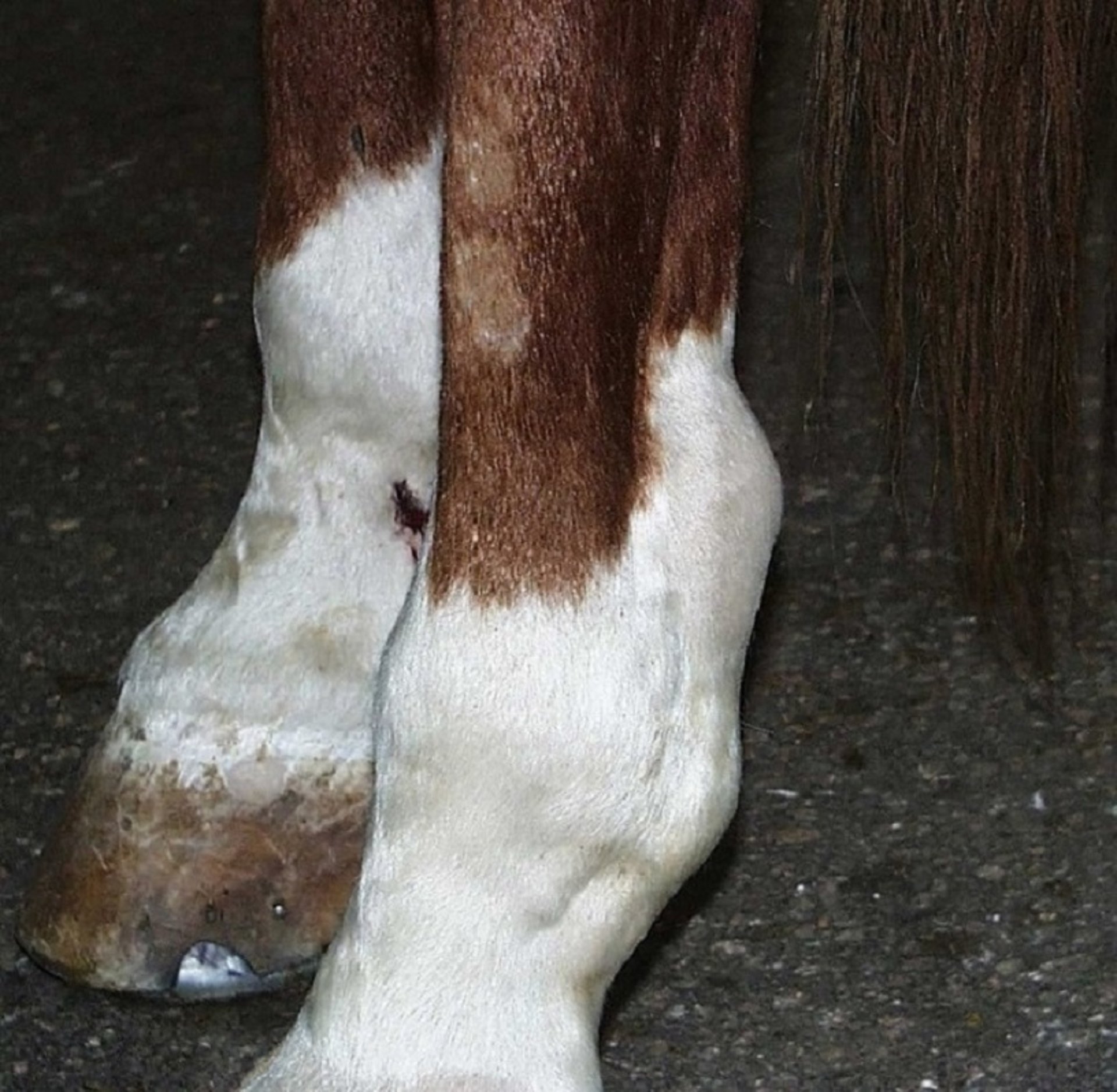
Courtesy of Dr. Stephen Adams.
The extent of synovial distention of the tendon sheath and lameness vary depending on the severity of tenosynovitis. Horses are markedly lame in septic tenosynovitis. In horses, chronic tenosynovitis is common in the tarsal sheath of the hock (thoroughpin) and in the digital sheath (tendinous windpuffs). These two entities must be differentiated from synovial effusion of the tarsocrural and fetlock joints, respectively. Evaluation of horses with signs of tenosynovitis should include a lameness examination, isolation of pain to the tendon sheath by use of local anesthetics, radiography (contrast radiographs can provide helpful information), and ultrasonography of the sheath and tendons enclosed within the sheath.
Treatment of Tenosynovitis in Large Animals
In idiopathic cases of tenosynovitis, no treatment is initially recommended. Acute cases with clinical signs may be treated symptomatically with bandages, cold packs, NSAIDs, and rest. More chronic tenosynovitis in which lameness is observed may respond to intrathecal corticosteroid or hyaluronic acid injections, combined with a rigorous rehabilitation program. Alternative treatment options include injecting the tendon sheath with platelet-rich plasma. When lesions in tendons are identified, extracorporeal shock wave therapy may be beneficial. Tenoscopic surgery may be used to treat tendon lesions and to remove synovial masses within the tendon sheath that give rise to clinical signs. Septic tenosynovitis requires local and systemic administration of antimicrobials, lavage, drainage, and tenoscopy to debride infected tissues. If adhesions develop between the tendon sheath and the tendon, persistent effusion and lameness are common.


