


Infraspinatus Contracture in Dogs and Cats
Infraspinatus contracture is a unilateral or bilateral fibrotic myopathy of the infraspinatus muscle that is usually secondary to trauma in hunting or working dogs but can occur in other animals as well.
Initial clinical signs of infraspinatus contracture include acute lameness, pain, and swelling in the shoulder region. The lameness subsides, but a gait abnormality develops 2–4 weeks after injury (see ), as muscle fibrosis and contracture progress.

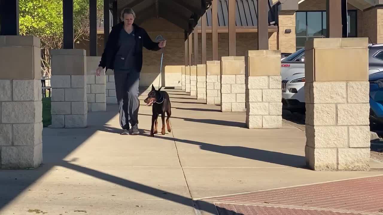
Clinical signs of infraspinatus contracture include characteristic adduction of the elbow, abduction of the forelimb, and external rotation of the carpus and paw. The limb is circumducted with each stride of the leg. Palpation of the shoulders reveals outward rotation of the humerus as the elbow is flexed.
Treatment of infraspinatus contracture consists of resection of the fibrous musculotendinous portion of the muscle, including tenotomy of the tendon of insertion. This surgical treatment immediately improves limb and joint functions (see ), and the prognosis for full recovery is excellent.

Tenosynovitis of the Biceps Brachii Tendon in Dogs and Cats
Tenosynovitis of the biceps brachii tendon of origin and associated synovial sheath can be unilateral or bilateral in small animals. Mature, large dogs are the most commonly affected. The mechanism of injury can be direct, indirect, overuse, or migration of osteochondral fragments (“joint mice”) from humeral osteochondrosis lesions.
Clinical signs of biceps tendon tenosynovitis include progressive or chronic, intermittent lameness that worsens after exercise and improves with rest. The range of motion of the shoulder joint is decreased, and there might be atrophy of the shoulder muscles. Acute pain can be elicited by applying digital pressure to the biceps brachii tendon during flexion of the shoulder and extension of the elbow joint.
Diagnosis of biceps tendon tenosynovitis can be confirmed by visualization of the damaged tendon via ultrasonography, arthroscopy, or MRI.
Radiography can reveal dystrophic calcification of the tendon, osteophytes in the intertubercular groove, or mineralized fragments within the tendon sheath.
Contrast arthrography can demonstrate filling defects and irregularities of the synovial sheath but is not often performed. Arthrocentesis might be inconclusive but can help rule out other diseases (such as immune-mediated arthropathy or infectious arthropathy.
Acute, mild cases of biceps tendon tenosynovitis in small animals can be treated with rest and oral NSAIDs. The duration of treatment depends on how long the clinical signs persist.
Activity restriction should be implemented for at least 6 weeks. If lameness resolves, activity can be very gradually increased over another 4–6 weeks. However, clinical signs might return as activity increases.
Acute, severe cases of biceps tendon tenosynovitis in small animals can be treated with a single peritendinous injection of methylprednisolone acetate (20–40 mg/dog or 10–20 mg/cat, IM), followed by 6 weeks of rest (1). The present author prefers to use a lower steroid dose, often 10 mg of methylprednisolone acetate in the joint.
Chronic bicipital tendon tenosynovitis cases refractory to medical management, or cases involving identifiable tendon tears (joint mice) can be treated by tenotomy or tenodesis (resection and attachment of the tendon to the proximal humerus) and osteochondral fragment removal.
Shoulder arthroscopy enables complete joint exploration and tenotomy of the biceps tendon under direct visualization. The prognosis for recovery is good.
Quadriceps Contracture in Dogs and Cats
Quadriceps contracture (also known as quadriceps tie-down or stiff stifle disease) in small animals is a serious fibrosis and contracture of the quadriceps femoris muscles that develops secondary to distal femoral fractures, to inadequate surgical repair (leading to excessive motion and callus formation), to excessive dissection, and to splinting the stifle joint in extension. Quadriceps contracture affects predominantly very young (ie, skeletally immature) animals. Dogs are more commonly affected than cats.
Adhesions develop between the bone, periosteal tissue, and quadriceps muscles, leading to replacement of muscle with fibrous scar tissue, permanent stifle joint extension, disuse, osteoporosis, degenerative joint disease, and bone and joint deformations.
Clinical signs of quadriceps contracture include stifle joint hyperextension without the ability to flex the joint and cranial displacement of the affected limb.
Surgery to resect fibrous tissues and increase motion of the stifle joint is typically not successful, so prevention is of the utmost importance. Prevention involves ensuring prompt, appropriate fracture stabilization and maintaining stifle joint range of motion after surgery.
Postoperative flexion bandages (such as the so-called 90-90 flexion bandage, which keeps the stifle and hock joints at 90° of flexion) have been used to help maintain stifle joint flexion after fracture repair (2). However, morbidity associated with these bandages has limited their use, and physical therapy with passive range-of-motion exercises is likely to be of greater benefit.
The prognosis for small animals with quadriceps contracture is guarded because the disease is irreversible. Affected animals might still be able to use the limb sometimes, particularly for balance while standing.
Amputation may be considered in cases of quadriceps contracture in which the animal's mobility is impaired, but it is not always necessary.
Achilles Tendon Disruption in Dogs and Cats
Injury to the common calcaneal tendon (Achilles tendon) in small animals can result from acute traumatic injury or chronic repetitive overloading.
Achilles tendon disruption can occur both in mature working and athletic dogs and in more sedentary animals. Injuries can occur anywhere along the length of the tendon, including at the musculotendinous junction, midtendon, and distally near the insertion on the calcaneal tuberosity. Ruptures can be partial or complete; in partial tears, the gastrocnemius tendon component is the most frequently affected.
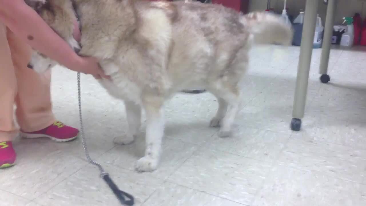
Clinical signs of Achilles tendon disruption can range from mild swelling of the tendon to severe non-weight-bearing lameness of the limb. Small partial tears might not result in any change to the tarsal joint angle. More substantial tears can lead to tarsal hyperflexion or a plantigrade stance.
Partial tears that do not involve the superficial digital flexor tendon can lead to hyperflexion of the hock, along with curling of the digits (see ). Palpation often reveals swelling in the area of the damaged tendon. A patient's ability to flex the hock while the stifle joint is fully extended helps to confirm the diagnosis of a substantial tear.
For complete tears of the Achilles tendon, torn or fibrotic tendon ends might be palpable. Orthogonal radiography of the affected tarsus might reveal avulsed bone fragments and soft tissue swelling in the area of the torn tendon.
Pearls & Pitfalls
|
Treatment of Achilles tendon disruption depends on the severity of the injury and clinical signs. Minor injuries that do not result in hyperflexion (or have very minimal hyperflexion) of the hock may be managed conservatively with several weeks of rest. Injection of platelet-rich plasma into the tendon, along with external support with an orthotic, can also be beneficial.
More severe Achilles tendon injuries resulting in tarsal hyperflexion and distinct areas of tearing usually require resection of scar tissue and surgical repair of the tendon ends (or reattachment of the tendon to the calcaneal tuberosity).
After surgery, the tarsus is immobilized for 4–6 weeks to protect the tendon during early stages of healing. Immobilization can be in the form of a bivalved cast, transarticular external skeletal fixator, or calcaneotibial screw. Long-term use of an orthotic to support the healing tendon might also be of benefit.
The prognosis in dogs with Achilles tendon injuries varies with the chronicity of the injury, success of the surgery, and expected performance of the dog.
Iliopsoas Muscle Trauma in Dogs and Cats
Trauma to the iliopsoas muscle or tendon of insertion can cause acute or chronic lameness in active dogs. Physical examination reveals focal pain at the proximal medial aspect of the thigh (attachment of the tendon to the lesser trochanter), especially during simultaneous hip joint extension and internal rotation.
Ultrasonography reveals disruption of muscle fibers, and radiography can reveal dystrophic calcifications at the region of tendon insertion.
Although iliopsoas tendinopathy can be a primary condition, it can also occur secondary to other abnormalities of the limb. In one study, 73.6% of dogs with iliopsoas strains had concurrent orthopedic disease (3).
Care should always be taken to fully evaluate the patient to make sure there are no other sources of pain (such as a cranial cruciate ligament tear or hip dysplasia).
For isolated injuries to the iliopsoas tendon, treatment with rest and NSAIDs is helpful. Initial rest periods should be at least 4 weeks. Initial treatment with NSAIDs for 1–2 weeks can help control clinical signs; thereafter, pain medications should be administered only as needed.
Limber Tail Syndrome in Dogs and Cats
Limber tail syndrome (also known as swimmer's tail) is a transient flaccidity of the tail. The syndrome is thought to be traumatic in origin (coccygeal muscle injury) and due to a compartment syndrome phenomenon.
Hunting dogs, primarily pointers and Labrador Retrievers but also setters and foxhounds, are most commonly affected by limber tail syndrome. It is more common in dogs but can occur in cats.
Strenuous exercise after a period of underconditioning, prolonged cage transport, or cold weather can predispose dogs to developing tail flaccidity.
Clinical signs of limber tail syndrome are sudden onset of tail flaccidity, with the tail either hanging from the tail base or projecting horizontally from the tail base for a short distance and then hanging downward. Some dogs show discomfort on palpation of the tail, most apparent approximately 8 cm distal to the base.
Limber tail syndrome is self-limiting, and complete recovery occurs within several days in most cases. NSAIDs are commonly recommended to minimize pain and distress associated with this syndrome; however, their use is anecdotal.
For More Information
Lane DM, Pfeil DV, Kowaleski MP. Synthesis of surgeon and rehabilitation therapist treatment methods of bicipital tenosynovitis in dogs allows development of an initial consensus therapeutic protocol. J Am Vet Med Assoc. 2023;262(2):1-8.
Also see pet owner content regarding exertional myopathy in dogs.
References
Bruce WJ, Burbidge HM, Bray JP, Broome CJ. Bicipital tendinitis and tenosynovitis in the dog: a study of 15 cases. N Z Vet J. 2000;48:44-52. doi:10.1080/00480169.2000.36157
Aron DN, Crowe DT. The 90-90 flexion splint for prevention of stifle joint stiffness with femoral fracture repairs. J Am Anim Hosp Assoc. 1987;23(4):447-454.
Sack D, Canapp D, Canapp S, et al. Iliopsoas strain demographics, concurrent injuries, and grade determined by musculoskeletal ultrasound in 72 agility dogs. Can J Vet Res. 2023;87(3):196-201. https://pubmed.ncbi.nlm.nih.gov/37397635


