


Imaging techniques allow vets to see inside a pet's body in ways that are noninvasive—that is, no instruments have to cut or go through the skin—and help diagnose many conditions. Some imaging equipment is large and costly and is available only in specialized facilities.
X-Ray Imaging
X-ray imaging (radiography) is the most common imaging method in veterinary clinics. Portable x-ray machines are used for large animals, like horses.
The procedure is painless, but pets may be sedated to decrease stress and help them stay still.
X-ray exposure is very brief and uses low doses of radiation. Lead shields protect the rest of the pet's body and the people taking the x-rays.
X-rays, table

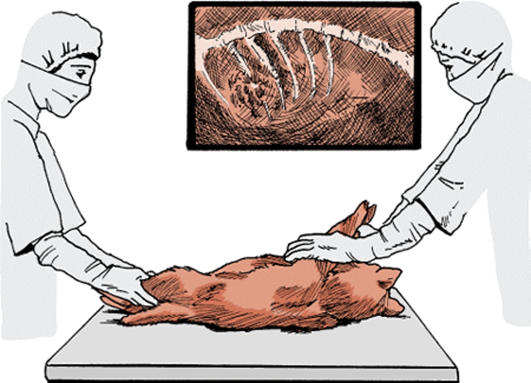
X-ray images (also known as radiographs) show bones, some foreign objects (like rubber or metal), and large body cavities. X-rays are good for detecting fractures, tumors, injuries, and deformities. Soft tissues do not show up well on standard x-rays, so your vet might use contrast procedures. A dye that blocks x-rays can be given IV or by mouth to outline organs like kidneys, heart, or the digestive tract. Sometimes air is used for “negative contrast.” Modern x-rays are usually made and stored digitally, allowing easy sharing with specialists for a second opinion.
Ultrasound
Ultrasound uses sound waves to create images from the echoes that bounce off tissues. Ultrasound is better than x-rays for showing soft tissues but can't image lungs, intestines, or bone well. The operator presses a probe against the body—usually the abdomen—and moves it to scan organs. Modern ultrasound provides real-time images that move as the probe moves. Ultrasound is painless, noninvasive, and safe. It is commonly used to examine the abdomen. Ultrasound of the heart is called echocardiography.
Computed Tomography (CT)
CT is a computer-enhanced x-ray that takes multiple cross-sectional images (slices) of the body. The animal lies on a motorized bed that moves through the CT scanner while x-rays are taken from different angles. A contrast dye might be injected to make some structures easier to see. CT gives detailed images of tissue density, which helps detect deep tumors and infections, blood vessel changes, and fractures. Because CT scans take time and require the animal to remain perfectly still, animals are usually anesthetized.
Magnetic Resonance Imaging (MRI)
MRI uses a very strong magnetic field and radio waves to create highly detailed images without x-rays. The animal is placed inside a tubular chamber and pulsed with radio waves. The tissues emit signals that a computer converts into images. MRI takes many repeated pulses and is longer than CT, so general anesthesia is used in most cases. MRI is especially useful for detailed imaging of the brain, spinal cord, and soft tissues.
Nuclear Medicine Imaging
Nuclear medicine imaging, also known as scintigraphy, uses a small amount of a radioactive element attached to a molecule that is taken up by a particular organ. A special camera detects the gamma rays emitted and creates images that show how the organ is functioning. This technique is most often used to evaluate lungs, kidneys, liver, thyroid, and heart. It can show changes in organ function over time.


