

Obstructive urolithiasis is a substantial cause of morbidity and death in male ruminants. Uroliths form from protein and mineral components of the diet. They develop primarily in the urinary bladder and lodge in the distal sigmoid flexure of cattle and small ruminants or the vermiform appendage (urethral process) of the small ruminant, causing obstruction. Early recognition and treatment are critical to achieving a positive outcome. Urolithiasis is a multifactorial disease, making prevention strategies challenging to develop.
For a more general introduction to urolithiasis, see Urolithiasis in Small Animals.
Etiology and Pathogenesis of Urolithiasis in Ruminants
Obstructive urolithiasis is most common in wethers, bucks, and rams, with commercial, exhibition, and pet animals at risk. Incidence of disease is lower in steers and bulls, with cattle in feedlots most at risk. Uroliths can also form in females; however, because females do not have the narrow urethral tract found in males, obstruction and subsequent pathology are much less frequent.
Early castrated males have the highest incidence of urethral obstruction; however, urethral obstruction is also common in intact male goats.
Blockage of the urethra is followed by urinary retention, which can lead to rupture of the urinary bladder or urethral perforation. The latter is common in feedlot steers.
Uroliths are solid crystalline formations composed of organic matrix (sugars, protein, and cells) and inorganic crystalloids, including calcium, magnesium, and phosphate. These components can come out of solution and bond in situations of supersaturation.
Four main types of uroliths affect ruminants:
phosphatic (including struvite, apatite, and amorphous magnesium calcium phosphate)
calcium carbonate
silicate
calcium oxalate
Matrix components can include uroepithelial cells from vitamin A deficiency, suture, tissue debris, blood clots, excess protein, or bacteria in the urine.
Urine supersaturation of mineral components occurs as a result of increased renal excretion, negative water balance, urine pH, and the presence or absence of crystallization inhibitors. The phosphatic and calcium carbonate stone types form in alkaline urine and in animals on high grain or legume diets. Urine pH likely has no impact on the formation of silicate or calcium oxalate uroliths; these form in animals grazing on siliceous pastures in the western US and Canada or on oxalate-containing plants.
Pearls & Pitfalls
|
Systemic illness and unpalatable water can result in decreased water intake, increasing urolith component concentration in urine. Diet and water intake are major contributors to the etiology of this disease; however, other factors, such as urethral diameter and individual metabolism, also likely contribute.
The anatomy of male ruminants does not contribute to stone formation, but rather to a predilection for obstruction. The urethra of male ruminants is long and sigmoid in shape, and small ruminants additionally have a 2- to 4-cm extension of the urethra beyond the glans penis called the vermiform appendage or urethral process. This is the most common site of obstruction in small ruminants, whereas the distal sigmoid flexure of the urethra is a common site in both cattle and small ruminants.
Uncommonly, uroliths form in the renal pelvis and lodge in the ureters, causing obstruction in females and males equally.
Clinical Findings and Diagnosis of Urolithiasis in Ruminants
Obstructive urolithiasis should be considered as a differential diagnosis in all sick male ruminants, particularly castrated sheep and goats and feedlot steers. Historical findings are important in investigating possible cases of obstructive urolithiasis, and specific questions regarding diet, age at castration, progression of clinical signs, any treatments given, and last observed urination normally provide valuable insight.
Clinical signs include the following:
anorexia
depression
weakness
bruxism
straining and stretching out
vocalization
ventral pitting edema ("water belly," most common in feedlot steers that have a ruptured urethra)
abdominal distention
Many affected animals exhibit bloat or straining to pass feces.
A thorough physical examination should be performed on presentation. Observations for grit on preputial hairs and pulsing of the urethra upon digital rectal examination are useful specific examination points for suspected urolithiasis cases.
A large plaque of ventral edema (pitting edema) surrounding or in front of the prepuce is indicative of a ruptured urethra, whereas a pear-shaped abdomen with bilateral ventral distention suggests a ruptured urinary bladder, both of which are associated with a worse prognosis. A grave prognosis for breeding should be given for any intact male if there is a urethral rupture, because penile adhesion formation is likely and will interfere with breeding.
Pearls & Pitfalls
|
If the animal does not urinate during examination or when placed in a clean stall, ultrasonography of the urinary bladder and/or exteriorization of the penis with examination of the vermiform appendage is recommended (see ). Transabdominal ultrasonography with a 3.5–7.5 MHz probe may reveal a distended urinary bladder (often > 8–10 cm [3.1–3.9 inches] in small ruminants) or free abdominal fluid in cases of rupture. Abdominocentesis may be performed, and a sample creatinine concentration greater than double the serum creatinine identifies the fluid as urine.
The penis in goats and sheep is best examined by positioning the animal in a "sitting" position.

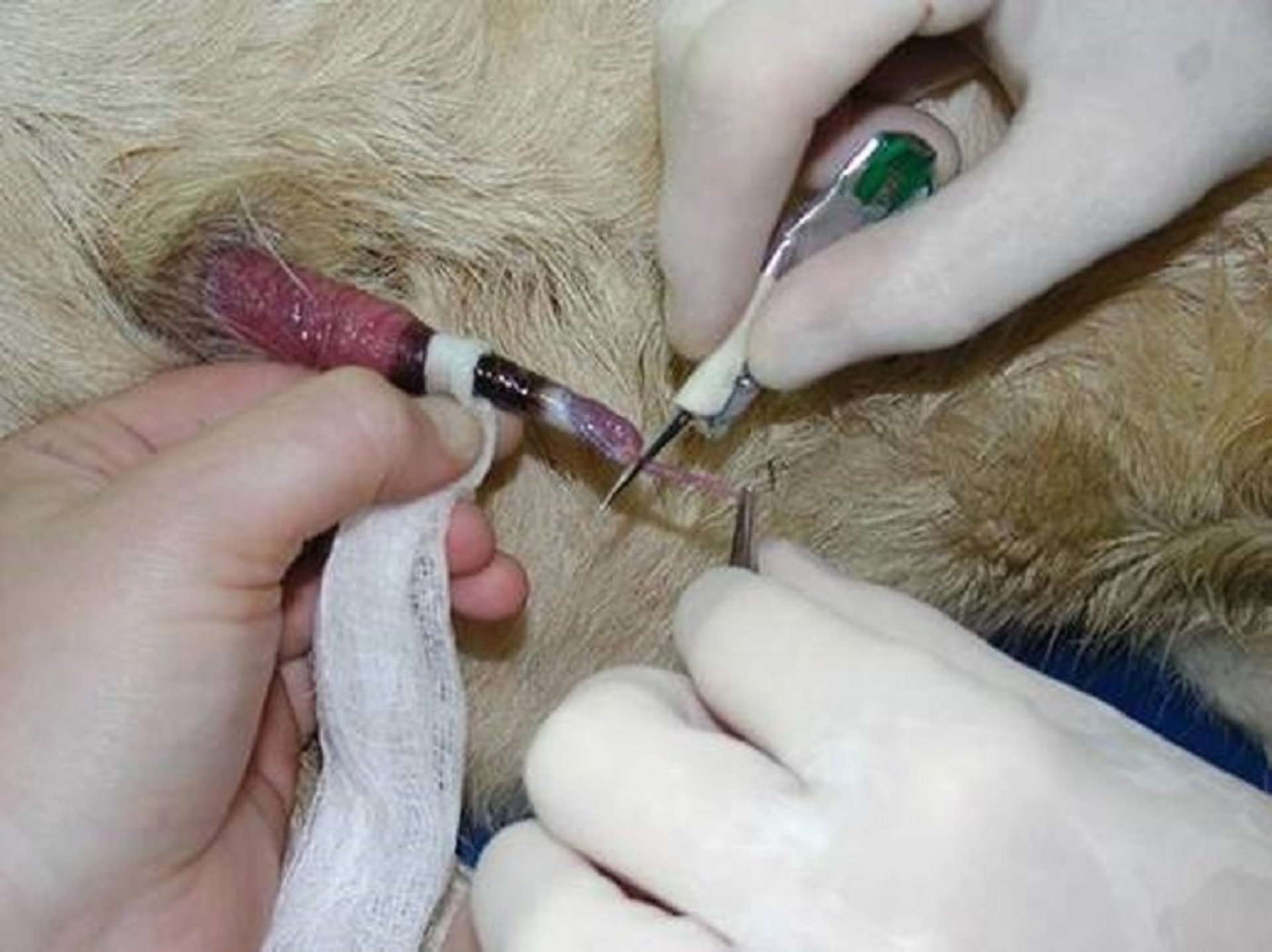
Exteriorization of the penis and removal of the vermiform appendage in a wether. The veriform appendage is being stretched with forceps prior to being cut with a scalpel blade.
Courtesy of Dr. Meredyth Jones.
Sedatives and local anesthesia can be useful to achieve restraint and exteriorization of the penis. Acepromazine (0.05–0.1 mg/kg, IV or IM), diazepam (0.25–0.5 mg/kg, slow IV; or 0.2–1 mg/kg, IM or SC), or midazolam (0.1–0.5 mg/kg, IV or IM) can serve to decrease anxiety and relax urethral tone (1). Another sedative protocol used by some veterinarians is a combination of butorphanol (0.05 mg/kg, IM) followed by midazolam (0.2 mg/kg, IV)) (2). Administration of all of these drugs constitutes extralabel drug use.
For patients with cardiovascular compromise, acepromazine should be avoided. Alpha-2 agonists produce analgesia and sedation in ruminants but also cause increased urine volume from transient hyperglycemia. This sudden increase in urine volume can cause rupture of a previously intact urinary tract and is therefore contraindicated in cases of urinary obstruction. Lumbosacral epidural administration of 2% lidocaine can also facilitate exteriorization of the penis.
Survey radiography is particularly useful in cases of calcium carbonate urolithiasis because the uroliths are quite radiopaque, allowing for determination of the extent of the obstruction and potential for reobstruction. Phosphatic stones, however, are less radiopaque and often missed on survey radiography because of the mass of the abdomen. The absence of uroliths on survey radiography does not rule out the presence of obstructive or nonobstructive uroliths.
Common findings on serum biochemical analysis include azotemia, hyperphosphatemia, and hypermagnesemia. Ruminants are able to manage BUN and potassium via the rumen and saliva, respectively, so these elevations in these components in blood are not as large as those observed in obstruction of monogastric animals. Creatinine concentration is therefore a more reliable indicator of impaired renal function.
Urinalysis may be performed if a sample can be obtained. Protein and blood can be present, along with crystals of the obstructive stone type; however, crystals are commonly absent from the urine of obstructed animals. Hemoglobinuria, myoglobinuria, and hematuria are causes of red urine. These should be differentiated by performing centrifugation of the urine, and differential diagnoses including copper toxicity, Brassica toxicity, cystitis, pyelonephritis, and leptospirosis should be considered.
Treatment of Urolithiasis in Ruminants
Catheterization
Supportive care
Surgical correction if needed
Treatment goals for ruminants with urolithiasis are to establish urethral patency, provide analgesia, correct fluid and electrolyte imbalances, decrease urethral inflammation, and prevent infection.
Catheterization and retropulsion is a mainstay of treatment for obstructive urolithiasis in many species. However, the presence of the urethral diverticulum, a dorsal outpouching of the urethra at the level of the ischial arch, prevents simple retrograde catheterization in male ruminants. Various curved catheters have been used with some success in accomplishing retrograde passage into the urinary bladder in male small ruminants. Hydropulsion can be performed using a tomcat catheter after instillation of 0.5–1 mL 2% lidocaine into the urethra. Hydropulsion with small volumes of saline solution (0.9% NaCl) should be attempted very carefully so as to not force fluid past the stone, further distending and possibly rupturing the urinary bladder or causing urethral perforation. Surgery is often necessary to relieve these obstructions.
Fluid therapy should be instituted based on clinical findings and the results of serum biochemistry testing. After or during the relief of the obstruction, diuresis is important to replace hydration deficits, manage azotemia, and flush remaining uroliths from the urinary tract. Normal (0.9%) saline solution makes an appropriate empirical first fluid for these cases. Additives should be used as indicated by serum biochemistry findings. Initial rates of fluid therapy should be slow until the obstruction is relieved and then increased to 80–100 mL/kg every 24 hours until the patient stabilizes.
Drug therapy can include the use of NSAIDs, opioids for pain management, and preoperative broad-spectrum antimicrobial therapy. Beta-lactam antimicrobial drugs make reasonable choices for these cases because of their spectrum of activity and their excretion via urine. These recommendations constitute extralabel drug usage, and drug labels should be followed for dosing and extended withdrawal times, even in pet animals.
Surgical techniques described for relief of urinary obstruction in ruminants include the following:
vermiform appendage amputation
tube cystotomy
urinary bladder marsupialization
urethrotomy
perineal (high), low (distal sigmoid), and prescrotal urethrostomy
If imminent bladder rupture is a concern, ultrasonographic guided placement of a Bonanno catheter will decompress the bladder, allowing time (12–24 hours) to stabilize the patient and decide on surgical treatment.
Pearls & Pitfalls
|
Vermiform appendage amputation can be performed successfully in small ruminants if it is the only site of obstruction. After exteriorization of the penis, the vermiform appendage is sharply removed at the glans penis. There are almost always more uroliths in these patients, however, and animals relieved of obstruction are at high risk of reobstruction.
Tube cystotomy is considered the gold standard treatment for cases of urethral obstruction because it provides an alternate route for urine flow while allowing healing of the urethra. Briefly, a paramedian cystotomy is performed and a Foley catheter placed to divert urine and allow the urethra to rest for 3–5 days. Urination generally occurs in uncomplicated cases in 7–14 days. Tube cystotomy is associated with a higher cost than other surgeries for obstruction but has been reported to have 76–90% short-term success at reestablishing urethral urine flow, with an 86% long-term success rate (2). The most common complication is the tube becoming displaced from the urinary bladder. Percutaneous and flank-approach placement of tubes have also been described and are an option to avoid general anesthesia. This is also the preferred treatment in valuable bulls intended for breeding.
Urinary bladder marsupialization creates a permanent or semipermanent stoma from the urinary bladder to the skin, providing urine outflow that bypasses the urethra. Via a paramedian incision, the apex of the urinary bladder is brought to the body wall and a stoma created for urine diversion. This procedure is associated with complications of urine scald, mucosal prolapse, and ascending urinary tract infections. Reported success rates with urinary bladder marsupialization in two studies were 66% and 94% (3, 4).
In a perineal urethrostomy, urination is established via a stoma created within the proximal urethra. The urethra is spatulated and sutured to the skin, creating the stoma. This procedure is associated with a high risk of stricture, which can be improved with excellent tissue handling and careful dissection to ensure that tension on the penis is relieved. Stricture is common in a majority of cases within 8 months, making this generally an undesirable option for most pets; however, some remain successful for years.
When stone material is obtained at or before surgery, urolith analysis is indicated to guide control and prevention measures. An initial presumption can be made based on appearance. Phosphatic stones occur in sandlike form or are easily crushable, whereas calcium carbonate uroliths are gold beads with a very stable structure (see images of and uroliths).
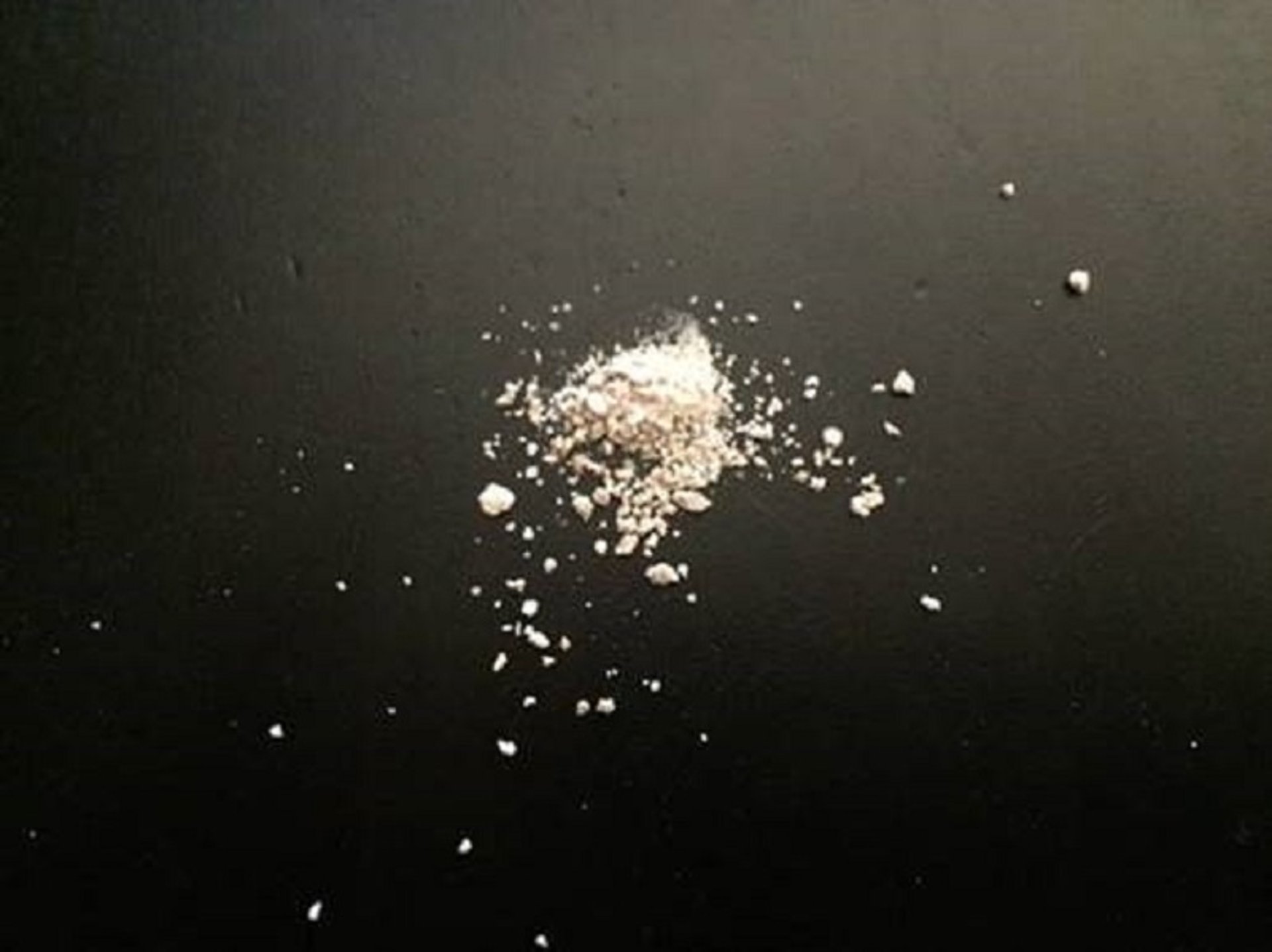
Phosphatic uroliths from a goat. Note the sandlike texture.
Courtesy of Dr. Meredyth Jones.
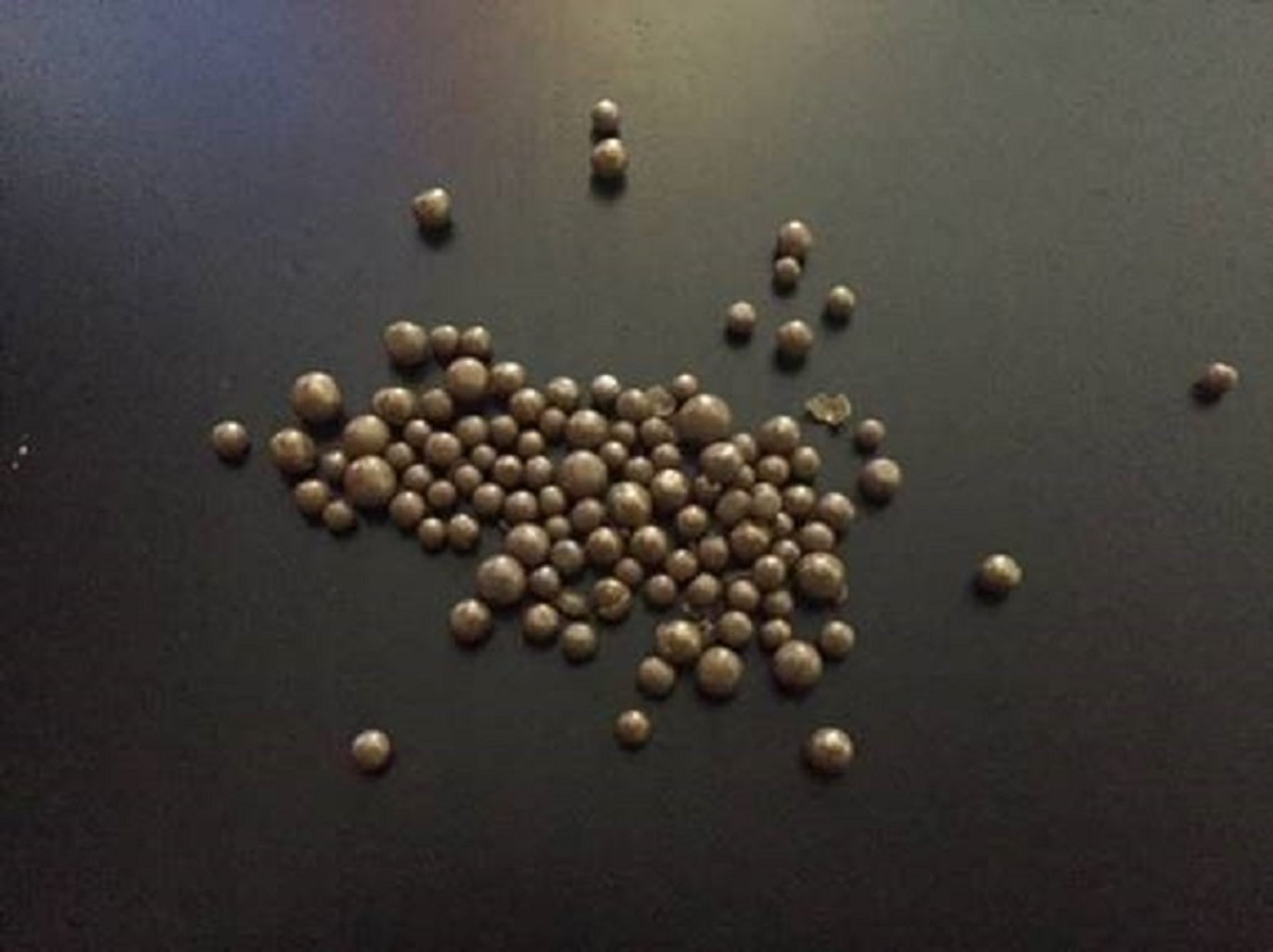
Calcium carbonate uroliths from a goat. Note the beadlike structure.
Courtesy of Dr. Meredyth Jones.
For pharmacological considerations, see the topics Pharmacotherapeutics in Urolithiasis, Controlling Urine pH, and Cysteine-Binding Agents Used to Treat Urinary Disease.
Control and Prevention of Urolithiasis in Ruminants
A thorough review of the diet and management of herds or flocks with cases of urolithiasis should be performed. Animal purpose, water source, and stone type are all important considerations.Availability and type of hay, grain, pelleted feed, pasture, and mineral supplementation must also be determined. In general, prevention focuses on four main aspects of the development of obstructive urolithiasis:
optimizing the anatomy of the urinary tract
increasing urine volume and dilution
decreasing matrix components
managing mineral components
The anatomy of the urinary tract in ruminants makes preventing and treating urolithiasis more challenging in these species. Two approaches can make the urinary tract less susceptible to obstruction:
Delaying castration until at least 8 weeks has been shown to have a positive impact on urethral diameter in calves and lambs, providing a larger outlet for any uroliths that form.
Prophylactic removal of the vermiform appendage removes the most common site of obstruction in small ruminants. The purpose of the vermiform appendage is to spray semen during copulation. No primary studies have evaluated whether removal negatively impacts fertility; however, for most experienced clinicians, this is not a concern.
Urine volume and dilution can be increased by increasing water intake. Providing clean, palatable, temperature-appropriate water, adding NaCl to the diet, encouraging grazing, and feeding a high-forage diet with limited grain and pelleted feeds all increase water intake. A high-roughage diet requires more water for mastication and digestion, therefore increasing urine output over that of concentrate diets.
Matrix components can be reduced by decreasing high-protein feeds and hays. Ensuring a solid trace mineral program with sufficient vitamin A decreases the likelihood of urinary tract metaplasia and cellular debris in the urinary bladder. Chloride salts, such as calcium chloride, sodium chloride, and ammonium chloride, can attach to the matrix binding sites and prevent formation of a nucleus.
Mineral component control is considered in light of the urolith type most likely to form in a particular species. Show animals and feeder animals are at highest risk for phosphatic stones. Pets and other animals consuming alfalfa and other legumes are at highest risk for calcium carbonate stones. Both of these stone classes form in alkaline urine. Urinary acidification can be achieved with ammonium chloride administered at 200–350 mg/kg, PO, every 24 hours (1). Administration should continue for a few days, along with diet adjustments, with the aim of a long-term decrease in calculogenic solutes in the urine. Animals on ammonium chloride should have their urine pH evaluated periodically 5–7 hours after feeding to determine whether adequate acidification (pH < 6.5) is occurring, with the dosage adjusted for the individual.
If clients insist on feeding grain or pelleted feed, the Ca:P ratio of the total ration should be held at 2–2.5:1 to limit phosphorus availability for phosphatic uroliths. An increase in this ratio predisposes to calcium carbonate uroliths, whereas a decrease predisposes to phosphatic uroliths. Loose trace minerals should be provided to all classes of animals.
Silicate and calcium oxalate urolith types are associated with specific plants on pasture. These should be identified and controlled, or males should have limited access, with females used to graze high-risk pastures.
Key Points
Obstructive urolithiasis is a major cause of morbidity and death in male ruminants.
Treatment requires urethral catheterization, surgical correction, and supportive care.
Prevention involves increasing urine output, adjusting urine pH, and making dietary changes to minimize urolith formation.
For More Information
Cook MJ. Urinary calculi of small ruminants. Vet Clin North Am Food Anim Pract. 2023;39(2):355-370.
Oman RE, Rivero L, Weaver LF, Simpson KM. Modified tube cystostomy technique for management of obstructive urolithiasis in small ruminants: procedure and outcome in 17 sheep and goats. J Am Vet Med Assoc. 2023;262(2):256-262.
Pongphitcha P, Chuchoed K, Thetsana T, Dachphun N, Sukhong P, Ratanapob N. Factors associated with success rate of oral force-feeding ammonium chloride administration to acidify urine in goats. Open Vet J. 2024;14(9):2310-2314.
Mejia S, McOnie RC, Nelligan KL, Fubini SL. Small ruminant urinary obstruction: decision trees for treatment. J Am Vet Med Assoc. 2022;260(S2):S64-S71.
References
Smith MC, Sherman DM. Goat Medicine. 3rd ed. Wiley-Blackwell; 2023:619-629.
Ewoldt JM, Anderson DE, Miesner MD, Saville WJ. Short- and long-term outcome and factors predicting survival after surgical tube cystostomy for treatment of obstructive urolithiasis in small ruminants. Vet Surg. 2006;35(5):417-422. doi:10.1111/j.1532-950X.2006.00169.x
May KA, Moll HD, Wallace LM, Pleasant RS, Howard RD. Urinary bladder marsupialization for treatment of obstructive urolithiasis in male goats. Vet Surg. 1998;27(6):583-588. doi:10.1111/j.1532-950x.1998.tb00534.x
May KA, Moll HD, Duncan RB, Moon MM, Pleasant RS, Howard RD. Experimental evaluation of urinary bladder marsupialization in male goats. Vet Surg. 2002; 31:251-258. doi:10.1053/jvet.2002.32441


