


Congenital abnormalities are conditions that an animal is born with; they are often referred to as “birth defects.” Some of these conditions are inherited and tend to occur within particular families or breeds, while others are caused by chemicals or injury during pregnancy. For still others, the cause is unknown. Some of the most common congenital abnormalities of the digestive system in cats are described below.
Mouth
A cleft palate or cleft lip (harelip) is caused by a defect in the formation of the jaw and face during embryonic development. It leads to a gap or cleft in the center of the lip, roof of the mouth (hard palate), or both. Often these gaps leave an opening between the roof of the mouth and the nasal cavity allowing food and liquids to pass into the breathing passages. These conditions have a wide range in severity. Usually the upper lip and palate are affected. Clefts in the lower lip are rare. The incidence of cleft lip is higher in Siamese cats than in other breeds.
Cleft palate or lip will usually be noticed shortly after birth when the kitten might have problems nursing. For example, milk might be seen dripping from the nostrils or the kitten might have difficulty suckling and swallowing. The veterinarian can readily identify the problem by examining the kitten’s mouth. Affected kittens require intensive nursing care, including hand or tube feeding and possibly antibiotics to treat respiratory infections. Surgical correction is necessary and is usually done when kittens are at least 12 weeks old to minimize further complications. Waiting until the kitten is older than 5 months or even an adult may improve the success of surgery. A variety of surgical techniques are used, and the success rate in cats is improving. However, large defects are usually associated with a poor outcome, even with surgery. The decision to perform surgery should be made carefully, and the affected animal should be spayed or neutered to prevent passing the defect on to its offspring.
Mandibular brachygnathia (or "overbite") occurs when the lower jaw is shorter than the upper jaw. It can be a minor problem or a serious defect depending on the degree of abnormality. Mild cases may cause no problems. More severe cases can cause damage to the hard palate (roof of the mouth) or restriction of normal jaw growth. The lower canine teeth are often removed or shortened to prevent this damage. Treatment early in life is recommended and improves the outcome.
Mandibular prognathia (or "underbite") occurs when the lower jaw is longer than the upper jaw. This characteristic is normal in some breeds (for example, Persian cats) and does not usually require treatment.
Teeth
In most animals, having too few teeth is rare. In cats, extra teeth sometimes occur and may lead to crowding and poor alignment of the teeth. Extra teeth that cause crowding should be extracted by a veterinarian as soon as they are discovered to prevent further dental problems.
Delayed loss of deciduous (baby) teeth is uncommon in cats. However, because teeth that do not fall out get in the way of the permanent teeth, any persistent deciduous teeth should be removed by your veterinarian as soon as possible.
Abnormal development of tooth enamel (the hard outer surface of the tooth) can be caused by fever, trauma, malnutrition, poisoning, birth defects, or infections. The damage to the enamel depends on the severity and duration of the cause and can range from pitting to the absence of enamel with incomplete tooth development. Affected teeth are prone to plaque and tartar accumulation, which lead to tooth decay. Resin restoration is sometimes used to cover defects, although careful dental hygiene and home care is critical in reducing the incidence of complications. Discoloration of the enamel may also occur. Giving tetracycline antibiotics to pregnant females or to kittens less than 6 months old may result in permanent brownish-yellow stains on the teeth.
Esophagus
The muscular tube that leads from the back of the mouth to the stomach is known as the esophagus. Some congenital abnormalities of the esophagus seen in cats include megaesophagus, vascular ring anomalies, and cricopharyngeal achalasia (see table: ). Signs of defects in the esophagus generally include regurgitation and problems with swallowing. These signs are especially noticeable when your cat starts to eat solid food. Regurgitated food can enter the lungs and cause frequent and severe pneumonia. These conditions are typically diagnosed with x-rays, usually done after the cat swallows a liquid dye that shows up on the x-ray. Other specialized tests may also be necessary. Surgical correction of some esophageal abnormalities (for example, vascular ring anomalies, in which abnormal blood vessels surround and restrict the esophagus) is effective if done early. If not, the esophagus can become permanently damaged by the stretching caused by trapped food. Special diets are often necessary for the life of the cat.
Congenital Esophageal Disorders of Cats
Type | Cause | Breeds Most Often Affected |
|---|---|---|
Congenital megaesophagus | Abnormal nerve development in esophagus; sometimes part of more widespread nerve problems | Siamese |
Vascular ring entrapment | Physical constriction of the esophagus by blood vessels | No breed tendencies reported |
Cricopharyngeal achalasia | Failure of the cricopharyngeal muscle (in the throat) to relax during swallowing | Rare; no breed tendencies reported |
Small pouches in the lining of the esophagus, called esophageal diverticula, will sometimes form. Clinical signs depend on severity and are seen in only 10% to 15% of cases. When they do occur, they may cause accumulation of food or become inflamed. In rare cases they rupture. Treatment (if necessary) is by surgical removal of the pouch.
Hernias
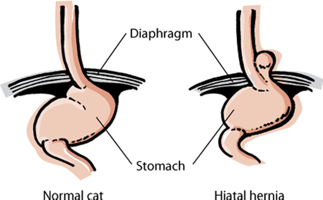
A hernia is the protrusion of a portion of an organ or tissue through an abnormal opening. These abnormal openings often occur in areas where the body wall does not close properly at birth (such as the umbilicus or "belly button"). Common congenital hernias involve an abnormal opening in the wall of the abdomen or the diaphragm (the sheet of muscle that separates the chest from the abdomen). The defect may allow abdominal organs to pass into the chest or bulge beneath the skin. Hernias may be congenital (present at birth) or result from injury. Signs of a hernia vary from none to severe and depend on the amount of herniated tissue and its effect on the organ involved. Hiatal hernias involve extension of part of the stomach through the diaphragm. These hernias may be “sliding” and result in signs (such as loss of appetite, drooling, or vomiting) that come and go. Hernias are diagnosed using x-rays; contrast studies (x‑rays that include special dyes to outline organs) are often needed. Endoscopy may be used to diagnose sliding hiatal hernias. In many cases, correction of a hernia involving the diaphragm requires surgery. However, the use of antacid preparations and dietary modification may control signs of a hiatal hernia, if they are mild.
Hiatal hernia

Hernias involving the abdominal wall include umbilical, inguinal, or scrotal, depending on their location (see table Types of Hernias below). Diagnosis of umbilical hernias is usually simple, especially if the veterinarian is able to push the hernia back through the abdominal wall (called “reducing the hernia”). These hernias are corrected by surgery in order to prevent the intestines from herniating in the future. Small hernias are often corrected at the same time that the cat is spayed or neutered. The tendency to develop hernias may be inherited.
Types of Hernias
General Area | Specific Type | Description |
|---|---|---|
Diaphragm | Peritoneopericardial | Abdominal contents extend into the sac surrounding the heart |
Pleuroperitoneal | Abdominal contents extend into the sac surrounding the lungs | |
Hiatal | Stomach and/or lower esophagus protrude through the esophageal opening (hiatus) into the chest cavity | |
Abdomen | Umbilical | Abdominal contents protrude at the site of the navel; most often genetic but sometimes caused by strain on the umbilical cord during or after birth |
Inguinal | Abdominal contents protrude into the groin, above the scrotum of male animals | |
Scrotal | Abdominal contents protrude into the scrotum (the sac surrounding the testes) of male animals |
Stomach
Besides hiatal hernia, another abnormality involving the stomach is pyloric stenosis. It is likely that pyloric stenosis is inherited. This condition results from muscular thickening of the pyloric sphincter (the “exit” of the stomach). The thickening of this opening slows or blocks the flow of digested food from the stomach to the small intestine. Because the flow of food out of the stomach is restricted, cats with this condition will often vomit food for several hours after a meal. Siamese cats are at higher risk of this condition. Poor weight gain, pneumonia, depression, and dehydration may also be seen. Diagnosis of the condition is often made using x-rays with contrast material (a special liquid that shows up on x-rays after being swallowed). It may also be necessary for your veterinarian to use a special camera (endoscope) to look inside the stomach. Treatment is through dietary modification, medication, and surgery.
Small and Large Intestine
Malabsorption occurs when nutrients are not properly absorbed into the bloodstream. These conditions often cause persistent digestive system problems, including vomiting, weight loss, diarrhea, or a combination of these signs. There are many potential causes of malabsorption. Some are inherited; some are acquired (for example, as a result of a viral infection). Most are associated with inflammation of the intestines called inflammatory bowel disease. Malabsorption is often treated with a combination of dietary changes and medication; the exact treatment will depend on the condition being treated. To provide the best life for a cat with these conditions, follow your veterinarian’s medication, diet recommendations, and other guidelines carefully.
Various malformations of the intestines can occur as birth defects, including duplication of sections of the intestine or rectum, failure of the rectum to connect with the anus, and openings between the rectum and other structures such as the urethra or vagina. Surgical correction is usually needed. The success rate depends on the extent of the malformation.
The inability to control urination and defecation (incontinence) is often seen in Manx cats as a consequence of spina bifida, a birth defect in which the spine does not properly close before birth.
Liver
The most common liver defect present at birth is a portosystemic shunt. In a healthy animal, blood coming from the intestines is processed by the liver, which removes toxins from the bloodstream before they reach the brain or other organs. In an animal with a portosystemic shunt, however, blood bypasses the liver through one or more “shortcuts” (shunts) and enters directly into the general circulatory system. Himalayan and Persian cats have an increased incidence of this condition. Signs of a portosystemic shunt include nervous system disturbances and a failure to grow and thrive. Affected cats may also salivate more than normal, have a poor appetite, vomit, have diarrhea, and be depressed. In the late stages, protein-containing fluid may accumulate in the abdomen, a condition called ascites. Your veterinarian may also notice enlargement of the kidneys and kidney stones. Cats with portosystemic shunts often have unique copper-colored eyes, although not all cats with this eye color have shunts. A definite diagnosis is made by using an opaque dye to highlight the blood vessels, followed by x-rays. This procedure can identify the location of the shunt and determine whether it is single or multiple. It also allows the veterinarian to assess whether surgical correction is possible. Animals with multiple shunts tend to do poorly.
Copper-associated hepatopathy is a defect that causes levels of copper to build up in the liver. This results in development of chronic hepatitis and cirrhosis of the liver. The condition is very rare in cats. Treatment involves the use of drugs that bind copper (chelators), low copper diets, and other supportive measures directed at helping animals with liver disease.
Other developmental liver disorders include hepatic (liver) cysts, which generally cause no signs. They are important mainly because they must be differentiated from abscesses in the liver. A veterinarian who finds a hepatic cyst will often want to examine the kidneys, because hepatic cysts often occur along with polycystic kidney disease, especially in cats.
Hyperlipidemia is an elevated level of certain types of fats in the blood. The condition is inherited in some families of cats. Affected cats can develop abnormal deposits of fat in the body and skin usually around the head and ears. The deposits can become severe enough to compress nerves and cause difficulty walking.
For More Information
Also see professional content regarding congenital and inherited disorders of the digestive system.


