


Abnormalities in Number of Teeth in Animals

Courtesy of Dr. Ben Colmery III.

Courtesy of Dr. Ben Colmery III.
Deviation from the dental formula has been observed in several species. Complete lack of the development of teeth, or anodontia, is rare.
Hypodontia (the absence of some teeth) and oligodontia (the absence of many teeth) have been described as inherited in a recessive manner in Kerry Blue Terriers and associated with X-linked hypohidrotic ectodermal dysplasia in other breeds. Most cases appear to affect the premolars.
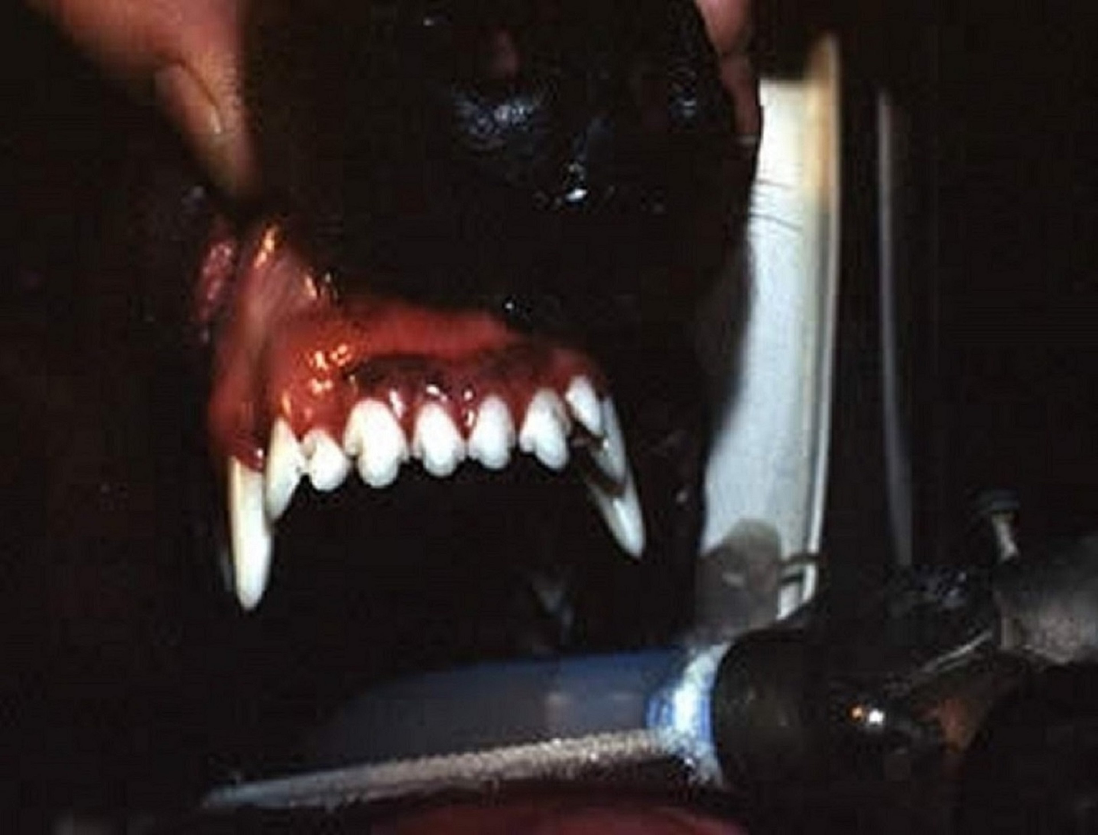
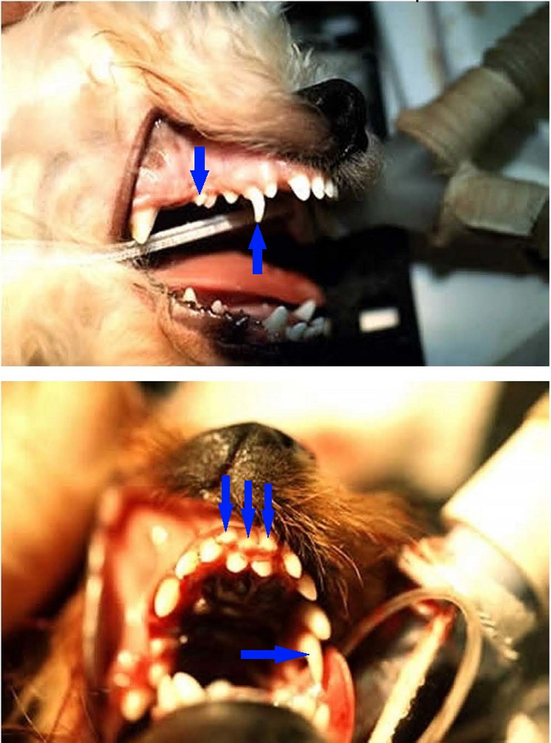
Hyperdontia, also called polyodontia—the existence of supernumerary teeth—occurs most often in the permanent teeth and can affect the incisors, premolars, and molars. In dogs, hyperdontia occurs most commonly on the maxilla and has a reported incidence of 7.6%. Presumably, these teeth arise from overproliferation of the dental lamina during development.
Supernumerary teeth tend to cause crowding and malocclusions, which can cause dysphagia, dental disease, and discomfort. In horses, supernumerary incisors are typically not extracted and are managed with regular reduction. For supernumerary cheek teeth, diastema and sinusitis due to oromaxillary sinus fistula formation are possible sequelae. Teeth are either extracted or reduced regularly to prevent complications.
Shedding Irregularities of Teeth in Animals

Courtesy of Dr. Gordon Baker.

Courtesy of Dr. Gordon Baker.

Courtesy of Dr. Ben Colmery III.
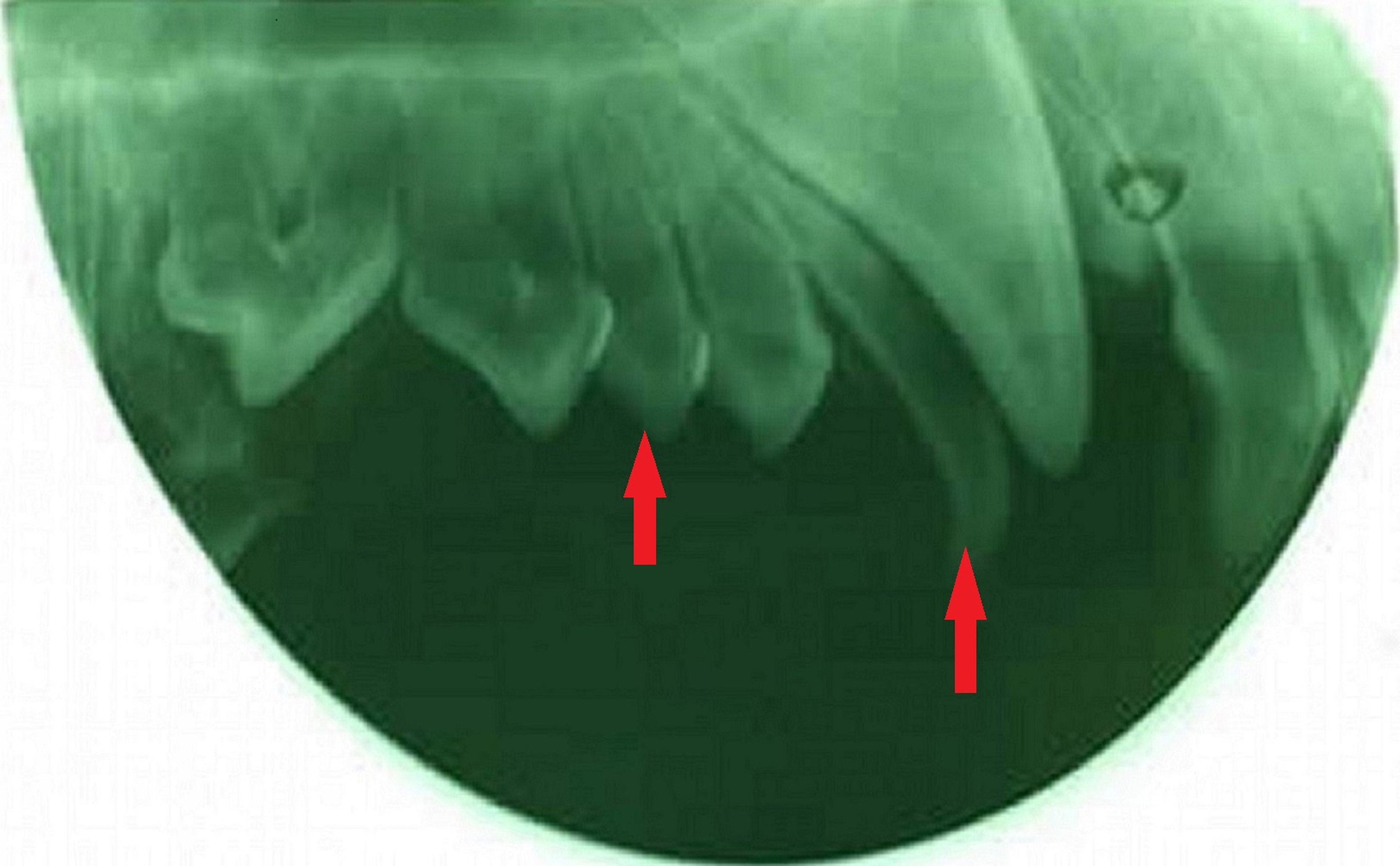
Retention of the deciduous teeth in horses is common. The incisors rostral to the permanent incisors tend to be retained; however, radiographs help to reveal their identity. Incisors retained in other orientations can result in malocclusion and/or displacement of permanent incisors. Retained cheek teeth are called caps, which are typically shed as the permanent teeth erupt underneath them. Loose caps can cause discomfort to the horse, manifested as head shaking, inappetence, weight loss, masticatory difficulty, quidding, and training problems. Caps can be extracted if they are loose, if the contralateral cap has already been shed, or if there is space between the cap and the permanent tooth below.
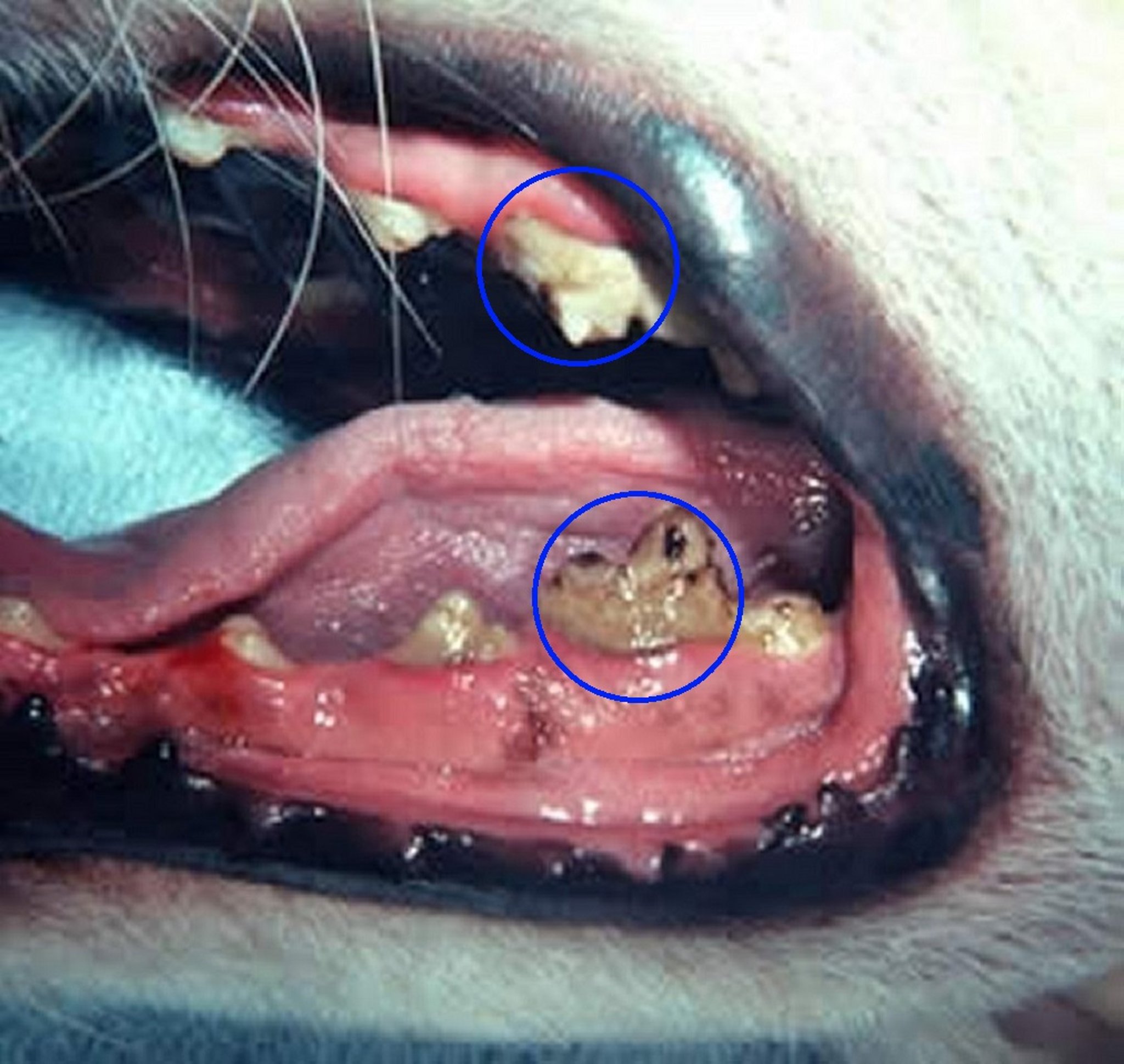
Retained deciduous teeth are common in dogs and secondary to the failure of the periodontal ligament to detach from the deciduous tooth, with the permanent canine teeth erupting rostrally. Small breeds are overrepresented, particularly Toy Poodles. Retention may lead to permanent tooth displacement, which can result in malocclusion or food entrapment and subsequent periodontal disease. Therefore, retained deciduous teeth should be removed as soon as possible, taking care not to damage the underlying permanent tooth bud.
Abnormalities in Tooth Position, Shape, and Direction in Animals
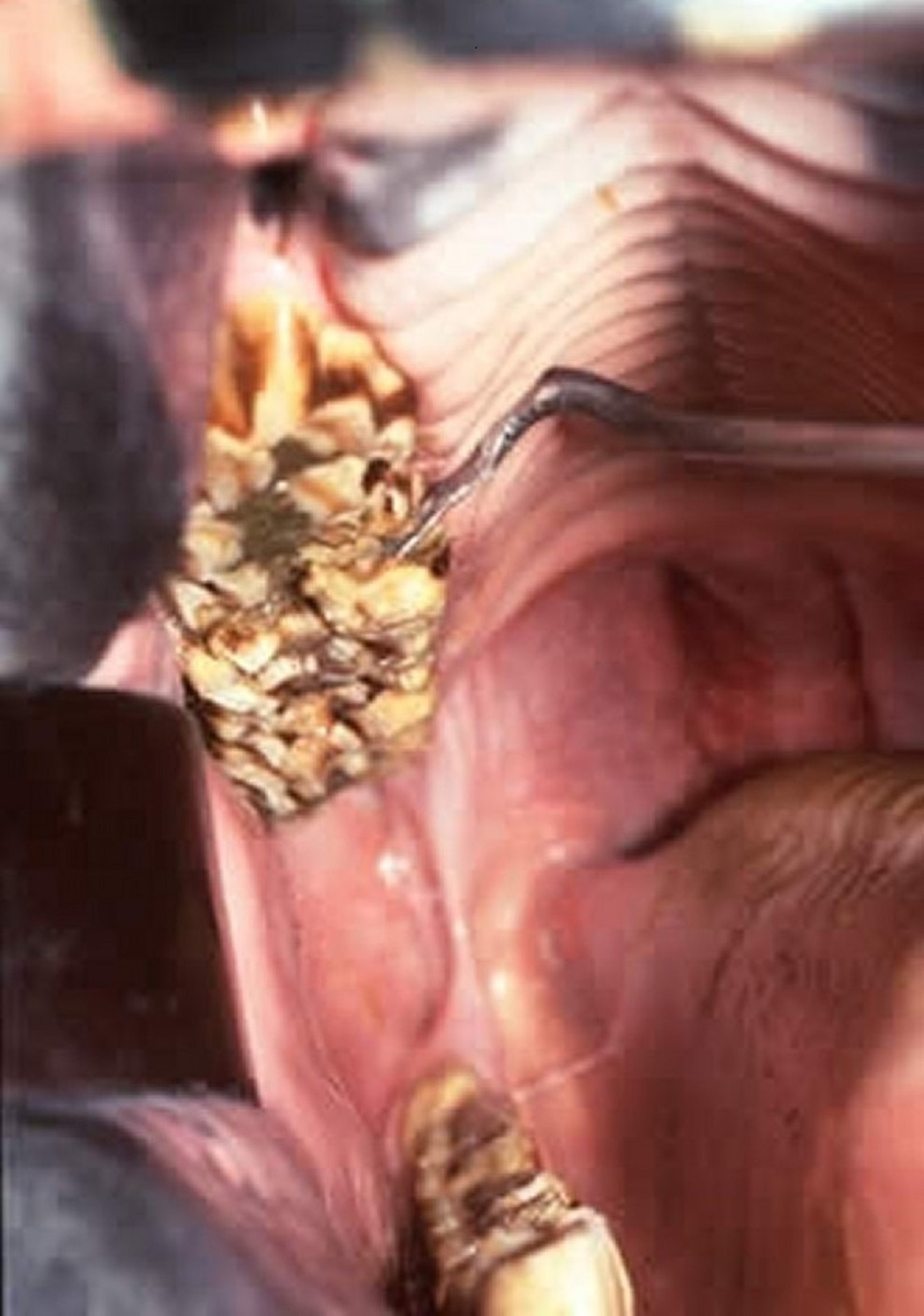
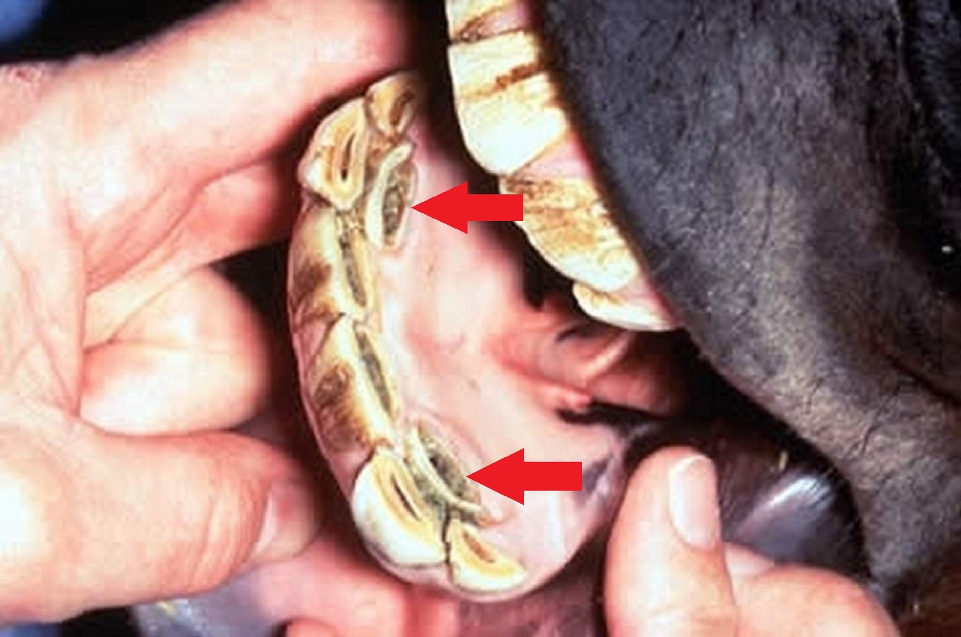
Displacement or rotation of teeth has been described in many species. In horses, the cheek teeth are affected more commonly than the incisors, and the permanent teeth are more often affected than the deciduous teeth. Most displacements are due to crowding during eruption. Sequelae include malocclusion, uneven wear and development of sharp points, and diastema with associated feed packing. Treatment includes regular floating of unopposed surfaces, or extraction if the condition is severe. Diastema can be addressed by mechanical widening. In dogs, rotation has been described commonly in brachycephalic and large breeds; the first mandibular premolar or upper third premolar is often affected. Abnormally located or directed teeth can result in malocclusions or affect the positioning of adjacent teeth. Extraction can be performed in severely affected patients; many cases are considered incidental findings.
Tooth Enamel Lesions in Animals

Courtesy of Dr. Ben Colmery III.
Enamel hypoplasia, hypomineralization, and dysplasia occur in both large and small animals. Common causes are pyrexia, trauma, malnutrition, toxicant or drug exposure during enamel development (eg, high concentrations of systemic fluoride, tetracycline), congenital disorders (eg, epitheliogenesis imperfecta in Saddlebred foals), and infections (eg, distemper in dogs or bovine viral diarrhea in calves) that affect ameloblast and odontoblast activity.
Lesions of enamel hypoplasia vary, depending on the severity and duration of the insult, from pitting of the enamel to the absence of enamel with incomplete tooth development. Affected teeth are prone to plaque and tartar accumulation, as well as subsequent bacterial penetration and the formation of caries. Teeth with exposed dentin, especially recently erupted teeth, may be more sensitive. Application of a dentin sealant or bonding agent may improve comfort in affected patients. In small animals, resin restoration has been used to cover defects, although diligent dental hygiene and home care is critical to decrease the incidence of complications. Enamel may also develop discoloration. In small animals, the administration of tetracyclines to pregnant females or to puppies < 6 months old may result in a permanent brownish yellow discoloration of the teeth. In ruminants, the enamel of some teeth may demonstrate flecks of varying color.
Ameliogenesis imperfecta, a type of hereditary enamel hypoplasia, has been documented in Swedish Standard Poodles, Italian Greyhounds, and Samoyed dogs.


