


Courtesy of UC Davis Comparative Ophthalmology Service.
Anterior uveitis or iridocyclitis is commonly diagnosed in dogs, cats, and horses but is observed in other species as well. It is often confused with other inflammatory conditions of the cornea and/or conjunctiva, so it is important to carefully assess patients for this condition.
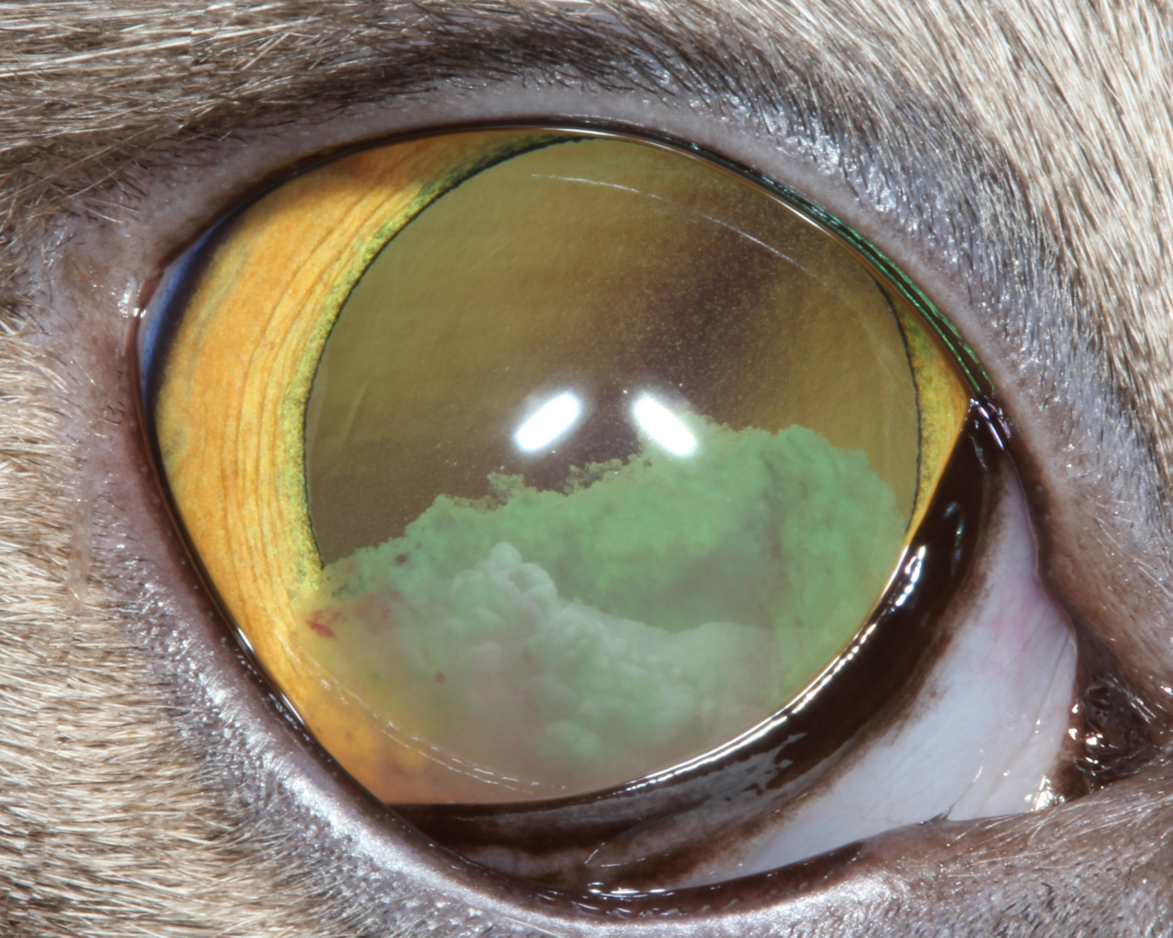
Clinical signs of acute anterior uveitis include:
blepharospasm
episcleral and/or conjunctival hyperemia
diffuse corneal edema
miosis
aqueous flare (indicative of protein and/or cells in the anterior chamber)
fibrin in the anterior chamber
hypopyon
hyphema
Chronic anterior uveitis, in addition, may exhibit anterior or posterior synechiae, keratic precipitates, dyscoria or irregular pupil shape, cataracts, and secondary glaucoma. Intraocular pressure (IOP) is typically low in patients with acute anterior uveitis but a normal or elevated IOP may be observed if aqueous drainage is decreased or obstructed by the inflammation. A fluorescein stain should always be performed to assess for concurrent corneal ulceration.
Anterior uveitis may be caused by other ocular disorders, including blunt or penetrating trauma, cataracts, a neurogenic reflex from corneal ulceration, lens instability, and intraocular neoplasia. Systemic diseases should be suspected, particularly when both eyes are involved. These include infectious and autoimmune diseases and metastatic neoplasia. Prognosis and therapy depends on the underlying cause.
Regardless of the diagnosis, therapy usually includes:
topical and/or systemic corticosteroids or NSAIDs
topical mydriatics
other drugs to target specific etiologies
Chronic or recurrent anterior uveitis (eg, uveodermatologic syndrome in dogs) is more challenging to manage because of the high likelihood of developing secondary cataracts, glaucoma, or phthisis bulbi.


