


Courtesy of UC Davis Comparative Ophthalmology Service.
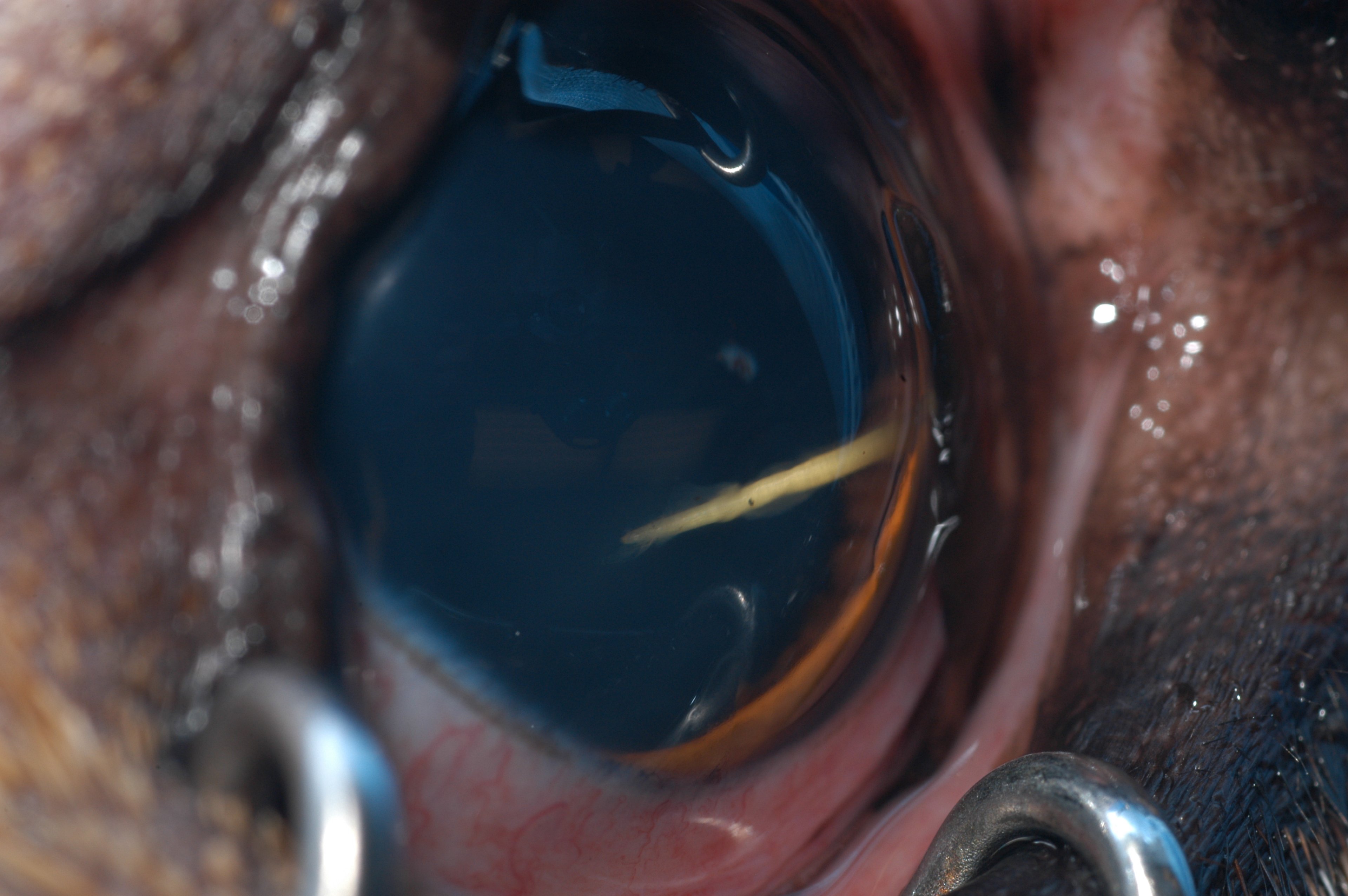
Penetrating intraocular injuries are seen most frequently in dogs and cats. They are often associated with cat claw injuries or plant foreign bodies (eg. cactus), but lead pellets and bullets that partially or totally traverse the ocular tunics can also result in a penetrating injury. Anterior lens laceration and rupture is a common sequela of cat claw injuries in young dogs. Thus, it is critical to assess for lens perforation, which can lead to cataract formation or severe, uncontrollable uveitis, particularly if bacteria are implanted within the lens.
Patients should also be assessed for vitreous and/or retinal hemorrhage, and retinal detachments are likely if the injury is from a lead pellet or bullet. Corneal cytology and aerobic bacterial and/or fungal cultures should be considered, particularly if the injury is chronic or demonstrates signs of infection, including stromal cellular infiltrate, stromal loss, and/or keratomalacia. A fluorescein stain should be performed to assess the size of the lesion and to determine whether active aqueous humor leakage is present (Seidel test). Ophthalmic ultrasonography and orbital radiology are helpful to assess pellet location and the integrity of the intraocular and orbital tissues.
Traditionally, it was thought that penetration of the anterior lens capsule (lacerations >2 mm) required lens removal as soon as possible, because escape of lens material causes gradually intensifying lens-induced uveitis that often progresses to secondary glaucoma and phthisis bulbi. However, a recent study suggests that dogs and even cats with large corneal lacerations and concurrent lens capsule ruptures can be successfully managed with medical therapy that includes topical and systemic broad-spectrum antibiotics, a topical mydriatic (eg, atropine), and systemic corticosteroid or NSAID.
The visual prognosis is guarded if the posterior segment is involved, particularly if the retinal detachment is large. If the penetrating injury only involves the cornea, then the prognosis for vision and globe retention is good. However, concurrent lens capsule rupture and posterior segment involvement carries a more guarded prognosis. In cats with lens capsule involvement, a discussion of the risk of traumatic lens-induced sarcoma is warranted, as well as the requirement for regular dilated ocular examinations.
Regardless of whether medical or surgical therapy is instituted, it is critical that the patient is kept quiet and an Elizabethan collar is placed to prevent self-trauma. Frequent rechecks will also be required to ensure proper healing is occurring and to assess for concurrent infection.
For More Information
Also see pet health content regarding penetrating eye injuries in animals.


