

Many management principles applied to other species are equally relevant for fish, including taking a good history, considering appropriate diagnostic techniques, and being cognizant of therapeutic options, water quality issues, and quarantine and biosecurity procedures.
Physiology
Fish are poikilothermic, and all physiologic processes are greatly influenced by water temperature. In freshwater, the internal tissues of fish are hyperosmotic to the environment, whereas in saltwater they are hypoosmotic. Surface injuries to the skin make osmoregulation more difficult and may be of serious consequence because of the loss of fluid balance and circulatory collapse.
The structure of the fish kidney varies with the species; generally it is divided into an anterior (head) kidney, which is usually located anterior to the gas bladder, and a posterior (caudal) kidney, which is retroperitoneal and ventral to the vertebral column. Hematopoietic, renal, and endocrine tissues are found in the kidney, with hematopoietic tissue located cranially and excretory tissue located caudally. Divalent ions are excreted principally via the kidney, and monovalent ions and nitrogenous excretions via the gills. Accordingly, lesions of the kidney and gills may seriously interfere with respiration, excretion, and fluid balance.
The gas bladder (also known as the swim bladder or air bladder) in bony fish, which originates as an appendage of the foregut, regulates body buoyancy and may also be used for sound production. Physostomous fish have an open connection between the gas bladder and the GI tract, whereas physoclistous fish do not. Although a single chamber in most fish, the gas bladder consists of dual chambers in cyprinids and three chambers in cod and suckers belonging to the genus Moxostoma.
A sensory lateral line system along the sides of the body and head receives stimuli from the aquatic environment and mediates adaptive responses through the CNS.
Fish depend on increases in environmental temperature for efficient antibody production during infections (or after vaccinations), when most pathogens are replicating at a more rapid rate. The optimal temperature for antibody production varies with the species of fish (tropical, temperate, or coldwater). Extremes in environmental temperature (above or below that of the natural habitat) inhibit antibody production. Like T lymphocytes in other vertebrates, those in fish are responsible for cell-mediated immunity. Immunity is not as age dependent in fish as it is in other animals; young fish are usually immunocompetent and can be vaccinated successfully. Antibodies are found in the mucus of the fish skin and GI tract.
Although vaccination of fish against specific diseases has been economically important in preventing losses, there is a need for improved methodology. Advances include increased use of autogenous vaccines; several companies will work with veterinarians and their clients to develop custom vaccines for specific situations. A few vaccines (eg, for Aeromonas salmonicida) are available or in development for pet fish, particularly koi.
Obtaining a History and Clinical Information
As with all species, a good history is critical to establish a diagnosis. Questions of particular interest for fish cases include the number of animals affected, whether one species or multiple, the chronicity of the problem, and a thorough description of animal housing and care, including the volume and design of the system, number and size of animals stocked, species, new additions, quarantine protocol, and previous medications.
Owners can be asked to bring fish and water samples into the clinic, or the practitioner may wish to visit the site. Site visits allow the system to be more accurately evaluated and the behavior of fish readily observed. If fish are brought to the clinic, the owner should provide an animal showing clinical signs of disease. A live animal can be transported in a cooler with a battery-powered aerator, or in a sturdy plastic fish bag with just enough water to cover it. A separate water sample should be provided in a plastic bag or bottle, ensuring no air bubbles are included with the sample, and transported on ice. A minimum of 500 mL of tank water should be requested for analysis.
If the animal must be anesthetized for examination, a larger volume of tank water should accompany the animal and is used in anesthetic recovery. Specimens that have been dead less than 24 hours and promptly stored at 4°C have diagnostic value and may be submitted to the veterinary clinic, or the owner may be directed to submit these to a laboratory experienced in fish necropsy and diagnostic testing. Water samples should be submitted with necropsy specimens.
Necropsy and Diagnostic Techniques
Although the same principles are used in necropsy of fish as in other animals, great emphasis is placed on an accurate and thorough history, premortem signs, fresh necropsy material, and direct microscopic examination of fresh tissue smears and squash preparations. Fish decompose quickly, and many saprophytic microorganisms reproduce rapidly in the decaying tissues, which complicates isolation of pathogens unless samples are collected immediately after death.

Dr. Denise Petty.
A general fish necropsy may include premortem blood collection; biopsy of gill, skin, fin tissues, and internal organs; bacterial or viral culture of internal organs; and histologic evaluation. A veterinary clinic or diagnostic facility familiar with fish necropsy protocols and aquatic microbiology should be used. Whenever possible, live fish should be submitted. If the fish has just died, the eyes should be clear and the gills normal in coloration and texture. There should not be the proverbial dead fish smell, as fish carcasses autolyze rapidly and are unusable for examination; however, some moribund fish may have a strong odor. Freshly dead fish should be placed in a sturdy plastic bag and submitted on ice. A water sample should always be submitted with the fish. An animal that has died and been placed in a freezer has limited diagnostic value, but freshly dead frozen fish may be useful for bacteriologic, virologic, or toxicological testing.
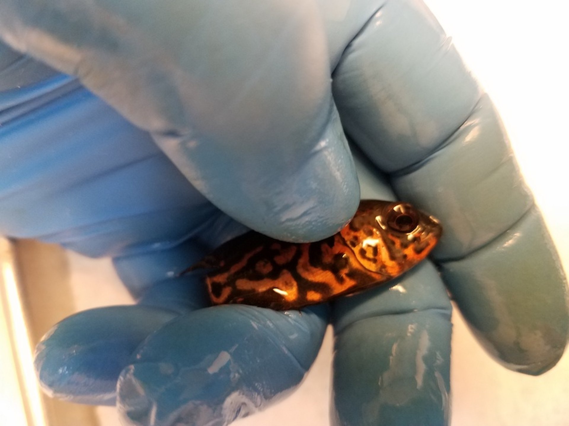
Moribund live fish can also be examined. It is possible to gently restrain some fish for some procedures. It is easier to restrain small fish than larger ones. When handling fish, nitrile exam gloves should be worn to prevent damage to the fish's epithelium. When holding fish in one's hand, only gentle pressure should be used. Stronger pressure often causes the fish to become more fractious. Unanesthetized fish should only be restrained for the few seconds it takes to obtain a skin mucus sample or fin biopsy or other nonlethal diagnostic technique. After sample collection, they should be returned to water immediately.
When it isn't possible to safely restrain fish for nonlethal diagnostic procedures, sedation should be used. The most common sedative agent used for fish is tricaine methanesulfonate (MS-222), which must be adequately buffered with sodium bicarbonate (at a ratio of 1:2 MS-222 to sodium bicarbonate). Unbuffered MS-222 should never be used; it is very acidic which can not only damage fish epithelium but can also cause some parasites to detach from the fish.
Fresh tissue samples of gill filaments, skin mucus, and fins should be collected, prepared as a wet mount, and examined under a light microscope at 40×, 100×, and 400×. Freshwater should be used to prepare wet mounts of external tissues from freshwater fish, and saltwater should be used to prepare wet mounts from marine fish. If uncertain, water from the tank or from the submitted water sample should be used. Ensuring that the salinity used to prepare mounts is similar to the salinity present in the environment should allow organisms to remain viable long enough for identification. Distilled water should not be used for tissue samples. Tissue should be examined for morphology and for the presence of parasites, bacteria, or fungal elements. Wet-mount examination of fish tissues is crucial for diagnosis of most parasites.
Selected Blood Collection Sites
Site | Technique and Landmarks |
|---|---|
Dorsal aorta | With the fish in ventral recumbency, insert the needle into the mouth and on the midline where the gill arches on each side meet until contact with bone is made. Slightly withdraw the needle until blood appears in the needle hub. This is easier to perform in a fish that has a large mouth opening. |
Ventral aorta | Insert the needle on the ventral midline just anterior to the heart. The heart is located in the most anterior ventral part of the body cavity. |
Cardiac | Insert the needle on the ventral midline into the heart (located in the most anterior ventral part of the body cavity). In small fish, this procedure may be lethal. |
Duct of Cuvier or common cardinal vein | Lift the operculum and insert the needle behind the 4th gill arch, directing the needle caudally so it enters the isthmus of the body. |
Gill arch | This technique is only suitable for large fish (total length, >700 mm). Insert the needle directly into a gill arch until an artery is reached. |
Caudal vessels | Lateral approach: Insert the needle at a 45° angle just below the lateral line of the caudal peduncle, and advance cranially until contact with the vertebral column is made. Then direct the needle ventral and lateral to the vertebral column as the syringe is gently aspirated. Ventral approach: Insert the needle on the ventral midline and advance dorsally until contact with the vertebral column is made, and then withdraw slightly. |
Fish should be sedated or anesthetized prior to blood collection. | |
Blood can be collected from a number of sites, and hematologic parameters measured. In fish >25–100 g, depending on species, the caudal vessels of the caudal peduncle, the duct of Cuvier (common cardinal vein), and the dorsal and ventral aortas are easily accessible. For smaller specimens that are to be euthanized, blood can be collected in a hematocrit tube immediately after euthanasia by severing the caudal peduncle and exposing the caudal vessels.
Use of hematologic evaluation and serum biochemical analysis is limited because normal values for many fish species are not readily available; however, the information may still be clinically useful. Lithium heparin is the anticoagulant of choice for most fish species, although EDTA is preferred for ictalurid catfish, and plasma may be used for biochemistry tests. Serologic testing may be diagnostic in certain cases (eg, heavy metal toxicoses). Whole blood (1–2 drops) can be incubated in brain-heart infusion broth at room temperature on an electric rotator. If cloudiness indicative of bacterial growth develops, a loop of the blood-broth mix can be used to attempt primary isolation of a systemic bacterial pathogen.
If not already dead, moribund fish should be euthanized and a necropsy performed under aseptic conditions. Microscopic examination of internal organs is highly recommended if the fish is dead. Unstained sections of stomach and intestine should be examined for presence of parasites, and the lower intestine should be examined for flagellates. Unstained sections of spleen, anterior and posterior kidneys, and liver should be examined for presence of parasites, granulomas, or other anomalies.
Specialized Culture Media
Culture Media | Pathogens |
|---|---|
Marine agar | Heterotrophic halophilic bacteria; used for external bacterial disease |
Thiosulfate-citrate-bile salts-sucrose agar (TCBS) medium | Vibrio spp |
Cytophaga agar | Flavobacteria (Flavobacterium columnare, F psychrophilum) |
Tryptone yeast extract salt agar (TYES) | Flavobacteria |
Hsu-Shotts medium | Flavobacteria |
Lowenstein Jensen medium | Mycobacteria |
Middlebrook media | Mycobacteria |
Sabouraud dextrose medium | Fungal isolation |
Low nutrient water agar | Saprolegnia |
A sterile swab of anterior kidney, spleen, or other organ of interest, may be shipped to a laboratory in transport media, but primary isolation directly onto an enriched media (eg, tryptic soy agar enriched with 5% sheep blood) is preferable. Specialized culture media can be used for the growth of certain pathogens See table Specialized Culture Media. If abscesses or other obvious anomalies are visible, those sites should also be sampled for culture.
In general, bacterial or fungal cultures taken from tropical fish should be incubated at room temperature (25°C) or no higher than 30°C. Some agents of concern will not grow at all at 37°C, the standard temperature for incubation of cultures taken from mammals. Agents of zoonotic concern, such as Mycobacterium, can be dual incubated at both 25°C and 37°C. An acid-fast stain should be available for benchtop staining of granulomatous material which, when positive, is strongly suggestive of Mycobacterium. If fish are seen spinning or showing other behavioral indications of neurologic disease before death, cultures on brain tissue are indicated.
Fish Diseases of Regulatory Concern in the US
Disease | Causative Agent | Susceptible Species | Clinical Signs and Pathology | Temperature Range | Status in US |
|---|---|---|---|---|---|
Viral hemorrhagic septicemia (Egtved disease) | Novirhabdovirus Family: Rhabdoviridae | Primary: salmonids (Oncorhynchus spp), turbo, herring and spat (Clupea spp), Japanese flounder Secondary: grayling, whitefish, pike, Atlantic and Pacific cod, haddock, and many freshwater, marine, and estuarine species | Acute form: nonspecific hemorrhaging (eyes, fins, or skin), darkening, exophthalmia, ascites Chronic form: few signs Neurologic form: spinning or flashing Gross: enlarged spleen, ascites, necrotic kidney Histologic: focal necrosis of kidney liver, spleen; hemorrhage in muscle | 48°–54°F (9°–12°C) optimal | Present in wild populations, sporadic, limited distribution; endemic in Pacific Northwest and Alaska (wild salmonids, haddock, and cod); emerging disease in Great Lakes region in a wide variety of fishes. |
Infectious hematopoietic necrosis | Novirhabdovirus Family: Rhabdoviridae | Primary: cultured salmonids (Oncorhynchus spp); lake trout and char (Salvelinus spp) are resistant | Rapidly increasing mortality (fish < 1 year), lethargic but sporadic bursts of rapid swimming occur, protruding vent, fecal casts, exophthalmic, pale gills, darkening, abdominal distention or ascitic fluid (possibly bloody) | 50°–54°F (10°–12°C) optimal; rare >15°C (59°F) | Present in western US, sporadic, limited distribution; endemic in Pacific Northwest and Alaska (wild salmonids); also present in parts of Europe and Asia |
Spring viremia of carp | Vesiculovirus Family: Rhabdoviridae | Primary: carp (including koi, goldfish), sheatfish (European catfish), orfe, tench | Nonspecific: darkening, exophthalmic, pale gills, distended abdomen, ascites, hemorrhage (gills, skin, or eye), petechiae in organs (including swim bladder), protruding vent with thick mucoid fecal cast Coinfection with Aeromonas or other bacteria common | 54°–72°F (12°–22°C) | US is free (last occurred in captive fish in 2004, wild fish in 2007); occurs in eastern Europe, Russia, China, and Middle East |
Epizootic hematopoietic necrosis | Ranavirus Family: Iridoviridae | Primary: redfin perch Secondary: rainbow trout (wild and farmed fish) | Acute and high mortality of redfin perch; darkening, ataxia, lethargy, hemorrhage around nares; morbidity and mortality of rainbow trout less severe; Histologic: necrosis, renal hematopoietic tissues | Redfin perch: 54°F (>12°C) Rainbow trout: 52°–63°F (11°–17°C) Experimental: 46°–70°F (8°–21°C) | Has never occurred in US; endemic in Australia Endemic in Australia |
Red sea bream iridoviral disease | Megalocytivirus Family: Iridoviridae | Red sea bream, many other estuarine species, other marine species | Severe anemia, lethargic, pale gills, enlarged spleen | Has occurred in imported tropical marine fish in US; occurs in Japan and Taiwan | |
Infection with HPR-deleted or HPR0 Infectious salmon anemia | Isavirus Family: Orthomyxoviridae | Atlantic salmon Brown trout, sea trout, rainbow trout | Pale gills, severe anemia (PCV < 10%), swollen liver (black or brown color), ascites, petechiae of viscera, mesenteric fat, swim bladder | In vitro: Maximum replication 59°F (15°C) No replication 77°F (25°C) | Present in northeast US, sporadic, limited distribution; endemic in Maine, New Brunswick, Scotland, and Norway Endemic in Maine, New Brunswick, Scotland, and Norway |
Infection with salmonid alphavirus (pancreas disease or sleeping disease) | Alphavirus Family: Togaviridae | Atlantic salmon, rainbow trout, brown trout | Necrosis of exocrine pancreas, heart and skeletal muscle changes | 54°–59°F (12°–15°C) | Has not occurred in US; detected in Ireland, UK, France, Germany, Italy, Spain, Switzerland, Poland, and Norway |
Koi herpesvirus | Cyprinid herpesvirus-3 Family: Alloherpesviridae | Common carp and hybrids, including koi and ghost carp | Severe necrosis gill tissue | 72°–78°F (22°–25.5°C) optimal* | Present in US, sporadic, widely distributed |
Epizootic ulcerative syndrome (mycotic granulomatosis) | Aphanomyces invadans Oomycetes (water mold) | Atlantic menhaden, striped mullet, many other freshwater and estuarine species; snakeheads, barbs (Puntias spp) sensitive; gouramis, goldfish and other ornamentals susceptible; tilapia resistant | Necrotizing deep ulcers (penetrate body wall), granulomatous tissue response; deep ulcers with red centers, white rims; invasive nonseptate hyphae (culture possible but difficult) | 77°F (< 25°C) (reduced salinity also contributes in brackish systems) | Present in US, sporadic, limited distribution; reported in North America, Africa, Asia, and Australia |
Gyrodactylus (Gyrodactylus salaris only) | Monogenea Gyrodactylus salaris only | Atlantic salmon, rainbow trout, brook trout, North American lake trout, brown trout, grayling arctic char | Has never occurred in US | ||
*Mortalities stop at 86°F ( >30°C) , but survivors remain carriers. | |||||
If viral disease is suspected, appropriate tissues may be collected. Specimens should include both fresh tissues placed in reagent ethanol and frozen tissues. Several viral diseases are of regulatory concern to veterinarians practicing fish medicine in the US ( see Table: Fish Diseases of Regulatory Concern in the US).
Tissue sections no larger than 1 cm3 should be placed in neutral-buffered 10% formalin for histologic evaluation. After 24 hours of fixation, they should be removed and placed in reagent alcohol in case molecular diagnostic techniques will be required.
Treatment Considerations
Treatments for pet and ornamental fish is often based on environmental management followed by the use of targeted therapy to control specific pathogens. Use of prophylactic medication in the absence of diagnostic testing is strongly discouraged and may contribute to resistant bacterial infection and other complications.
Drug therapy can be provided via several routes of administration, including exposure by bath (adding medication to water), medicated feed, injection, or topical administration. Generally speaking, bath and topical treatments are most useful for external infections, whereas medicated food and injection are most appropriate for internal infections.
Using a bath treatment requires accurate measurement of the volume of water to be treated (see sidebar for formulae on tank volume calculations).
Some medicated feeds can be purchased commercially for aquaculture or pet fish. Custom-made medicated feeds can be prepared for use in ornamental fish. Flake, pellet, or gel diets can be used as a base for medicated food for pet fish. Cooking oil spray is an effective binder for use with pelleted or flake foods for ornamental fish. The addition of medication to commercially available gel diets is easily done as the gel cools. In general, medication should not be added when the gelatin is hot, because some medications, particularly oxytetracycline, are heat labile.

Courtesy of Dr. Denise Petty.
Courtesy of Dr. Denise Petty.
Courtesy of Dr. Denise Petty.
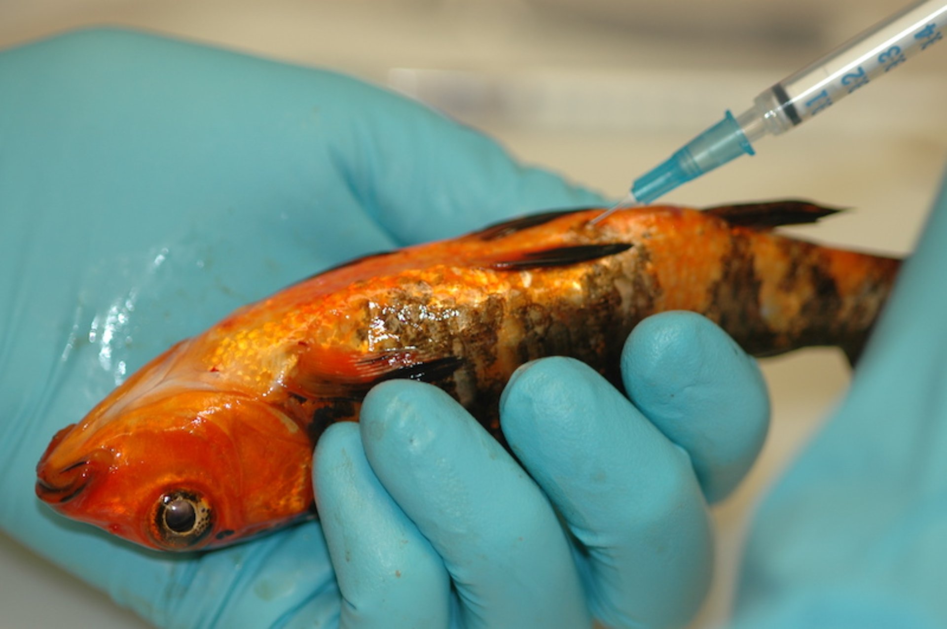
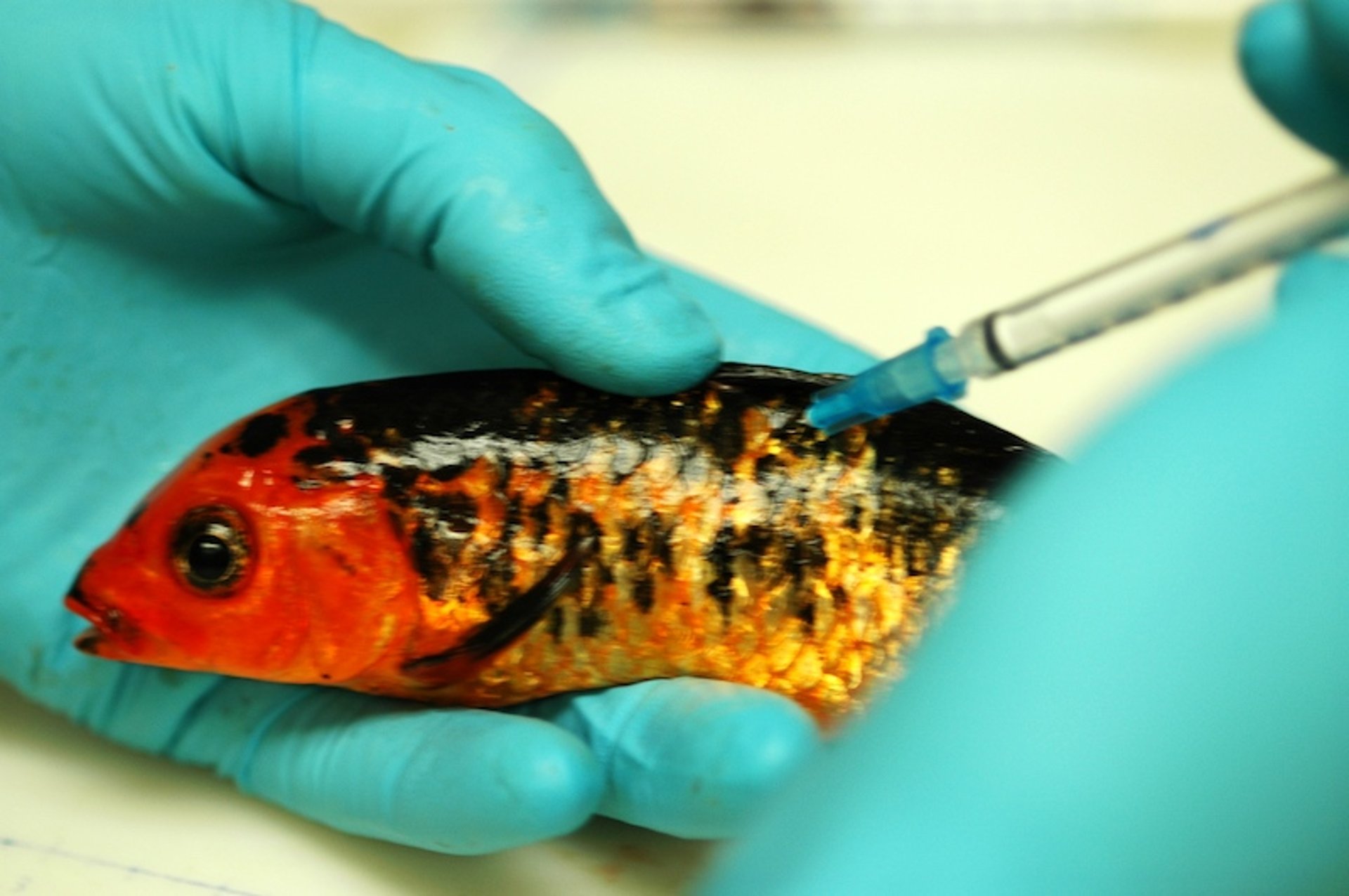
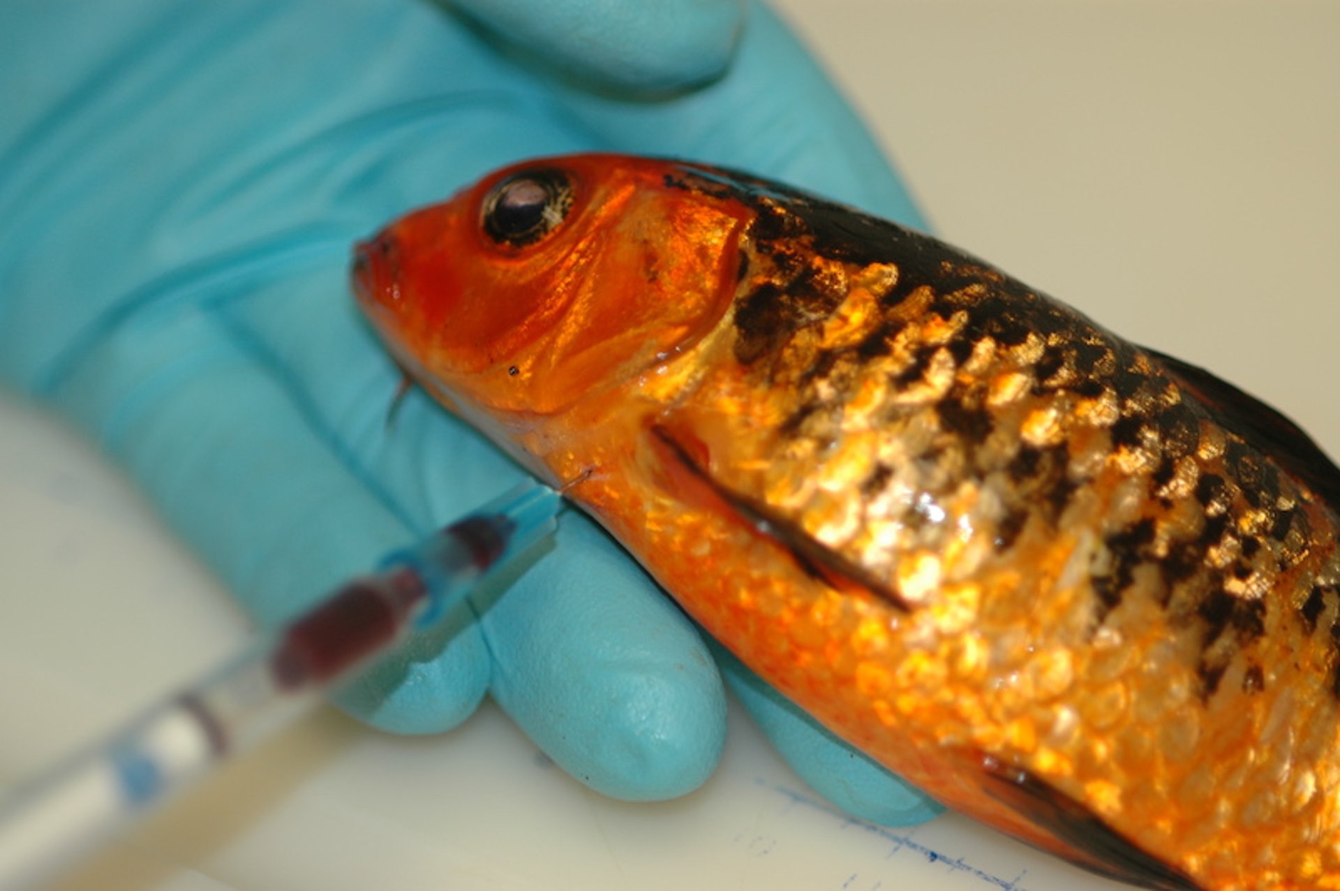
Injections can be given by either IM or intracoelomic (ICe) routes. Intramuscular injections are given in the epaxial muscles, lateral to the dorsal fin. Injection of some drugs can cause muscle necrosis, so it is important to alternate injection sites and to limit injections to every 3 days unless required more often. To administer ICe injections, fish should be placed head downward in dorsal recumbency to move internal organs away from the injection site, which should be anterior to the anus, just off the ventral midline. Topical treatments, usually in ointment form, should be applied directly to the external lesions. The fish can be manually restrained, usually without removing the entire animal from the water. The area to which the treatment is applied should be held out of the water for several seconds (< 1 minute) to allow some absorption before returning the fish to the water. Repeated applications may be necessary.
Sunburn can occur in surface-swimming fish or can be induced (even in bottom-dwelling species) by feeding photodynamic drugs such as phenothiazine, although ultraviolet light penetrates water poorly. Affected fish will have visible lesions along the dorsal surface. Providing shade solves the problem.
FDA-approved Drugs and Regulatory Concerns for Aquarium Fish
In many cases, therapeutic management of fish other than catfish or salmonids requires extralabel use of drugs as approved therapeutic options in fish are limited. The FDA website provides current information on the status of drugs and chemicals. Drug indexing has been developed by the FDA to allow for legal use of unapproved drugs in minor species, including ornamental fish. The FDA publishes a list of indexed drugs that are not FDA approved but are considered legally marketed if used exactly as labeled. The following information is intended for use in pet fish medicine.
Antimicrobial Use in Aquarium Fish
Antimicrobials can be delivered to pet fish through any of the treatment routes listed; however, medicated food is most common and usually most effective. Common antimicrobials used in pet or ornamental fish include oxytetracycline, potentiated sulfa drugs, and enrofloxacin (in koi and exhibit fish). For approved dosages and withdrawal times, see the FDA website. Oxytetracycline can be fed at a dosage of 55–83 mg/kg/day for 10 days to control many gram-negative bacterial infections, including columnaris disease. Because oxytetracycline has been on the market for many years, there may be significant resistance to it in some bacterial isolates. Sulfadimethoxine and ormetoprim also is effective against many gram-negative organisms, with less resistance. Palatability can be a problem when feeding medicated food to sick fish, which may have a poor appetite.
Florfenicol is available ommercially for use in medicated feed ( See also page Aquaculture).
Erythromycin is not approved by the FDA for use in fish but is an excellent antimicrobial for fish infected with gram-positive bacteria, particularly Streptococcus. Erythromycin can be fed at a dosage of 100 mg/kg/day, for 14 days. Palatability may be a concern with erythromycin-medicated feed. Erythromycin has been used in management of bacterial kidney disease of salmonids and streptococcal infections in food and nonfood species. Permission from the FDA is required for use in food animals in the US.
Delivery of antimicrobials in a bath treatment is not generally recommended because of unknown or limited efficacy and damaging environmental effects (ie, antimicrobials tend to kill the nitrifying bacteria in the biofilter). Oxytetracycline (100–400 mg/L for 1 hour, daily for 10 days) has some efficacy when delivered in a bath. Oxytetracycline in a bath treatment is chelated by hard water and is therefore ineffective in marine systems. Enrofloxacin has been used as a bath at 2.5–5 mg/L for 5 hours, daily for 7 days. Water changes are recommended after the 5-hours contact time, and the drug may be chelated by hard water. Kanamycin has also been used as a bath treatment at dosages of 50–100 mg/L for 5 hours, repeated every 3 days for three treatments, with water changes recommended after the 5-hour contact time. Nephrotoxicity is a concern in fish treated with aminoglycosides. Many other options are discussed in the literature.
Injection is the most effective way to control the amount of antimicrobial delivered to a fish. The intracoelomic (ICe) route is preferred over the IM route. Enrofloxacin can be delivered ICe at a dosage of 5–10 mg/kg, and the higher dosage can be repeated every third day, minimizing handling. Three treatments are generally recommended. When using enrofloxacin, the less concentrated dose (22.7 mg/mL) is recommended, even for use in large fish, because of adverse tissue reactions with the concentrated dose. If injection volume is excessive, more than one injection site can be used. Other injectable antimicrobials include amikacin (5 mg/kg, IM, every 3 days for a total of three treatments). As with other aminoglycosides, nephrotoxicity may be a concern. Erythromycin (10 mg/kg/day, IM, for 3 days) can be used to treat gram-positive infections in large fish.
Use of topical ointments containing antimicrobials can be practical in pet fish. Some seem to absorb fairly quickly, but if treating a substantial wound, it may be appropriate to cover it with a waterproof compound. Frequent application of antimicrobial ointment (eg, twice a day) can work well to treat superficial ulcers in pet fish.
Parasiticide Use in Aquarium Fish
Several brands of formalin (37% formaldehyde in aqueous solution, not 10% formalin used for fixation of tissues for histology) are FDA approved as parasiticides for use in finfish and penaeid (saltwater) shrimp (see the FDA website). These products are usually used as a prolonged bath at concentrations of 15–25 mg/L. Vigorous aeration during formalin treatment is essential. A concentration of 25 mg/L is equal to 2 drops/gal. or 1 mL/10 gal. (useful for delivering formalin to aquarium fish).
Salt is categorized as of low regulatory concern by the FDA and has many uses in fish medicine, including destruction of protistans and management of osmoregulation. Seawater typically has a salinity of 32–37 g/L. By increasing or decreasing the amount of salt to which a freshwater or marine fish is exposed, osmoregulatory stress can be minimized and many external parasites eliminated (see sidebar on using freshwater or saltwater dips). A salt solution of 3–5 g/L is recommended for transportation of freshwater fish; most species will tolerate this concentration for several hours or days. Permanently increasing salinity to 2–3 g/L in a freshwater system can minimize some parasitic protistans. Lowering salinity to 16–18 g/L can be very useful when treating some parasitic diseases, particularly Cryptocaryon.
Use of copper in freshwater systems can be dangerous if the dose is not carefully calculated. The total alkalinity of the water to be treated must be known, as it is part of the equation used to calculate the concentration of copper that is needed (see the information on copper sulfate in Aquaculture: Other Chemotherapeutic Drugs). In saltwater systems, copper is sometimes applied in a chelated form, because it stays in concentration longer. Chelated compounds may be difficult to use safely and require careful monitoring. Copper sulfate (CuSO4) can be used to treat marine fish, but the concentration of active copper ions (Cu2+) must be closely monitored (test kits are available) and should be maintained at 0.18–0.2 mg/L for up to 3 weeks. When using over-the-counter products in marine aquaria, label directions should be followed. The Cu2+ concentrations should be tested at least once a day, but note that copper test kits must be selected for their ability to test the form of copper being used. Copper is extremely toxic to invertebrates and plants, which must be removed before the water is treated. Finally, copper will adversely impact the nitrifying bacteria in biofilters, and a transient increase in ammonia and nitrite should be expected for weeks to months after treatment. Monitoring of ammonia and nitrite until measurable concentrations subside is recommended.
Organophosphates have been used in nonfood fish practice for decades to control monogeneans, parasitic crustaceans, and leeches. Organophosphates can be used in freshwater systems, for ornamental fish only, at concentrations of 0.25 mg/L as an indefinite bath. Most compounds are sold as 37.3% active ingredient liquid. Toxicity and efficacy are affected by pH, with more acidic pH resulting in increased toxicity. For this reason, an increased dosage may be necessary in marine ornamental fish systems (pH 8–8.3), with concentrations up to 1 mg/L used by some facilities. Some veterinarians add atropine (0.1 mg/kg, PO, IM, or ICe) to the food of sensitive freshwater fish and marine exhibit fish before treatment with organophosphates. Because of environmental concerns, organophosphates should not be used in outdoor ponds, unless specific provisions for such use exist in state law and are followed by the veterinarian. After treatment with organophosphates, most facilities hold water for up to 96 hours before allowing any discharge, and divers are usually restricted from entering exhibit tanks for at least 48 hours. Organophosphates may not be used in food fish in the US.
Diflubenzuron is a chitin synthesis inhibitor effective against anchor worms (Lernaea), fish lice (Argulus), and other crustacean parasites in aquarium fish only. It is used as a prolonged bath at concentrations of 0.03 mg/L. It has a fairly long half-life (>1 week), and treated water should be retained for 28 days and then run through a carbon filter before discharge.
Metronidazole is used to control intestinal protistans and can be delivered in a medicated food or as a bath when fish are anorectic. Although very effective against Spironucleus spp, metronidazole does not seem to be effective against gastric infections with Cryptobia iubilans. A concentration of ~7 mg/L (~250 mg metronidazole dissolved in 10 gal. of water) can be administered daily for 5 days. A daily water change a few hours after treatment is recommended. Metronidazole can be administered in medicated feed at 50 mg/kg, PO, for 5 days. Metronidazole may not be used in food fish in the US.
Several anthelmintics have been used to control intestinal nematodes in fish; however, efficacy and safety is not known for many species. Fenbendazole has been used at a dosage of 25 mg/kg, delivered in food for 3–5 days. When fenbendazole has been used in a bath treatment or via gavage, high mortality has occurred. Consequently, this use is not recommended. Levamisole administered in a bath treatment at a concentration of 2 mg/L is also used by some clinicians. Administration has been described in a sandbar shark (10 mg/kg, IM) as well as in feed, although the dosages varied widely, ranging from 5-10 mg/kg up to 300 mg/kg. Though ivermectin has a low safety margin and must be used with caution, it has often been used in feed at the rate of 0.05 mg/kg. Emamectin has also been used in feed at 35 mg/kg once a day for 14 days. It is reported to have a higher safety margin than ivermectin. None of these anthelmintics are legal for use in food fish in the US.
Praziquantel is selective for flatworms, so consequently it is used to control cestodes and external monogeneans. The most common use of praziquantel is as a prolonged bath in large marine aquaria for control of capsalid monogeneans. It is applied at a concentration of 5 mg/L and may remain active for several weeks. Treated water should be run through an activated carbon filter before discharge. Praziquantel can also be administered at a dosage of 35–125 mg/kg, PO, for up to 3 days or as a short-term bath treatment at a concentration of 10 mg/L for 3 hours. Efficacy has been demonstrated in yellowtail kingfish fed praziquantel at a dosage of 50 mg/kg/day, for 7 days. Use of praziquantel is not permitted in food fish in the US.
Chloroquine has been used to control Amyloodinium spp in ornamental marine fish. It is applied as a prolonged bath at concentrations of 10 mg/L. Efficacy in recirculating systems seems to be very good; however, there are essentially no data on treatment intervals, effects on biofilters, or other basic husbandry effects. Weekly examination of fish, including biopsy of infected tissue, is recommended to assess treatment efficacy. Some aquarists have used chloroquine (10–15 mg/L for 7 days; follow-up with 10 mg/L may be required) coupled with decreased salinity (16–18 g/L) as a treatment for Cryptocaryon in marine fish. Results are mixed, but advantages include decreased labor insofar as intensive water testing is not required (as is the case for Cu2+). Use of chloroquine is not permitted in food fish in the US. Treated water should be run through a carbon filter before discharge.
Dips to Remove Ectoparasites
Dips are useful to remove some ectoparasites, especially when moving fish from one location to another. Regardless of whether they are used for freshwater or marine fish, pH and temperature of the dip water should be the same as the tank water. Freshwater fish are usually exposed to a 30 g/L dip for 0.5–10 min, depending on species. Less information is available on lowering salinity for marine fish; however, freshwater dips ranging from 0–4 g/L have been used. If fish show signs of distress, commonly manifested by rolling on their side, they should be immediately removed from the bath. Use caution when working with a new species. |
Anesthetic Use in Aquarium Fish
Tricaine methanesulfonate (MS-222) is the most commonly used anesthetic for fish and is used to sedate, anesthetize, or euthanize fish. An FDA-approved product is available. It should always be buffered with baking soda (sodium bicarbonate) at a ratio of 2 parts baking soda to 1 part MS-222.
Eugenol and clove oil (an over-the-counter product, usually 84% eugenol) have become popular with pet fish enthusiasts as anesthetics. These products are not FDA approved for use in fish, and use in food animals is prohibited in the US. When eugenol at concentrations of 50, 100, and 200 mg/L was compared with MS-222 in red pacu, it resulted in effective immobilization of fish; however, there was concern about analgesia, prolonged recovery, and a narrow margin of safety, especially at higher concentrations. Use of both MS-222 and eugenol resulted in hypoxemia, hypercapnia, respiratory acidosis, and hyperglycemia in red pacu. A 10% eugenol aquatic anesthetic product1 is indexed in the US as an anesthetic for ornamental fish. The active ingredient is isoeugenol, but evidence of carcinogenicity in laboratory animals has caused the use of this product in any food fish to be strictly prohibited. The US Fish and Wildlife Service has an active Investigational New Animal Drug program; under this exemption authorization, freshwater and marine fish that are sedated for field applications with this drug may be immediately released. All other uses have a 72-hour withdrawal period.
References
AQUI-S, AQUI-S New Zealand Ltd, Melling, Lower Hutt 5010, New Zealand.
Euthanasia of Aquarium Fish
Veterinarians should follow the AVMA Guidelines for Euthanasia of Animals. When using overdoses of MS-222 for euthanasia, the pH should be carefully ascertained before use; the sodium bicarbonate buffering can be variable because of the type of water used to make up the euthanasia solution.
Surgical Considerations
Increasingly, surgery is an option for management of some medical problems in pet or exhibit fish, including neoplastic disease, failure to ovulate (ie, egg-bound fish), and gas bladder repair for buoyancy problems. Fish skin does not have the subcutaneous tissue that provides flexibility to domesticated animal tissue; therefore, wounds are not usually treated by surgical closure but instead allowed to heal by second intention. The clinical evaluation before surgery is similar to that in other species, although there may be more emphasis on imaging and less on bloodwork. Radiography and ultrasonography work very well in fish, and use of these techniques before an invasive surgical procedure is recommended.
Descriptions in the literature show how a fish may be positioned for a surgical procedure. For smaller animals, such as koi, a foam bed is easily constructed and can be covered with something as simple as clear plastic wrap so that the fish does not lose skin, scales, or mucus from direct contact with the foam. The bed can be positioned over an aquarium using a plastic tray with holes to allow water drainage. A pump can move water containing an anesthetic solution (usually MS-222) from the aquarium through a small tube or catheter and across the fish’s gills. Ideally, dissolved oxygen concentration, ammonia concentration, and pH in the anesthetic solution should be monitored. The fish can be covered with a clear plastic surgical drape (avian drapes work well), followed by fairly minimal preparation of the incision site. Plucking scales along the incision line and carefully cleaning the area with a sterile swab soaked in sterile saline (0.9% NaCl) solution may be all that is required. Very dilute solutions of disinfectants such as povidone-iodine or chlorhexidine can be used, but if the fish is clean this is probably not necessary (or recommended) because the normal mucus has substantial antibacterial properties.
Absorbable sutures are generally not recommended in fish because they may persist for long periods (>1 year in some cases). Monofilament material and a needle with a cutting edge generally work well. The simple interrupted suture pattern works well to close fish skin, but other techniques can be used successfully. Skin sutures should be removed when the incision site has healed, generally at 3–4 weeks. Surgical staples have been used successfully in fish. Results using cyanoacrylate tissue adhesive have been mixed because of a pronounced inflammatory reaction seen in some species. If postoperative antimicrobials are used, enrofloxacin delivered via ICe injection at 5 mg/kg is a good option but can only be given to non-food fish. Butorphanol given at 0.1–0.4 mg/kg, IM, and meloxicam at 0.15 mg/kg, IM, have been used for postoperative pain control in non-food fish. Increasing the salinity in freshwater systems to 1–3 g/L is strongly recommended during the recovery and healing periods. Most freshwater fish should be able to tolerate a salinity of 3 g/L on an almost indefinite basis.
Quarantine and Biosecurity
Quarantine is strongly recommended for pet fish and certainly for animals intended for display in public aquaria. A 30-day period is the minimum time for quarantine, but longer periods may be necessary. Quarantined animals should be handled only when contact with all other animals is finished for the day. Quarantined animals should have their own designated equipment (nets, buckets, siphons, etc) and be kept separate from other animals. Disinfectant should be used on equipment and in footbaths at entry points to the quarantine area.
When receiving fish, a thorough history should be obtained regarding previous treatment or disease outbreaks. Fish should be examined early in the quarantine period; a visual examination may suffice, but for valuable specimens a full clinical examination, including recording the weight of the animal and completing gill, skin, and fin biopsies, is recommended. Moribund fish should be examined, and dead fish necropsied. Prophylactic use of antimicrobials may be warranted in some shipments, especially recently imported marine specimens. Treatment with praziquantel for monogeneans is often prudent with marine fish as well. Goldfish commonly have significant monogenean infestations, and treatment with formalin or praziquantel may be appropriate. Koi should be quarantined to prevent introduction of koi herpesvirus (KHV), a serious and reportable disease, to established populations. Koi should be quarantined for a minimum of 30 days at a temperature of 24°C (75°F). Fish that become ill during quarantine should be tested for koi herpesvirus.
Quarantine is not useful for some pathogens, but that does not mean it should not be done. Examples are mycobacteria, which at this time cannot be detected with nonlethal tests, and many viruses, microsporidia, and myxozoans. These diseases are usually detectable using necropsy techniques if quarantined animals that die are thoroughly examined. Quarantine is most useful for detection of external parasites and some internal parasites that can be detected by examination of feces.
Biosecurity Considerations for Fish in the Home Aquarium
For a modest investment, a hobbyist can set up a quarantine tank using an inexpensive 10-gal. tank, sponge filter, small aeration pump, and heater. Once fish have cleared quarantine, the tank and equipment should be disinfected and stored dry until needed again. During the quarantine period, separate nets and siphon hoses should be used exclusively for the quarantine tank. Before purchasing new fish, the hobbyist can run the previously disinfected sponge filter in an established tank that is free of disease to inoculate the sponge with nitrifying bacteria. This practice will help avoid the common water quality problems that occur in newly set up aquaria.
Common Water Quality Problems in Hobbyist Aquaria
New tank syndrome is a water quality problem that usually occurs within the first 6 weeks after a new fish hobbyist sets up a tank. Owners may report everything was fine for several weeks, and then the fish suddenly become lethargic, anorectic, and frequently die. Although parasites may be present in newly purchased fish, it is critical to perform a complete analysis on a water sample from the affected tank. Often total ammonia nitrogen (TAN) or nitrite, or both, are high enough to result in toxicity. Treatment should include decrease in feeding, water changes, the addition of chloride for nitrite toxicity, and evaluation of adequate biofiltration. It can take up to 8 weeks for a biofilter in a tropical fish tank to become established. New tank syndrome can be avoided by several methods. One method, known as fishless cycling, is to completely set up the tank but with no fish addition; ammonia is added to achieve a concentration of at least 1–5 mg/L. This will initiate the cycling process, which should be monitored by regular testing for ammonia, nitrite, and nitrate. Once no ammonia or nitrite is present, fish can be added safely. Another method is to slowly add several fish to the tank over several months, although this can result in high ammonia or nitrite concentrations if not done carefully, and this practice can be inhumane.
Old tank syndrome can occur when water changes are small and infrequent. In some cases, no water is exchanged, and water is added to the tank to replace what has evaporated (the practice of topping off). Although this can occur in any tank, it is a common problem in tanks that are inhabited by large, carnivorous fish. These fish eat large, high-protein meals, frequently in the form of other fish, and as a result excrete large amounts of TAN. Consequently, the biofilter uses up bicarbonate to transform TAN to nitrite and nitrate, and the bicarbonate depletion decreases total alkalinity. This results in either a gradual or dramatic drop in pH. If the pH decline is gradual, the fish will adapt to the slow change often with no outward sign of distress. If the pH decline is rapid, the result could be death for the resident fish (see sidebar for the typical aberrations of water quality parameters seen in this problem).
Old tank syndrome must be treated carefully. Often TAN will be high because of inadequate bicarbonate for the nitrogen cycle, but it is in the non-toxic form because of the low pH. If the fish are still alive, the water quality parameters should be returned to normal by performing daily small water changes to avoid pH shock and ammonia toxicosis that can occur as the pH increases. From that point on, the tank should receive regular, large water changes to prevent a recurrence of the problem.
For More Information
Also see pet health content on special considerations for fish.


