


Fur chewing (clipping) and tail biting are common abnormal behaviors of mink and may be caused by boredom resulting from relatively barren captive environments. Fur chewing decreases value of the pelt, and chronic tail biting may result in infection or fatal hemorrhage. Enhancing the pen environments of mink with shelves and various manipulable resources may help to reduce stress and boredom associated with captivity. Mink demonstrating these behaviors may need to be culled. Differential diagnoses include fighting between cagemates, dermatophytosis, and ectoparasite infestations, and pelts should be examined for evidence of fungal infections and ectoparasites, such as lice (ie, Stachiella larseni).
Starvation and chilling may cause death in mink fed inadequate fat or provided with too little feed during the winter and early spring. Affected mink are thin and may die peracutely. Such deaths usually occur after the environmental temperature decreases suddenly, especially in the early spring when mink are being brought into breeding condition. Postmortem examination reveals emaciation and an absence of body fat, in some cases accompanied by hepatic lipidosis and gastric ulceration. This is preventable with proper management of mink.

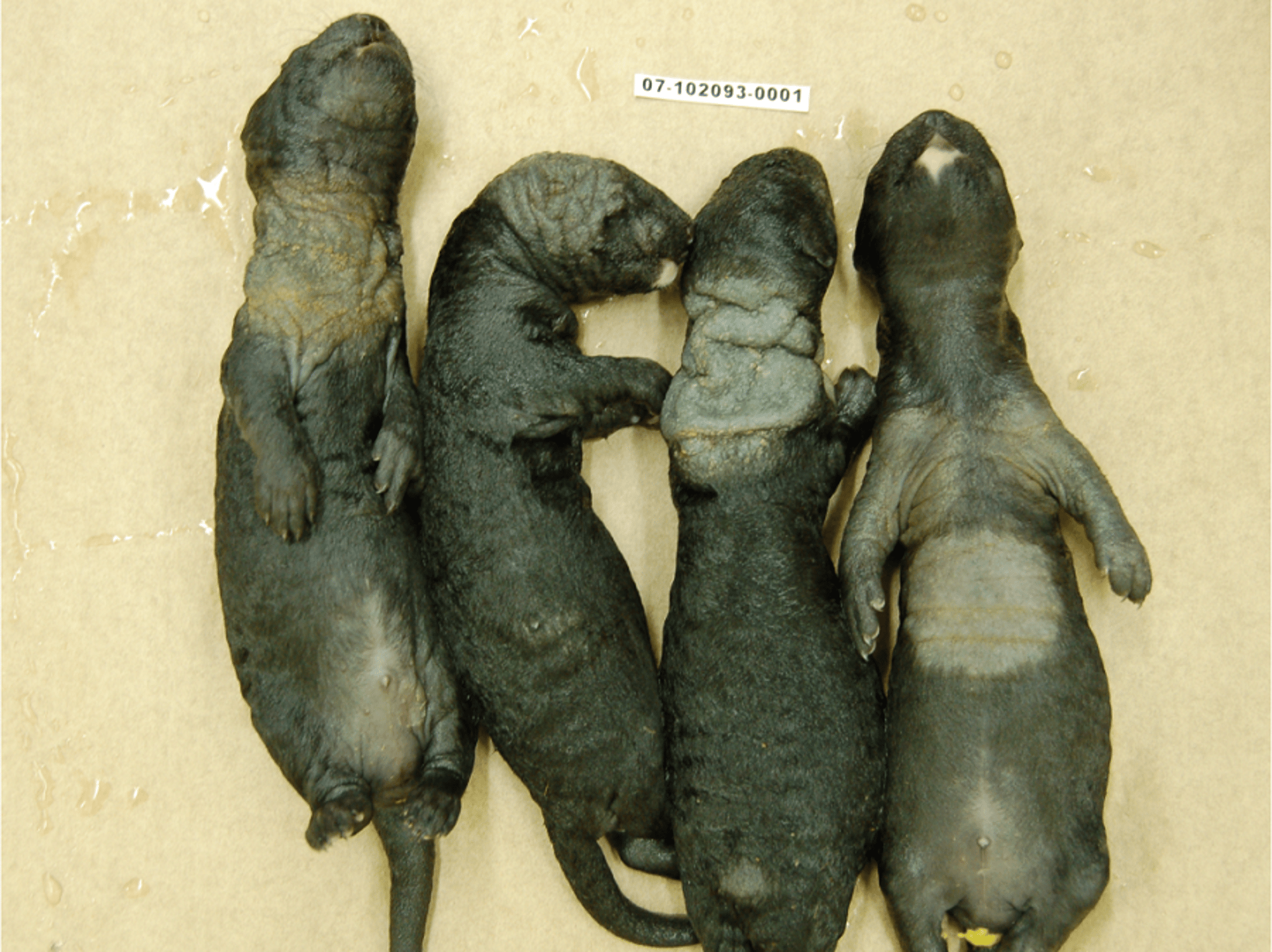
Dermatophytosis in mink kits occurs as patchy areas of hair loss with thickening of the affected skin.
Courtesy of Dr. Marina Brash.

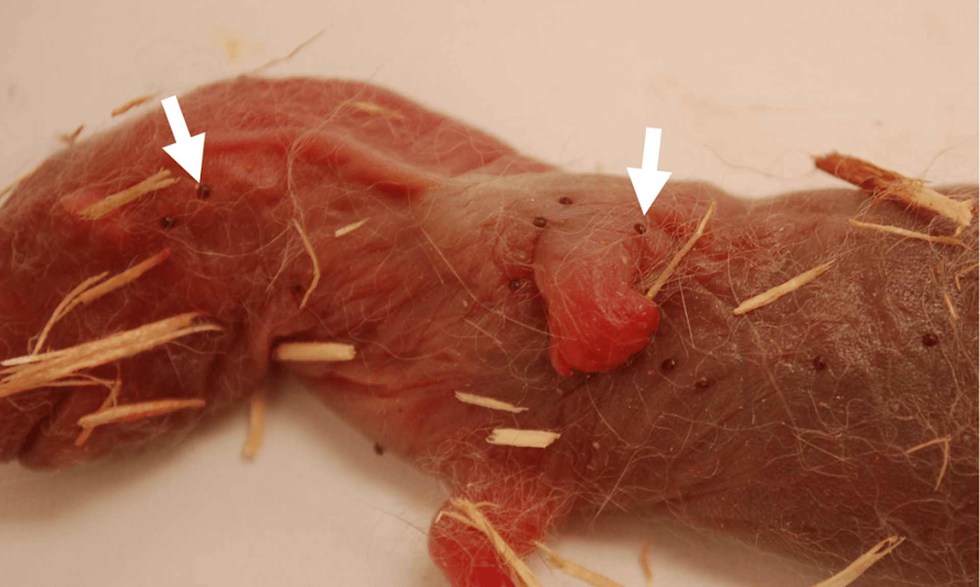
Mink kit heavily infested with ectoparasites (indicated by white arrows), which are swollen with blood.
Courtesy of Dr. Patricia Turner.
Dermatophytosis (ringworm) occurs sporadically in mink as patchy alopecia with crusting. Fungal culture of crusts or hair from affected areas and microscopic evaluation of punch biopsies for fungal hyphae may be used to confirm the diagnosis. Both Trichophyton and Microsporum spp may be isolated. Domestic animals, such as cats, are common carriers of ringworm and should not be permitted to enter mink sheds. Dermatophytosis should be differentiated from dermatitis arising from sarcoptic mange or other ectoparasite infestations, such as lice.
Gastric ulcers are commonly seen at postmortem examination in mink and are a nonspecific indication of stress. Red or dark intestinal contents and tarry feces may be seen concurrently in animals with gastric ulceration. Management factors, including adequacy of diet and maintenance of optimal body condition, should be evaluated as a preventive measure.
Hereditary diseases include hydrocephalus, hairlessness, the abnormal behavioral pattern termed screw neck, bobbed tails, Ehlers-Danlos syndrome, hemivertebrae, and tyrosinemia. Culling the sire, dam, and littermates of affected mink is necessary for control.
Coccidiosis, caused by infection with either Eimeria or Isospora spp, occasionally causes losses in young mink shortly after weaning, when maternal antibody concentrations are low. Affected mink have diarrhea, blood-tinged feces, dehydration, and weight loss. Diagnosis may be confirmed by examination of the feces or intestinal contents for oocysts or via histologic evaluation of intestinal tissue. Coccidiostats may be administered to control outbreaks. Infection can be prevented with good sanitation and regular manure removal. It may be seen in conjunction with mink viral enteritis outbreaks.
Myiasis can develop in mink when Wohlfahrtia spp flies lay maggots directly on the skin of kits. The larvae penetrate the skin and produce inflammation and lesions resembling abscesses. Affected kits become restless, lose body condition, and may die. Malathion dust (5%) placed beneath the litter in the nest boxes beginning a few days before the flies appear may help prevent infestation. The dust should not be used before whelping or until the kits are 1 week of age. Treatment may be repeated once after a 2-week interval. Myiasis is rare in mink. (Also see Cuterebra Infestation in Small Animals.)
Tangled umbilical cords may occur occasionally at whelping, such that kits are tightly associated and cannot be separated. Umbilical cords can be cut with sterile scissors or scalpels to separate kits and return them to their dam.


