


Physical restraint of ratites is different for each species. An ostrich’s head can be caught by hand or hook, and a hood can be placed over its head. Once hooded, an ostrich's head should be maintained below the level of its body to restrict it from kicking its legs forward. An ostrich kick can seriously injure a handler and, once captured, the bird will quickly try to back up and kick anyone in front of its body. Because an ostrich will back up when its head is held, an assistant should be positioned behind the bird to push as the person holding its head leads the ostrich forward. Emus must be handled from behind, grasping the wings and lifting the bird slightly upward and back. The front of the bird should be avoided to prevent being injured by the claws on the feet, because, as with all ratites, emus will kick forward to try to escape. Rheas are handled much like emus. Some handlers use hoods to facilitate handling emus and rheas, although this technique is not as effective as it is with ostriches.
Juvenile birds from 4 months of age to yearlings are best handled by slowly and calmly herding them into an enclosed barn. If they cannot see out of an enclosure and are crowded into a corner, they will often sit if not startled. When calmly herded into a confined area, young birds can easily be sexed, banded, and administered antiparasitic medication, and diagnostic samples (eg, blood) can be collected. If there is no enclosed area, portable panels with plywood or plastic applied can be used to herd the birds into a desired space.
Physical and Laboratory Examination of Ratites
Before a bird is restrained, it should be examined from a distance while walking/running in its enclosure for conformation, gait, body condition, respiration rate and character, and behavior-related problems.
The enclosure should be inspected for fresh droppings and urine. Green urates may be an indication of hepatitis, whereas dry, hard, fecal material is common in birds that are dehydrated or suffering from gastrointestinal impaction. The droppings should be examined for tapeworm segments and collected for fecal flotation and direct parasite evaluation.
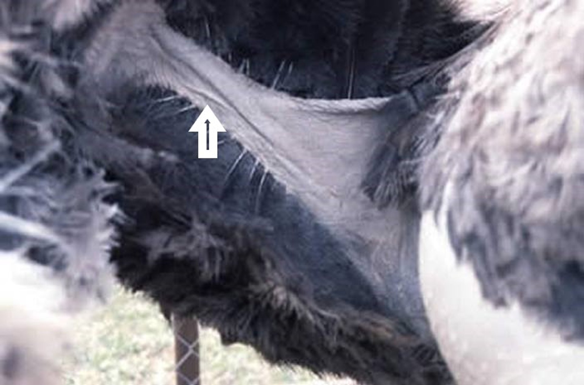
The eyes and sinuses are examined for any discharge or swelling, and the beak and oral cavity are examined for any lesions. The neck is palpated, especially in the area of the thoracic inlet, for any swellings.
Overall body condition should be noted and is determined by the epaxial muscle mass, rated on a scale of 1–9, with 1 being emaciated and 9 obese. The bird's feathers and skin should always be examined for feather-destructive behavior, external parasites, and lesions. Evaluation of the cranial coelomic cavity should be accompanied by auscultation of the heart and lungs/air sacs. Heart rhythm and rate are determined during auscultation, as well as listening for any abnormal lung and air sac sounds while the animal is breathing. The caudal coelom is palpated from the ventriculus, which lies immediately caudal to the breastplate, to the proventriculus, located between the legs. The caudal coelom is palpated and balloted for any evidence of coelomic fluid and/or retained eggs.
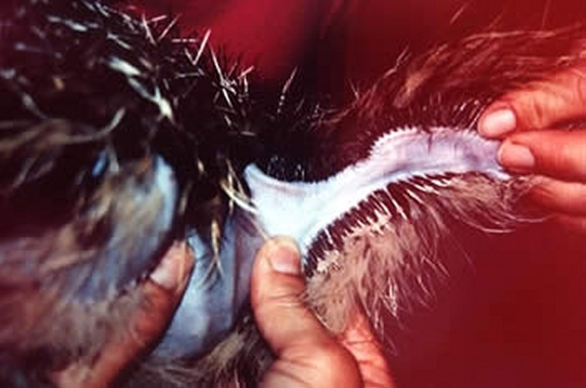
Finally, a cloacal examination is performed to verify normal anatomy. When indicated, samples for microbial culture can be collected from the trachea, cloaca, and caudal oviduct, and blood can be collected for a CBC and serum chemistry panel. (For hematologic and serum biochemical reference ranges for ostriches, see Table # Hematologic Reference Ranges in Ferretsa and see Table # Serum Biochemical Analysis Reference Ranges). Sodium heparin is the preferred anticoagulant for both the CBC and serum chemistry panel. A slide should be prepared immediately for cytologic evaluation.

Courtesy of Dr. Karen Hicks-Alldredge.

Courtesy of Dr. Karen Hicks-Alldredge.

Courtesy of Dr. Karen Hicks-Alldredge.
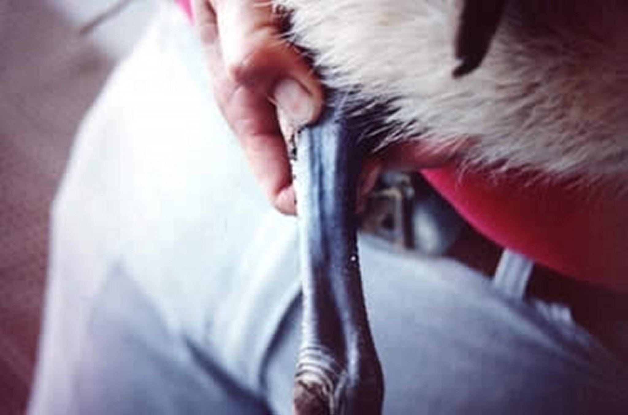
Recommended sites for venipuncture and catheterization in ostriches and rheas are the cutaneous ulnar veins on the ventral side of the wings and the medial metatarsal veins. In emus, the jugular vein and medial metatarsal vein are the blood collection sites of choice. Venipuncture of the jugular vein is more difficult in ostriches because of their size and because sudden movement by the bird can result in lacerations and exsanguination. In debilitated ratites, even ostriches, catheterization (adults, 14-gauge, 13-cm catheter) of the jugular vein is easy and provides a readily accessible port to the vascular system. The right jugular vein is more developed than the left.
Anesthesia in Ratites
Chemical restraint for handling and surgical procedures in ratites involve typical veterinary drugs. Anesthesia may be induced in younger birds with isoflurane or sevoflurane using the same procedures as with small animals. Intubation for maintenance is recommended; however, the cuff should not be inflated, because of the possibility of pressure necrosis (birds have complete tracheal rings that do not expand with the tracheal cuff). Xylazine and ketamine combinations are commonly administered for short procedures on induction, followed by intubation and gas anesthesia. Xylazine administered at 2.5 mg/kg, IV, followed, after sedative effect is noticed, by ketamine at 1 mg/kg, is an effective protocol. However, xylazine has notable cardiorespiratory depressant effects and should not be administered to severely ill birds. Also, xylazine is not recommended as a premedication for inhalation anesthesia, because it may greatly enhance the cardiodepressant effects of gas anesthetic agents.
For chemical sedation and restraint, the following protocols have been successful in ostriches. As an initial sedative, acepromazine (0.25 mg/kg, IM) or xylazine (1 mg/kg, IM) can be administered. After waiting approximately 20 minutes for the sedatives to take effect, the anesthetic agent can be administered. If acepromazine is used as the preanesthetic agent, either propofol (4 mg/kg, IV) or ketamine (5 mg/kg, IV) and diazepam (0.25 mg/kg, IV) or tiletamine/zolazepam (3 mg/kg, IV) is administered to achieve full sedation. If xylazine is used as the preanesthetic agent, then ketamine (5 mg/kg, IV) and diazepam (0.25 mg/kg, IV) or tiletamine/zolazepam (3 mg/kg, IV) is effective for most short-term procedures that need to be performed on the bird.
Recovery may be smoother if diazepam is administered at 0.2 mg/kg, IV or IM. Tiletamine/zolazepam is an induction alternative for ratites, dosed at 2–10 mg/kg, IM, or 1–3 mg/kg, IV, with the higher end of the dose range recommended for emus and rheas. Once the bird has been induced, padding should be placed under the body during the entire procedure to minimize neuropathy and myositis. Ostriches may develop peroneal nerve paralysis when placed in lateral recumbency, without padding, for < 1 hour. Sternal recumbency does not appear to interfere with respiration while anesthetized; however, the large body size of ratites may restrict their ability to breathe. Therefore, manual or mechanical respiration is recommended during general anesthesia to reduce the cardiorespiratory depressant effects of both gas anesthetic agents and physical compression of air sac volume.
The neck should be straight and the head elevated slightly above the body during general anesthesia. Vital signs of ratites should be monitored while under anesthesia along with body temperature (esophageal) to maintain normothermia; intravenous access should be established for drug and fluid administration. During recovery, the bird should be placed in a dark, quiet, preferably padded enclosure in sternal recumbency. Wrapping the body with the legs immobilized under the animal while in sternal recumbency will often allow the bird to recover with minimal struggle.
Surgical Procedures in Ratites
Surgical procedures in ratites generally are related to the GI tract, orthopedics, and trauma repair. Proventriculotomy for foreign body removal and impactions in ratite species is a common surgical procedure. In young birds, incisions should be made carefully, because the abdominal wall is very thin. To perform a proventriculotomy, the bird is positioned in right lateral recumbency, with the left pelvic limb abducted and supported caudally in a stand. In ostriches, the ventriculus is located caudal to the proventriculus in the coelomic cavity; therefore, the surgical incision to access this part of the GI tract should be through a left paramedian approach beginning ~15 cm caudal to the keel.
Yolk sac removal due to nonabsorption and/or infection is common. Egg retention in hens is addressed surgically, and multiple eggs are often removed. Orthopedic surgeries are addressed as in other species with pins, plates, and transfixation casting as needed for fracture repair. Laceration repair is performed as in other species. Upper esophageal tears from attempted hooking (capture) will often heal by second intention and, unless severe, do not require surgical repair.
As in all production animals, the cost of surgical correction of many conditions might be greater than the commercial value of the individual animal presented. Producers should be made aware of cost considerations.
Nutrition in Ratites
To date, there has been little reliable research about ratite nutrition. The formulations of commercial diets available are based on studies in Africa and Australia and on extrapolations from available poultry information. Current trends are to feed 14%–20% protein from the time chicks hatch to 3 months of age, reducing the protein level after 3 months of age. Ostriches and rheas are hindgut fermenters and have the ability to digest fiber from a young age. For optimal health and production results, appropriate ratite feed formulated for ostriches, emus, and rheas is recommended. Ostriches, emus, and rheas have significantly different digestive systems and specific nutritional requirements for optimal growth, reproduction, and health. High-quality grass can be used as a grazing supplement for ostriches and rheas. Chicks allowed to hatch in the nest are coprophagous, eating feces from the parents for the first few weeks of life.
Vaccination of Ratites
When indicated in a specific flock, autogenous bacterins for Salmonella, Escherichia coli, and clostridial diseases have been used; however, this is not a common practice, and these products may be difficult to obtain. For emus, equine encephalitis vaccination is recommended. Tissue-culture propagated, inactivated bivalent Eastern and Western equine encephalomyelitis (EEE, WEE) vaccine prepared and licensed for use in horses, and that may contain equine tetanus antitoxin, has effectively protected emus against EEE and WEE infection. A vaccination protocol is to administer intramuscularly an initial full equine dose at 6 weeks of age, followed by booster vaccinations at 10 weeks of age and at 5- and 6-month intervals thereafter and before and after breeding season (April and September). A booster should be administered when EEE or WEE is identified within 10 miles of the production facility or before transfer to an endemic area. The recommended vaccine protocol (emus) for Clostridium chauvoei (blackleg) using the ruminant vaccine with or without tetanus antitoxin is to administer an initial vaccine at 2 months of age, followed by a booster vaccination at 3 months and then annually after the end of breeding season in April.


