


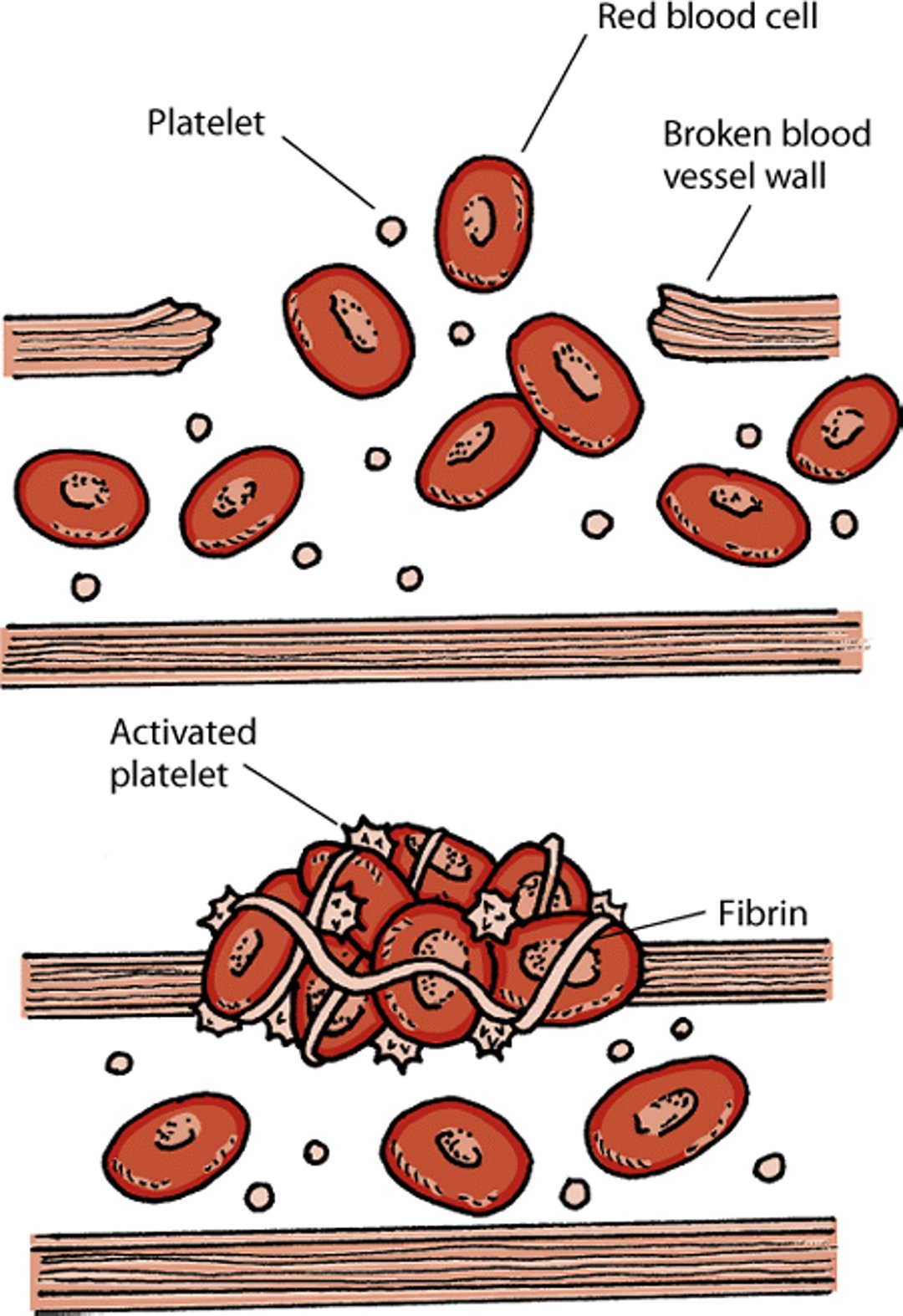
When bleeding occurs in an organ or body part, a process is set in motion to stop the bleeding. This is called hemostasis. In order to work, hemostasis requires an adequate number of platelets, the right amount of blood clotting proteins (often referred to as factors), and blood vessels that constrict properly. When an injury occurs, the wall of the blood vessel breaks. The affected blood vessel will narrow so that blood flows more slowly, allowing the clotting process to begin. Platelets also rush to the broken wall where certain proteins change the platelets’ shape from round to spiny so that they can stick to blood cells, the broken vessel wall, and to each other. Other proteins form long strands called fibrin. These fibrin strands form a net that traps and helps hold together the platelets and blood cells, creating a clot that plugs the break in the vessel wall. After the clot has formed and stabilized, other proteins stop the clotting process and eventually dissolve the clot.
Hemostasis

Bleeding disorders may be present at birth (congenital) or occur later. Defects in blood clotting proteins usually show up as delayed bleeding and bruising deep in tissues, while platelet defects usually show up as superficial small bruises, nosebleeds, black stools caused by bleeding into the bowels, or prolonged bleeding at injection and surgery sites.
Excessive clotting leading to blocked arteries may be inherited disorders of anticlotting proteins or acquired disorders. Conditions that cause excessive clotting are called hypercoagulable states. Acquired clotting diseases are more common in animals than are inherited disorders.
Blood clotting tests can help identify animals with defective clotting proteins. However, the tests are not very sensitive, so an animal must have a severe deficiency for the tests to find the problem.
Congenital Clotting Protein Disorders
Many different proteins are involved in the clotting process. Deficiencies of any of these proteins can cause bleeding disorders. Congenital clotting protein disorders are present at birth. In a severe deficiency or defect of clotting proteins, signs will appear at an early age. Severe defects are usually deadly. Animals may be stillborn or die shortly after birth. Lack of clotting proteins or vitamin K (which is also part of the clotting process) in a newborn animal may make a clotting defect worse. If the amount of any particular clotting protein is 5 to 10% of normal, the newborn may survive, but will usually show signs of illness before 6 months of age. It is during this time, when numerous routine procedures (for example, vaccination or gelding) are usually done, that a bleeding tendency may be noticed.
Hemophilia A (Factor VIII deficiency) has been reported in several breeds of horses, including Arabians, Standardbreds, Quarter Horses, and Thoroughbreds. Usually, females carry the gene for the disease without showing any signs, whereas males show signs. Carrier animals have higher levels of Factor VIII (40–60% of normal), and the results of their clotting tests are usually normal. Affected animals may bleed spontaneously or bleed excessively after an injury or surgery. The diagnosis is harder to confirm in animals less than 6 months old because the livers of even normal foals may not yet have produced enough of the clotting proteins, meaning affected foals are harder to identify. Treatment requires repeated blood transfusions until bleeding has been controlled.
Acquired Clotting Protein Disorders
Most clotting proteins are produced in the liver. Therefore, liver disease can lead to decreased levels of clotting proteins, particularly Factors VII, IX, X, and XI, and proteins that break up clots. The decrease in clotting proteins can range from small to large. Animals with liver disease do not usually bleed spontaneously (without injury) unless they have another underlying condition.
Disseminated intravascular coagulation (DIC) is a condition in which small blood clots develop throughout the bloodstream, blocking small blood vessels and consuming the platelets and clotting factors needed to control bleeding. It usually develops after numerous triggering events, such as severe infections, heat stroke, burns, tumors, or severe injury. In many cases, the signs are uncontrolled bleeding and the inability to form a normal clot. Death is caused by extensive blood clots or collapse of circulation, leading to the failure of one or several organs. If the animal survives this crisis, a longterm form of DIC can occur. Your veterinarian will determine and attempt to correct the underlying problem causing this condition. The underlying cause must be treated promptly and thoroughly. Intravenous fluids are extremely important for maintaining normal circulation. DIC is a very serious disorder and is often fatal.
Platelet Disorders
Disorders of platelets include having too few platelets (called thrombocytopenia) or having platelets that do not work properly. Each type of disorder can be either congenital (present at birth) or acquired later in life. Congenital platelet disorders are rare in horses. Several acquired thrombocytopenias causing decreases in platelets in the bloodstream are reported in horses. Numerous causes have been identified, most involving the immune system. Thrombocytosis (having too many platelets in the blood) is rare.
Immune-mediated thrombocytopenia (also called idiopathic thrombocytopenia or idiopathic thrombocytopenic purpura) is a reduction in the number of platelets in the blood caused by immune system dysfunction. It occurs when the immune system makes antibodies that destroy platelets or platelet-producing cells in the bone marrow. It has been seen in horses. At diagnosis, the horse typically will have a severely decreased number of platelets in the blood. Signs include tiny, purplish red spots on the gums or skin, bruising, bleeding into the bowels resulting in black stools, or nosebleeds. An evaluation of the bone marrow may be necessary to help determine whether circulating platelets or the platelet-forming cells are targeted by the antibodies. Corticosteroids are the usual treatment, although other drugs are sometimes used. The affected horse should be kept at rest. Blood transfusions may be necessary if anemia occurs.
Thrombocytopenia caused by drugs has been reported in horses. Some drugs and classes of drugs (including estrogen and some antibiotics) suppress the production of platelets in the bone marrow. Other drugs (including aspirin, acetaminophen, penicillin, and others) destroy platelets circulating in the bloodstream. Drug reactions are rare and unpredictable. Platelets usually return to normal shortly after the drug is discontinued. Drug-induced bone marrow suppression may last longer, however. If your horse is taking one of these drugs, your veterinarian will likely monitor the blood count to check for any serious reductions in the number of platelets.
Von Willebrand disease is a congenital platelet function disorder that has been reported in horses. It is caused by a defective or deficient von Willebrand’s factor, a protein that regulates the first step in clot formation. Affected horses usually have a normal number of platelets and clotting factors. However, they may bleed spontaneously (for example, nosebleeds) or only after an injury or surgery. Affected horses may require a blood transfusion after an episode of bleeding. Drugs that can interfere with clotting (such as aspirin) should not be used in these animals.
Glanzmann thrombasthenia, previously called thrombasthenic thrombopathia, has been diagnosed in Thoroughbred-cross, Quarter horse, and Oldenburg filly horses. Affected horses have prolonged bleeding times and form large bruises easily. A large number of oddly shaped, giant platelets are seen in blood tests. Platelets from horses with this disorder do not clump together or separate as they normally should. There is no specific treatment. In cases of severe bleeding, blood transfusions can be given.
Acquired platelet dysfunction is associated with some diseases. This can affect platelets and reduce their ability to form a blood clot. Longterm kidney disease can decrease the ability of platelets to stick together. A rare cancer called multiple myeloma can cause platelet defects and poor clotting. Many drugs can also impair platelet function; however, the problem may not be noticed unless another clotting disorder is also present.
Blood Vessel Disorders
Abnormal or weakened blood vessels can result in inappropriate bleeding.
Hereditary equine regional dermal asthenia (HERDA), also known as cutaneous asthenia, is caused by a defect in type 1 collagen (a protein connective tissue in the skin) that is present at birth. This causes weak structural support of blood vessels and can lead to blood clots and easy bruising. The disorder has been reported in horses but is rare. The most striking clinical abnormality is loose skin that stretches to a greater than normal degree and tears easily. This disease is caused by a genetic defect in Quarter Horses and related breeds; genetic testing is available to confirm the diagnosis or to identify unaffected horses that carry 1 copy of the abnormal gene (known as “carriers”, or “heterozygotes”), so that these horses are not used for breeding.
Excessive Blood Clotting Disorders
Excessive blood clotting (known as hypercoagulability) is the uncontrolled clotting of blood. Blood clots can break free and travel through the blood to block smaller arteries. Inherited disorders of anticlotting proteins are not known to occur in animals. However, there are several diseases that can cause acquired clotting disorders. Coagulation screening tests performed by a veterinarian can identify which clotting protein is affected. Disorders most relevant to horses are described below.
Blood clots have been seen in horses with generalized inflammatory diseases such as colic (a digestive disease), laminitis (inflammation of the hoof), or equine ehrlichial colitis (an infection of the colon). Catheters inserted in the jugular vein in the neck for long periods and treatment with drugs that irritate the blood vessels may also cause blood clots to form.
Horses with colic associated with endotoxemia (the presence of bacterial toxins in the bloodstream) have decreased activity of enzymes that break down blood-clotting agents, such as fibrin. These horses have increased death rates and a higher risk for blood clot formation. Laminitis is thought to be the result of several diverse whole system disorders. Tiny blood clots form in the blood vessels of the hoof in the early stages of laminitis. One theory is that bacterial toxins in the bloodstream directly affect the blood vessels and activate the clotting system. Swelling, blood vessel compression, and possibly the shifting of blood flow to the upper part of the horse’s hoof increase the damage to the blood vessels. When blood flow is restored, it releases toxins into the bloodstream, which causes blood clot formation.
The best treatment for an animal with blood clots is diagnosing and treating the underlying disease, along with providing good supportive care. Maintaining blood flow to the tissues is critical. Your veterinarian may prescribe medication to dissolve or prevent clots. In other cases, transfusions may be the most effective treatment.
For More Information
Also see professional content regarding bleeding disorders


