


The structural and functional unit of skeletal muscle is the motor unit. It consists of a ventral motor neuron with its cell body in the central horn of the spinal cord and its peripheral axon, the neuromuscular junction, and the muscle fibers innervated by the neuron. Each of these components must be functionally intact for the muscle to contract properly. The ventral motor neuron is the final common pathway conducting neural impulses from the CNS to the muscle.
The transmission of a nerve impulse at the neuromuscular junction involves massive release of acetylcholine from small synaptic vessels, where it is stored. The acetylcholine fills the synaptic cleft between the nerve terminal and the muscle fiber membrane, where most of it is destroyed by cholinesterase within a fraction of a second. This short period of activity is sufficient to excite the muscle fiber membrane, which results in a significant increase in membrane permeability to sodium ions and allows rapid influx of sodium into the muscle fiber. The sodium ion increases the endplate potential, which elicits electrical currents that spread to the interior of the fibers, where they cause a release of calcium ions from the sarcoplasmic reticulum. The calcium ions initiate, in turn, the chemical events of the contractile process. When this occurs in all the muscle fibers innervated by each motor neuron (possibly thousands), muscle contraction results.
Normal muscle, comprising many motor units, is dynamic, and its function and structure can be influenced by many diseases. Complete paralysis, paresis, or ataxia may be caused by primary muscular dysfunctions of infectious, toxic, or congenital origin. However, in most instances the primary disorder can be attributed to the nervous system (eg, tetanus, rhinopneumonitis, canine distemper, protozoal myelitis), with the muscular system merely representing the effector organ. Disorders that affect the neuromuscular junction (eg, myasthenia gravis, hypocalcemia, hypermagnesemia) can result in muscle fatigue, weakness, and paralysis. The neuromuscular junction can also be affected by muscle-relaxing drugs (eg, curare, succinylcholine, M99), certain antibiotics, and toxins (eg, botulism, tetanus, venoms).

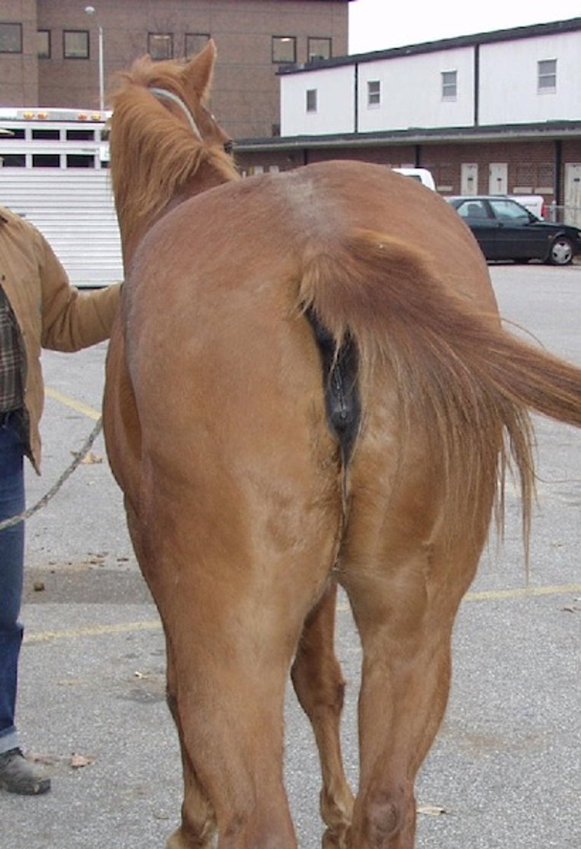
Courtesy of Dr. Stephen Adams.
Disorders primarily of the muscle membrane and, to some extent, of the actual muscle fibers are called myopathies. Muscle membrane disorders may be hereditary (eg, myotonia congenita in goats) or acquired (eg, vitamin E and selenium deficiencies, hypothyroidism, and hypokalemia). Myopathies involving the actual muscle fiber components include muscular dystrophy, polymyositis, eosinophilic myositis, white muscle disease, and exertional rhabdomyolysis. Various laboratory tests, eg, histopathologic examination, determination of serum enzyme levels, electromyographic studies, thermography, and determinations of conduction velocity, are very useful in confirmation of a specific diagnosis.
Trauma to muscles is common in horses and also occurs in other species. The trauma can be from external injury or from intense athletic activity, during which muscle tears and ruptures may occur. Fibrotic myopathy in the rearlimb of horses is a mechanical lameness caused by trauma and subsequent fibrosis or ossification of the muscle.
For More Information
Also see pet health content regarding disorders of muscle in dogs, disorders of muscle in cats, and disorders of muscle in horses.


