

Colibacillosis is caused by infection with a strain of Escherichia coli. Syndromes associated with colibacillosis can vary and include acute fatal septicemia, airsacculitis, pericarditis, perihepatitis, peritonitis, and lymphocytic depletion of the bursa and thymus. Diagnosis is usually made by isolation of a pure culture of E coli, consistent with colibacillosis, from the lesion of a bird. Most bacterial isolates are resistant to multiple antimicrobials, so prevention of exposure through good management is recommended.
Colibacillosis results in a localized or systemic infection caused by avian pathogenic Escherichia coli (APEC). Colibacillosis can manifest in diverse ways, including acute fatal septicemia, subacute pericarditis, airsacculitis, salpingitis, peritonitis, and cellulitis. In laying hens, peritonitis and salpingitis are common, whereas disease in young chicks may include omphalitis (yolk sac infection) or swollen head syndrome.
Colibacillosis is one of the most commonly occurring and economically devastating bacterial diseases of poultry worldwide, resulting in multimillion dollar losses annually that affect many facets of poultry production.
Etiology and Pathogenesis of Colibacillosis in Poultry
Escherichia coli is a gram-negative rod-shaped bacterium, a strain of which causes colibacillosis. It is normally found in the intestine of poultry and other vertebrates. Though many E coli are not pathogenic, some have acquired virulence factors, greatly increasing their capacity to cause disease.
Most cases of colibacillosis appear to be due to E coli that have acquired a number of virulence genes clustered together on plasmid-borne pathogenicity islands (PAIs). These PAI-containing plasmids are said to be the defining feature of the APEC pathotype.
Other cases of colibacillosis are due to infection with commensal E coli that gain access to birds weakened by some predisposing condition, such as the following:
poor air quality
other environmental stresses
Previously, most APEC isolates were assigned to three main serogroups: O1, O2, and O78; however, it has been shown that there is great diversity in the serogroups of APEC causing colibacillosis. A high percentage of APEC isolates cannot be grouped using current methods and require extensive analysis (eg, serogrouping or whole genome sequencing) to conclusively identify a strain. Therefore, no single E coli serogroup used as a bacterin is likely to provide full protection against all of the serogroups that cause colibacillosis.
Virulence factors associated with APEC pathogenesis include possession of large virulence plasmids and the following characteristics:
resistance to phagocytosis and serum killing
acquisition of iron under the low-iron conditions within the poultry host
adherence to host structures
APEC isolates generally do not produce toxins.
Large numbers of E coli are maintained in the poultry house environment through fecal contamination. Initial exposure to APEC may occur in the hatchery from infected or contaminated eggs.
The portal of entry into birds varies but can include the respiratory tract, areas of skin trauma, cloaca, damaged intestinal mucosa, and navel. From these entry sites, E coli can extend locally or gain access to the bloodstream to cause colisepticemia, which may progress from acute septicemia to death.
Infection can also extend to serosal surfaces to cause subacute polyserositis and chronic granulomatous inflammation.
Clinical Findings and Lesions of Colibacillosis in Poultry
Clinical signs of colibacillosis are nonspecific and vary with age, organs involved, and concurrent disease.
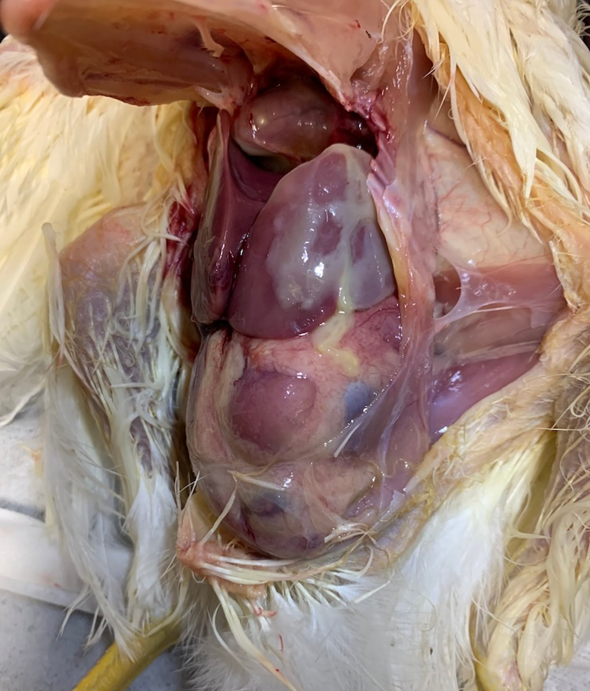
Young birds dying of acute septicemia may have few lesions on necropsy except for an enlarged, hyperemic liver and spleen with increased fluid in body cavities. Birds that survive septicemia can develop subacute fibrinopurulent airsacculitis, pericarditis, perihepatitis, and lymphocytic depletion of the bursa and thymus (see and images). Unusually pathogenic salmonellae produce similar lesions in chicks.

The air sac is slightly more opaque than normal and contains some serous exudate.
Courtesy of Dr. Luke Baldwin.


Perihepatitis in a chicken. Note the caseous material on the liver surface.
Courtesy of Dr. Catherine M. Logue.
Although airsacculitis is a typical lesion of colibacillosis, it is unclear whether it results from primary respiratory exposure or from extension of serositis.
Sporadic lesions include pneumonia, arthritis, osteomyelitis, peritonitis, and salpingitis.
Diagnosis of Colibacillosis in Poultry
Isolation of E coli in pure culture
Determination of virulence factors using multiplex PCR assays
Unlike pathogenic E coli associated with illnesses in other animal species, avian isolates used in diagnosis of colibacillosis are generally nonhemolytic on agar with 5% sheep blood.
Isolation of a pure culture of E coli from heart blood, liver, bone marrow, or typical visceral lesions from a fresh carcass indicates primary or secondary colibacillosis. Consideration should be given to predisposing infections and environmental factors.
Pathogenicity of isolates is established either by using multiplex PCR panels for plasmid-mediated and chromosomal-associated virulence genes, such as iron-related genes (sitA, iroN, iutA, and etsB), toxin- or bacteriocin-related genes (hlyF), and protectin genes (iss), or when parenteral inoculation of young chicks or poults results in fatal septicemia or typical lesions within 3 days.
Pathogenicity can also be detected by inoculation of the allantoic sac of 12-day-old chicken embryos. Resulting gross lesions include cranial and skin hemorrhages in addition to encephalomalacia in embryos inoculated with virulent isolates.
Treatment and Control of Colibacillosis in Poultry
Good management practices
Antimicrobials contraindicated
Prevention of colibacillosis relies on good management to decrease exposure of birds to APEC and lessen the impact of stress and predisposing infections on the susceptibility of birds to APEC infection. Best management practices include the following:
sanitation of water lines and closed water systems
adequate ventilation to decrease ammonia and dust buildup
moisture control of litter
pest control (rodents, flies, beetles, etc)
In addition, experimental and commercial vaccines of various types have been used to prevent colibacillosis, to mixed effect. Often the vaccines may protect against specific serogroups; however, cross-protection against different serogroups of E coli is not always possible due to the diversity of APEC isolates causing disease.
Treatment of colibacillosis with antimicrobial agents is problematic due to widespread multidrug resistance among APEC isolates and restrictions on antimicrobial use in poultry imposed by regulation and public concern. If treatment is pursued, it should be based on antimicrobial susceptibility testing.
Most isolates are resistant to tetracyclines, streptomycin, and sulfa drugs, although success can sometimes be achieved with tetracycline. However, the vast majority of clinical isolates are resistant to tetracycline, with most APEC isolates resistant to five or more antimicrobials. Extra-label use of fluoroquinolone in poultry is now banned in many countries, including the US.
APEC isolates also show widespread resistance to disinfectants, including certain heavy metal compounds, further complicating control of colibacillosis. Alternative approaches such as prebiotics and probiotics show potential, but their efficacy is currently not well known.
Key Points
Colibacillosis is a major cause of morbidity, death, and economic loss for all types of poultry worldwide.
Avian pathogenic E coli (APEC), the causative agent of colibacillosis, encompasses a diverse grouping of E coli, most of which harbor large virulence plasmids.
Control of colibacillosis is problematic due to widespread antimicrobial resistance among APEC isolates, restrictions on use of antimicrobial agents in poultry, and the lack of vaccines to provide protection against all types of APEC isolates causing colibacillosis.
For More Information
Nolan LK, Vaillancourt J-P, Barbieri NL, Logue CM. Colibacillosis. In: Swayne DE, ed. Boulianne M, Logue CM, McDougald LR, Nair V, Suarez DL, associate eds. Diseases of Poultry. 14th ed. WileyBlackwell; 2020:770-830.
Nolan LK, Barnes HJ, Abdul-Aziz TA, Logue CM, Vaillancourt JP. Colibacillosis. In: Brugere-Picoux J, Vaillancourt J-P, Shivaprasad HL, Venne D, Bouzouaia M, eds. Manual of Poultry Diseases. French Association for the Advancement of Science; 2015:301-315.
Johnson, TJ, Wannemuehler Y, Doetkott C, Johnson SJ, Rosenberger SC, Nolan LK. Identification of minimal predictors of avian pathogenic Escherichia coli virulence for use as a rapid diagnostic tool. J Clin Microbiol. 2008;46(12):3787-3996. doi:10.1128/JCM.00816-08


