


Trichomoniasis in cattle is a venereal disease characterized by embryonic loss. The disease typically results in variable interestrus intervals, decreased pregnancy rates, and occasionally abortion. Affected herds frequently demonstrate an increased prevalence of postcoital pyometra. Diagnosis relies on isolation of the pathogen, most often from the bull's prepuce. Currently, there is no effective treatment for trichomoniasis in cattle. Vaccination, where available, is unreliable. Control is via bioexclusion principles and removal of infected bulls.
Trichomoniasis in cattle is a venereal disease that typically leads to subfertility. It is due to infection with the protozoan Tritrichomonas foetus, an obligate venereal pathogen. The disease has a worldwide distribution and is primarily found in extensive pastoral systems. Incidence varies widely depending on geographical location (1, 2). Trichomoniasis is transmitted primarily via coitus; however, artificial insemination with infected semen can also lead to disease transmission. Infected bulls are typically carriers without overt clinical signs, and most infected cows mount an effective local immune response.
Etiology and Epidemiology of Trichomoniasis in Cattle
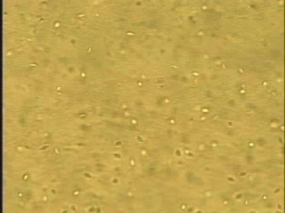
The causative agent of trichomoniasis is Tritrichomonas foetus, a protozoan of the subkingdom Protozoa (now classified in the kingdom Protista). T foetus is usually piriform in shape but is pleomorphic. It is 10−25 mcm long and has three anterior flagella (hence the name) and a long, trailing flagellum extending beyond an undulating membrane (see T foetus with bovine spermatozoon image).


Courtesy of the Drost Project and the Visual Guides of Animal Reproduction (visgar.vetmed.ufl.edu).
After copulation, T foetus invades the cow's vagina and migrates to the cervix and uterus. Infection has little impact on conception and early embryonic development. However, the organism induces an inflammatory response that can lead to endometritis, cervicitis, vaginitis, and possibly salpingitis, all of which can result in embryonic and early fetal death. Fewer than 1% of cows become chronic carriers.
Trichomoniasis occurs worldwide, wherever cattle are present. The three epidemiological features typically associated with trichomoniasis are the following:
The disease is transmitted via coitus (or by artificial insemination with infected semen).
Typically, infected bulls are clinically normal carriers.
Females are infected at the time of breeding, and most mount an immune response and clear the infection after two or three estrous cycles.
Clinical Findings of Trichomoniasis in Cattle
Trichomoniasis results in decreased herd fertility and irregular calving distribution. Infection of a cow by an infected bull is extremely efficient, whereas bulls are not readily infected by cows. Trichomoniasis can be transmitted mechanically via artificial insemination; however, this method is not a major contributor to disease transmission.
The most common indicator that T foetus has infected a herd, especially in naive populations, is that a large proportion of cows show clinical signs of returning to estrus as a result of embryonic death; however, embryonic loss can occur after maternal recognition of pregnancy, resulting in long interestrus intervals. Conceptus losses early in the breeding season result in an overall decrease in pregnancy rates, especially in a restricted breeding season, and in an increase in late-season pregnancies. The prevalence of postcoital pyometra is increased in herds infected by T foetus. Although the main effect of trichomoniasis is conceptus losses early in the breeding season, late-gestation abortions can occur, and live calves can be born to infected cows.
Typically, infected bulls do not show clinical signs of trichomoniasis.
Diagnosis of Trichomoniasis in Cattle
Observation of decreased pregnancy rates
Collection of samples, typically from bulls
Definitive: organism isolation and identification
History and clinical signs can be suggestive of trichomoniasis and other venereal diseases, including campylobacteriosis and possibly mycoplasmosis or ureaplasma infection. Patients with a lack of cyclicity due to immaturity, poor body condition, metabolic disease, or infertility (in the case of bulls) have a similar history and clinical signs. (For more on differential diagnoses for reproductive loss in cattle, see Embryonic and Fetal Death, Abortion, and Abnormal Fetal Development in Cattle.)
Definitive diagnosis of trichomoniasis relies on the identification of the T foetus organism via direct microscopic observation, culture, or PCR assay. Importantly, trichomonads that resemble T foetus can be found in feces, so fecal contamination must be minimized via appropriate aseptic techniques.
The most reliable source for isolation and identification of T foetus is the bull. The organism inhabits the epithelial folds of the bull's prepuce and penis, and it has been found in the urethra. T foetus is fastidious and therefore difficult to collect and culture. Procedures for collecting, storing, and transporting samples for T foetus testing must address these challenges to enhance the likelihood of a successful diagnosis. Cooling, heating, or exposure to sunlight can adversely affect the viability of the sample collected for identification. Working closely with relevant laboratories is essential to maximizing diagnostic outcomes.
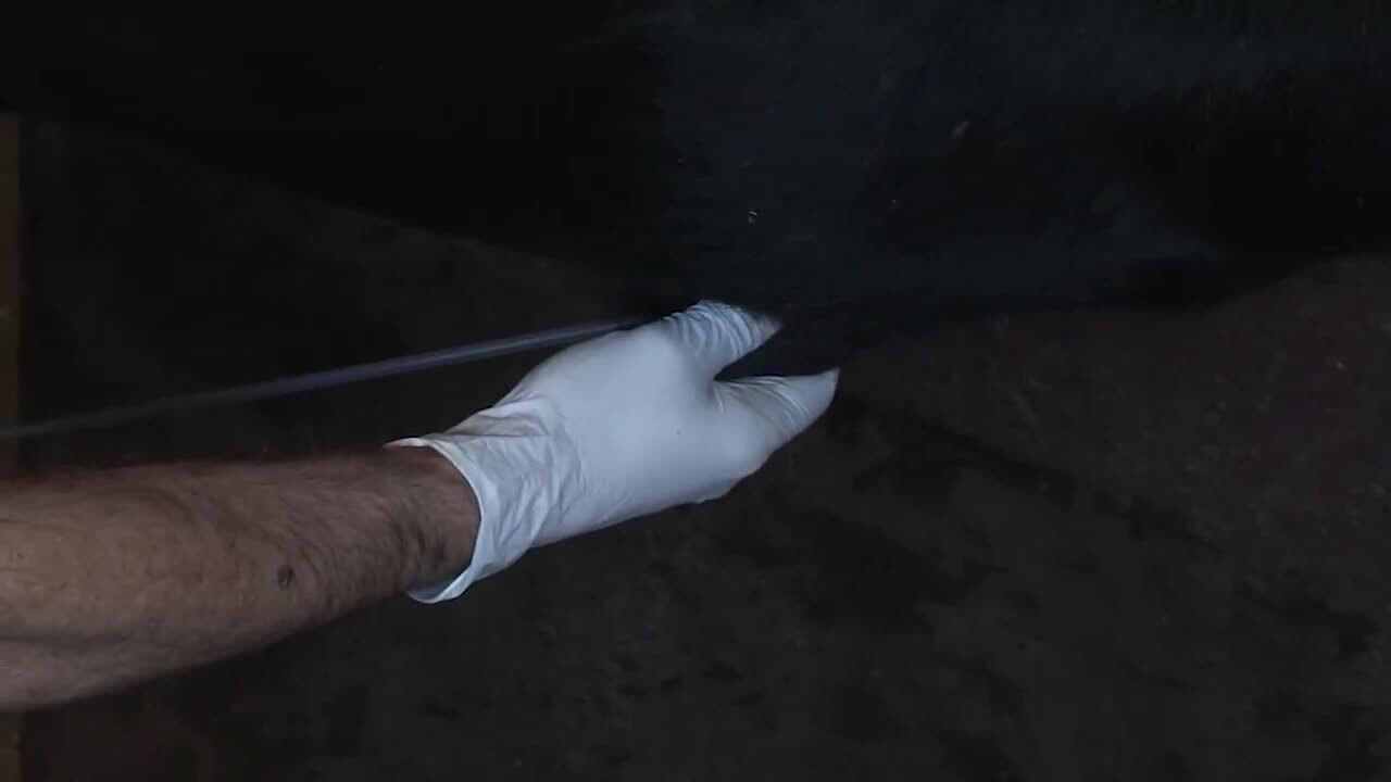
Because of the epidemiological features of trichomoniasis, diagnostic efforts are best directed at the bull. Penile and preputial mucosal samples are collected using either the "wet" or "dry" method.
For the wet method, an infusion pipette approximately 60 cm long is attached to a syringe containing 20 mL of phosphate-buffered saline or other appropriate fluid (various media are available, depending on the laboratory being used). The pipette is inserted into the prepuce and moved in a back-and-forth motion, while the fluid is simultaneously expelled out of and aspirated into the syringe (see prepuce sampling image). Some veterinarians advocate placing a bevel on the distal end of the pipette to enhance mucosal exfoliation. Alternatively, the sampling fluid is introduced to the prepuce, the preputial orifice is closed off, and the sheath is vigorously massaged. The pipette is then reintroduced, and the fluid is aspirated from the prepuce.


Courtesy of Dr. John Campbell.
For the dry method, an instrument (various brands available) approximately 60 cm long, with a radially ribbed end approximately 6 cm long, is used to exfoliate the penile and preputial mucosa with a back-and-forth motion. The smegma from the ribbed end is collected in the sampling fluid (see placement in transport medium image).

Courtesy of Lew Strickland.
In cows, samples from the vaginal mucosa can be collected by lavaging with phosphate-buffered saline or by scraping the mucosa with an instrument similar to the one described for use in bulls. Aseptically aspirated fetal stomach contents can be collected from aborted fetuses.
When the wet method of preputial sampling is used, or when samples are taken from cows or aborted fetuses as described, the aspirated fluid should be placed in the transport medium provided by the diagnostic laboratory. On-site microscopic examination of fluid for the characteristic motion of T foetus may enhance diagnosis (see T foetus movement video).
For an in-depth demonstration of sampling processes for T foetus and other obligate venereal pathogens in cattle, see sampling processes video.
Collecting three samples (one per week at 1-week intervals) after 2 weeks of sexual rest can increase the probability of a positive test result for T foetus, especially in bulls. Cows should be tested in accordance with the epidemiology of the disease and the clinical signs in those animals.
Most cows mount an effective immune response to T foetus infection and clear the infection within a few estrous cycles. They can be reinfected on subsequent exposure. The immune response does not induce a reliably measurable antibody titer.
Treatment and Control of Trichomoniasis in Cattle
No effective treatment available
Prevention: bioexclusion
Control: bioexclusion and biocontainment
Currently, there is no effective registered pharmaceutical treatment for trichomoniasis in cattle.
Trichomoniasis is controlled by detecting and removing infected cattle using strict biosecurity measures, especially bioexclusion principles. Herds can be managed by keeping potentially infected cattle separate from noninfected cattle. In addition, the particular risk to a given herd is assessed on the basis of trichomoniasis test results and clinical outcomes.
Vaccination against trichomoniasis is an option in some countries; however, it does not appear to be reliable or effective. Hence, the disease can still be transmitted within a herd. Reported vaccination data is not always usefully extrapolated to field conditions, and efforts to develop a safe and effective vaccine are ongoing.
Typically, trichomoniasis is notifiable; veterinarians should check their local jurisdiction requirements.
Herd Biosecurity of Trichomoniasis in Cattle
The primary means of controlling trichomoniasis in a herd is through the application of strict biosecurity principles, especially bioexclusion (see Principles of Biosecurity and Biosecurity in Beef Cattle). To prevent the introduction of T foetus into a herd, every new herd addition should be tested before it is allowed to commingle with the established herd. If an infected bull is introduced to a farm and then moved to other areas of the operation, trichomoniasis will be rapidly transmitted across groups of breeding cows. A neighbor's trespassing bull and leased or borrowed bulls are common sources of inadvertent transmission of T foetus infection.
Artificial insemination can be used to control venereal pathogens such as T foetus, provided the semen is not contaminated. A veterinarian should be employed to develop protocols for herd-entry testing and herd culling to protect against trichomoniasis. Biocontainment principles, together with veterinary advice, can help prevent T foetus from being transmitted in an infected herd.
Zoonotic Risk of Trichomoniasis in Cattle
T foetus infection is not generally zoonotic; however, there is a zoonotic risk of trichomoniasis for some humans: the World Organisation for Animal Health reports that T foetus infection has led to meningoencephalitis and peritonitis in immunocompromised humans (3).
Key Points
Trichomoniasis in cattle is due to infection with Tritrichomonas foetus, an obligate venereal pathogen, and typically leads to decreased herd fertility.
Fertility is typically affected by the loss of embryos and fetuses early in pregnancy.
Diagnosis is confirmed by isolation and identification of T foetus, most often from the bull.
There is no effective treatment for trichomoniasis in cattle.
Vaccination is of limited benefit and is restricted to the few countries with licensed vaccines.
ForMore Information
Michi AN, Favetto PH, Kastelic J, Cobo ER. A review of sexually transmitted bovine trichomoniasis and campylobacteriosis affecting cattle reproductive health. Theriogenology. 2016;85(5):781-791.
Ondrak JD. Tritrichomonas foetus prevention and control in cattle. Vet Clin North Am Food Anim Pract. 2016;32(2):411-423.
Waters K, Gard J. Infectious agents: Tritrichomonas. In: Hopper RM, ed. Bovine Reproduction. 2nd ed. John Wiley & Sons; 2021:725-732.
References
Dąbrowska J, Karamon J, Kochanowski M, Sroka J, Zdybel J, Cencek T. Tritrichomonas foetus as a causative agent of tritrichomonosis in different animal hosts. J Vet Res. 2019;63(4):533-541. doi: 10.2478/jvetres-2019-0072
Martin KA, Henderson J, Brewer MT. Bovine trichomonosis cases in the United States 2015-2019. Front Vet Sci. 2021;8:692199. doi: 10.3389/fvets.2021.692199
Trichomonosis. World Organisation for Animal Health. Accessed March 27, 2024. https://www.woah.org/en/disease/trichomonosis/


