


Equine dysautonomia (grass sickness) is a disease characterized by degeneration of autonomic neurons in the brain, ganglia, and enteric nervous system. Clinical signs include GI hypomotility, ptosis, rhinitis sicca, anorexia, and cachexia. The etiology is unclear but is associated with access to green grass. Diagnosis is helped by the phenylephrine eye drop test and can be established with a rectal or small intestinal biopsy. There is no treatment other than supportive measures, particularly careful feeding of high-energy food. Some mildly affected cases can survive.
A frequently fatal dysautonomia of unknown etiology, equine grass sickness causes a marked decrease in GI motility due to widespread degeneration within the autonomic nervous system. The majority of cases are reported in the UK; however, there have been cases worldwide, particularly in northern Europe and South America, and especially in Argentinian Patagonia, where the disease is known as mal seco.
Grass sickness occurs at any age after weaning and at any time of year; however, incidence is highest in spring and in horses 2–7 years old. The majority of cases are younger horses kept solely at grass. All Equidae appear susceptible. The disease occurs in acute, subacute, and chronic forms; the first 2 are invariably fatal.
Clinical signs of acute and subacute equine dysautonomia relate mainly to alimentary tract dysfunction (dysphagia, gastric distention with fluid, decreased intestinal motility, colonic impaction, and colic), but skeletal muscle tremors and sweating are also evident. However, chronic cases can survive for weeks or months, and some cases have fully recovered.
The disease almost always occurs in grazing horses and is strongly associated with particular premises. While single cases often occur, outbreaks are also common. Work done in Hungary suggests that equine dysautonomia may have a genetic causative factor with an X-linked recessive inheritance pattern (1).
Etiology of Equine Dysautonomia
The cause of equine dysautonomia is unknown. An association with toxicoinfection with Clostridium botulinum type C has been suggested, although a cause-effect relationship remains unproven. Equine dysautonomia is associated with alterations in the GI microbiome; however, it is unclear whether that is causative.
Clinical Findings of Equine Dysautonomia
Horses with equine dysautonomia are afebrile and show tachycardia, ileus, and colic. Patchy sweating and fine muscular fasciculations are often observed over the shoulders and flanks, and penile prolapse may develop.
Within a few days of onset, horses adopt a "tucked-up" stance (see chronic grass sickness image). In grass sickness, the horse may rest on one limb; that would not occur in cases of equine motor neuron disease. In contrast to feline dysautonomia, pupillary light reflexes and tear production are normal.

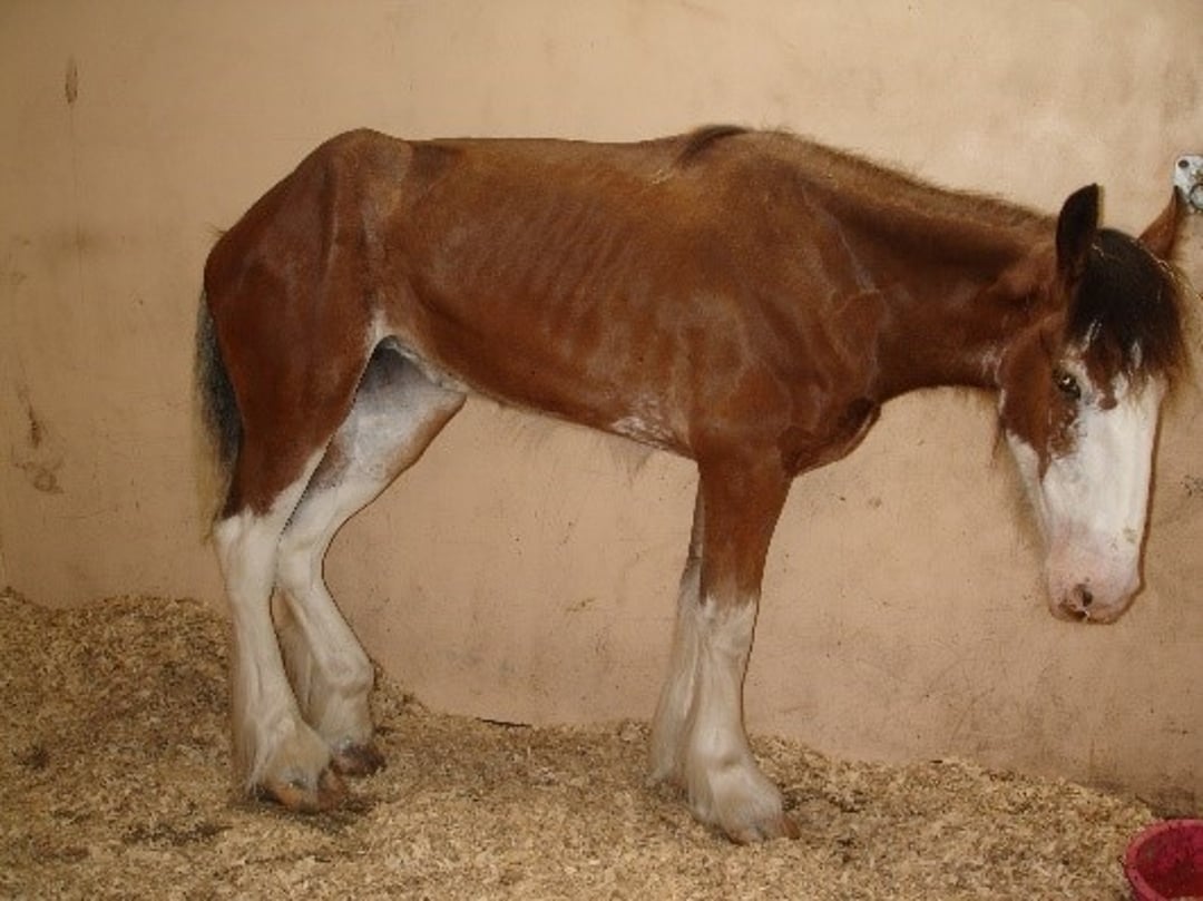
Courtesy of Dr. Caroline Hahn.
Ptosis, with droopy eyelashes, tends to be prominent because of smooth muscle paresis. Rhinitis sicca (dry nose) commonly develops in chronic cases and is considered to indicate a poor prognosis (see grass sickness after eye drops image). Affected horses often have dysphagia and esophageal dysfunction, which cause drooling, difficulty passing a stomach tube, nasal reflux of gastric contents, and pooling of barium contrast in the thoracic esophagus.

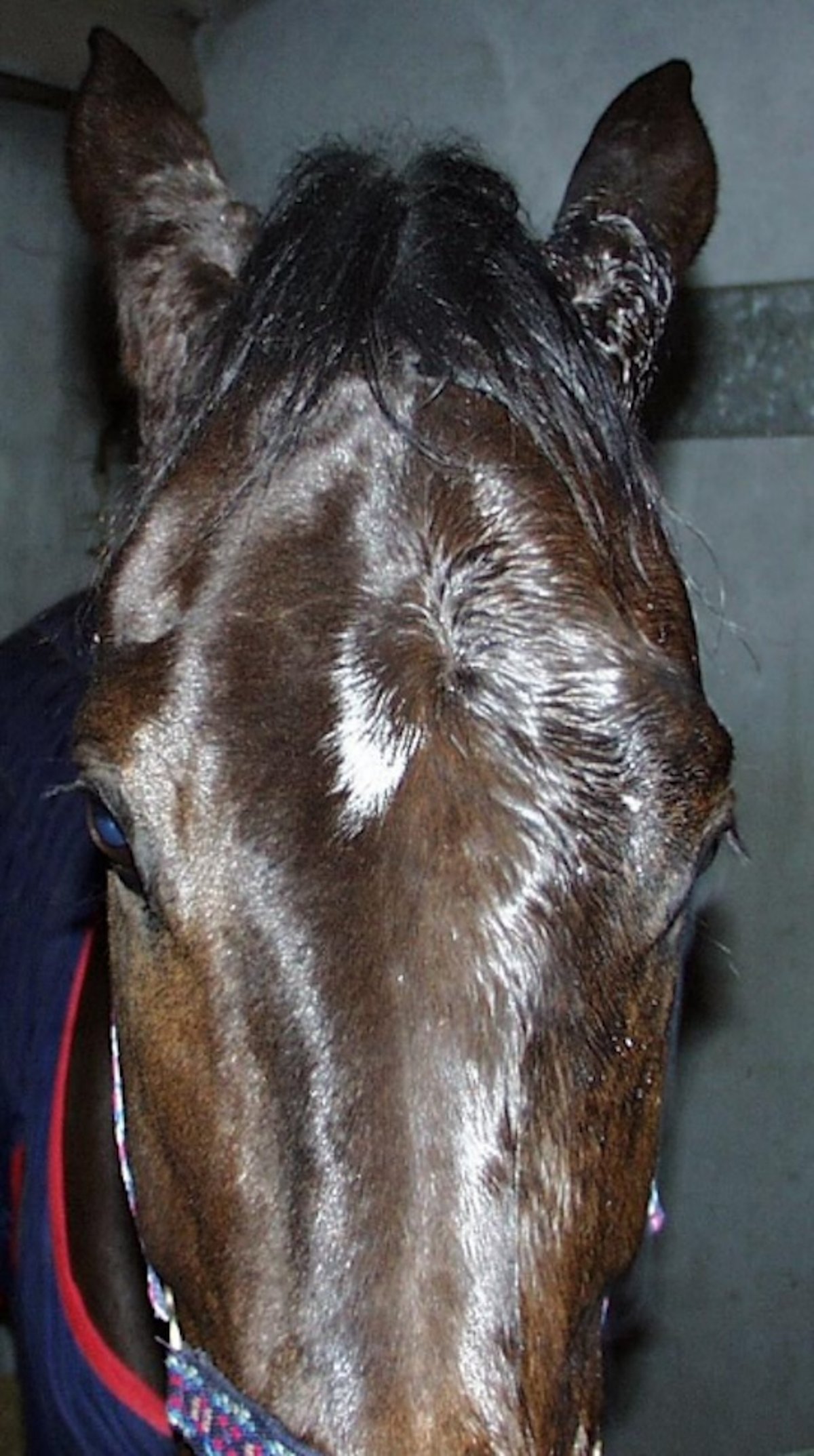
Courtesy of Dr. Caroline Hahn.
On rectal palpation, the mucosa is dry and tacky, and feces are scant and hard. Distended loops of small intestine and an impacted large colon are observed in the more acute cases. Secondary ileal dilation and impaction and displacement of the large colon can be confusing features.
Cachexia can be profound in chronic cases; in fatal cases, animals show a more rapid and greater overall body weight loss than survivors. The rapidity and magnitude of body weight loss in chronic grass sickness cases has been found to be associated with a poor prognosis, and it is worthwhile weighing these cases at regular intervals as a further indication of the severity of disease (2).
Diagnosis of Equine Dysautonomia
Patient history and clinical signs
Eye drop test and biopsy
Diagnosis of equine dysautonomia is based on the patient's history. Clinical signs such as dysphagia, tachycardia despite few signs of pain, decreased GI tract motility, a tucked-up stance (chronic cases), and ptosis are useful in diagnosis.
Eye Drop Test
Administration of dilute (0.5%) phenylephrine to one eye should, within 20 minutes, result in marked decrease in ptosis (most easily observed as a decrease in the angle of the eyelashes to the head when viewed from the front).
Biopsies
A pathologist experienced at examining grass sickness lesions can confirm diagnosis with biopsies of ileal and rectal tissue (1-cm-long, formalin-fixed biopsies preferred). Standing gustatory papillae biopsy has also been used as a confirmatory diagnostic test (3). Cytological examination of cranial cervical ganglion cytological scrapings at postmortem examination can be submitted for a rapid diagnosis in suspected equine dysautonomia cases so that management changes can be implemented sooner at the affected property, decreasing the chance of further cases occurring.
Postmortem confirmation of diagnosis depends on histological examination of autonomic ganglia. The cranial cervical ganglia are the most accessible ganglia on postmortem examination and can be found in a fold of mucosal tissue in the caudal wall of the medial compartment of the guttural pouch. Although the CNS changes are typical, the pathologist must be skilled in neuropathology.
Treatment of Equine Dysautonomia
Supportive care for chronic cases
Acute and subacute cases of equine dysautonomia do not survive and should be euthanized on humane grounds.
Treatment of chronic cases is limited to dedicated nursing care. A wide variety of high-energy, high-protein feeds that are easily swallowed should be offered to encourage feed intake. Appetite often varies markedly from day to day, both in quantity and food preference, and “good” days are often followed by “bad” days.
Many cases show nasal crusting, which makes nasal breathing difficult and discourages patients from eating. The accessible part of the nostril should be cleaned with warm water several times a day.
Unlike data from the 1980s when the recovery rate was ~55% (4), the recovery rate for carefully selected chronic cases at a large teaching hospital in Scotland is now ~70%, of which ~40% are back to work and the other 25% of more recent cases were still gaining weight (5). This represents a major improvement in the prognosis for such cases compared with the situation before the late 1980s.
Lesions
In acute cases, the stomach and small intestine are markedly distended with fluid (which can result in gastric rupture), and the large intestine is impacted. In chronic cases, the GI tract is usually empty. All forms may show linear ulceration of the esophagus and hard, tarry fecal balls. Neuronal degeneration of pre- and postganglionic sympathetic and parasympathetic neurons is characteristic. A specific distribution of chromatolytic autonomic and somatic lower motor neurons is found in the brainstem and spinal cord.
Recovered cases show evidence of substantial neuronal loss in prevertebral and paravertebral ganglia and in the enteric plexuses of the small intestine, particularly the ileum. However, the interstitial cells of Cajal networks are mainly intact in the muscularis externa of the small intestine, which may contribute to the maintenance of intestinal motility in such cases.
Prevention of Equine Dysautonomia
Keys to decreasing the risk of equine dysautonomia include the following:
Minimize exposure to pastures with a history of cases.
Stable at-risk stock for part of the day.
Minimize any pasture and soil disturbance (eg, plowing, mechanical feces removal, construction work).
Minimize soil exposure (eg, close grazing or poaching of fields).
Maintain consistent diet (quantity and feed type).
Additional measures may include co-grazing with ruminants, keeping pasture grass cut, removing feces manually, and supplementary forage feeding (hay or haylage).
Key Points
Equine dysautonomia is a disease in which central and peripheral sympathetic and parasympathetic neurons degenerate.
Clinical signs include GI hypomotility, ptosis, rhinitis sicca, anorexia, and cachexia. The etiology is unclear but is associated with access to green grass.
Diagnosis is helped by the effect of administration of dilute (0.5%) phenylephrine to one eye.
Definitive diagnosis requires a rectal or small intestinal biopsy.
There is no treatment other than supportive measures. Some mildly affected cases can survive.
For More Information
Equine Grass Sickness Fund. Moredun Foundation.
Role of fecal bacteria in equine grass sickness. Kentucky Equine Research: Equinews Nutrition & Health Daily. Updated May 26, 2016.
Hunter LC, Miller JK, Poxton IR. The association of Clostridium botulinum type C with equine grass sickness: a toxicoinfection?Equine Vet J. 1999;31(6):492-499. doi:10.1111/j.2042-3306.1999.tb03857.x
Also see pet health content regarding dysautonomia in horses.
References
Vincze B, Varga M, Kutasi O, et al. Family aggregation analysis shows a possible heritable background of equine grass sickness (dysautonomia) in a Hungarian stud population. Acta Vet Hung. 2020;68(3):263-268. doi:10.1556/004.2020.00038
Jago RC, Handel I. Hahn CN, et al. Bodyweight change aids prediction of survival in chronic equine grass sickness. Equine Vet J. 2016;48(6):792-797 doi:10.1111/evj.12551
Quéré E, Volmer C, Mespoulhès-Rivière C. Standing gustatory papillae biopsy procedure for antemortem diagnosis of equine grass sickness. J Am Vet Med Assoc. 2023;262(2):201-208. doi:10.2460/javma.23.07.0403
Wood JL, Milne EM, Doxey DL. A case-control study of grass sickness (equine dysautonomia) in the United Kingdom. Vet J. 1998;156(1):7-14. doi:10.1016/s1090-0233(98)80055-5
Pirie RS, Jago RC, Hudson NP. Equine grass sickness. Equine Vet J. 2014;46(5):545-553. doi:10.1111/evj.12254


