


Thrombosis (clot formation within a blood vessel), embolism (process by which unattached material such as a blood clot, fat or cholesterol deposit, gas, tissue, or foreign material travels within the bloodstream and occludes flow within a vessel), aneurysm (dilatation or outpouching of a blood vessel wall), and dissection (partial- or full-thickness tear in the arterial wall layers) are pathological abnormalities that can occur within the vasculature. Clinical signs, diagnostic testing, and treatment vary depending on the classification, location, severity, chronicity, and associated or concurrent comorbidities. Typically, mainstays of treatment include addressing the underlying disease process and administering antithrombotic drugs.
Thrombosis, embolism, aneurysm, and dissection are pathological abnormalities that can occur within the vasculature in animals.
Etiology and Pathophysiology of Thrombosis, Embolism, Aneurysm, and Dissection
Thrombosis is the process of formation of a thrombus, an intravascular aggregation of blood cells, platelets, and fibrin (ie, a blood clot).
The three main conditions that contribute to thrombosis are termed the Virchow triad:
hemodynamic changes such as blood stasis or turbulence
endothelial injury or dysfunction
a hypercoagulable state
A thrombus can develop in a cardiac chamber and be attached to the endocardial wall (mural) or, less likely, free-floating (ball); or it can originate in situ within a blood vessel, where it can cause partial or complete obstruction to blood flow.
Thrombi can be classified by their location and the clinical signs they produce (eg, jugular venous thrombosis associated with venous catheterization in large animals, pulmonary arterial thromboembolism associated with heartworm disease in dogs).
Clinically important vascular thrombosis commonly affects animals with underlying diseases that result in a hypercoagulable state, such as systemic inflammation, endotoxemia, neoplasia, endocrinopathies (eg, hyperadrenocorticism), immune-mediated hemolytic anemia (IMHA) in dogs, or antithrombin deficiency (eg, protein-losing nephropathy or enteropathy).
Poor IV injection or catheterization techniques, along with inferior catheter material, can also result in vascular thrombosis.
If left untreated or uncontrolled, systemic thrombosis can result in hemorrhagic diathesis or DIC, a life-threatening disorder of hemostasis with deposition of microthrombi and consumption of coagulation factors that results in concurrent hemorrhage.
Thrombi can form in both large and small arteries and veins. Horses and cattle are more likely to develop venous thrombi; in dogs and cats, arterial thrombi appear to be more important clinically. However, aortic-iliac thrombosis has been reported in horses and critically ill calves, and thrombosis of the cranial vena cava has been reported in dogs.
Arterial thrombosis or embolization leads to decreased or occluded blood flow within a vessel, resulting in ischemia of the tissues and infarction (tissue death or necrosis) in cases of prolonged ischemia (eg, in cats with cardiac disease and subsequent arterial thromboembolism).
Systemic arterial thromboembolism is more important in cats. Dogs and large animals appear to develop in situ arterial thrombosis more commonly.
Thrombosis of limb arteries, causing lameness and gangrene, has been reported in adult horses and foals. This type of thrombosis occurs secondary to hypercoagulation and systemic inflammation (eg, septicemia in foals).
A high risk of thrombosis has been identified for dogs with IMHA, protein-losing nephropathy, protein-losing enteropathy, or clinical dirofilariasis; for cats with cardiomyopathy and associated risk factors; and for dogs and cats with more than one disease or risk factor for thrombosis (1, 2). Arterial thrombosis in dogs is most commonly associated with protein-losing nephropathy and neoplasia; however, idiopathic thrombosis also occurs.
Embolism is the process by which unattached material (an embolus) travels within the bloodstream and occludes flow within a vessel.
All or part of a thrombus can break off and be carried through the bloodstream as a thromboembolus that lodges distally at a point where the size of the embolus exceeds the vascular diameter.
Alternatively, an embolus can be composed of fat or cholesterol deposits, gas, tissue, or foreign material.
Emboli from infective conditions such as endocarditis are classified as septic (containing bacteria). Septic emboli can result in bacterial dissemination and infection of distal capillary beds. Neoplastic emboli can also occur and can contribute to metastasis.
Air emboli occur when air or other gases enter the venous or arterial system. Air emboli have been described in association with IV injections or catheterizations, certain dental procedures, thoracic spinal surgery, cystoscopy, and laparoscopy.
Air that enters the venous circulation can eventually find its way to the arterial circulation via a right-to-left shunt or by overwhelming the filtering capacity of the lungs. Therefore, signs of venous air embolism are similar to those of direct arterial embolism: primarily cardiovascular collapse and CNS injury.
An aneurysm is a vascular dilatation caused by weakening of the tunica media of blood vessels. The weakness might be primary or caused by degenerative or inflammatory changes progressing from an intimal lesion.
Dissection occurs when the weakened walls of a blood vessel (eg, aorta or pulmonary artery) tear, causing blood to accumulate between the arterial wall layers, which causes a localized collection of blood called a false aneurysm or pseudoaneurysm. A dissection that occurs through all three layers of the arterial wall is termed a "rupture."
Disruption of the endothelium associated with a true aneurysm can cause the formation of a thrombus with subsequent embolization; thus, aneurysms, dissections, thrombi, and emboli can be recognized simultaneously. In humans, the presence of an aneurysm increases the risk of a dissection or tear within the same vessel.
Aneurysms are rare in domestic animal species; however, they have been reported in dogs, cats, horses, primates, turkeys, reptiles (eg, aortic aneurysms in bearded dragons), and other exotic species.
Athough aortic and pulmonary artery dissections are generally rare, they have been reported with increasing frequency in dogs. There are reports of spontaneous aortic dissections in dogs (3), as well as iatrogenic pulmonary arterial dissections after balloon pulmonary valvuloplasty procedures in dogs with severe pulmonic stenosis.
Clinical Findings and Diagnosis of Thrombosis, Embolism, Aneurysm, and Dissection
In cases of thrombosis, embolism, aneurysm, or dissection, the clinical signs vary depending on the classification, location, severity, and chronicity, as well as associated or concurrent comorbidities.
Acute onset of dyspnea is often associated with pulmonary thromboembolism; however, some animals can develop hemoptysis. Hemoptysis is usually associated with pulmonary arterial disease, such as that resulting from heartworm infection.
Septic cardiac thrombi are associated with endocarditis; nonseptic cardiac thrombi are associated with myocardial disease (most commonly in cats) or, rarely, with cardiac or pulmonary neoplasia. Clinical signs associated with infarction within the genitourinary system can include hematuria, abdominal pain, and splinting. Splanchnic infarction usually results in abdominal pain, with vomiting, in small animals.
Aneurysms cause no clinical signs, unless a dissection or hemorrhage occurs or an associated thrombus develops. Except for hemorrhagic vasculopathy of turkeys, aortic rupture in horses, hemorrhage associated with guttural pouch mycosis in horses, or pulmonary arterial aneurysm in cattle, spontaneous aneurysmal hemorrhage is rare, and clinical signs usually relate to thrombosis.
An aneurysm of the abdominal aorta and its branches in large animals can be palpated rectally as a fixed firm swelling with a rough, irregular surface that pulsates in rhythm with the heartbeat. Fremitus can be present. In excess thrombus formation, the pulse can be delayed distally and have a slow rate of rise in pressure, or it can be absent.
In addition to history and physical examination findings, relevant diagnostic modalities can include routine laboratory tests (eg, CBC, serum biochemical panels), screening for hypercoagulability (eg, thomboelastography, prothrombin time [PT], activated partial thromboplastin time [aPTT]), and diagnostic imaging (ultrasonography, CT, infrared thermography, and angiography).
Clinical Findings and Diagnosis in Cattle
In cattle, thrombosis of the caudal vena cava commonly occurs in association with rumenitis. Rumenitis leads to hepatic abscess formation, which can result in a thrombus in the caudal vena cava if the vessel wall is infiltrated by the abscess (see ). Usually, the thrombus is visualized within the hepatic portion of the caudal vena cava; however, it can also be found in the perirenal, subphrenic, or intrathoracic portion.


Gross pathology photograph of a thrombus within the caudal vena cava of a bovid. Note the dark red to black, smooth, glossy clot occluding the lumen of the incised caudal vena cava, which is held open with forceps. Note also the pale color of the lungs, with areas of hemorrhage.
Courtesy of Dr. Sameeh M. Abutarbush.
Typical clinical signs of thrombosis of the caudal vena cava in cattle include the following:
poor general condition
chronic weight loss
intermittent fever
Embolic pneumonia with secondary pulmonary abscess formation, thromboembolism, and pulmonary arterial aneurysms are common sequelae of thrombosis of the caudal venal cava.
Signs in affected cattle with pulmonary involvement include coughing, tachypnea, dyspnea, and abnormal lung sounds.
Aneurysms in pulmonary arteries that contain septic emboli can rupture and cause intrapulmonary hemorrhage, or pulmonary abscesses can erode into bronchi and result in hemorrhage into the airways. Possible sequelae include epistaxis, hemoptysis, and death.
Clinicopathological data usually support a diagnosis of thrombosis of the caudal vena cava, but they are not specific. Increased fibrinogen, anemia, and, in cases with an active abscess process, increased liver enzyme activity can occur.
In affected cattle, ultrasonographic evaluation of the hepatic portion of the caudal vena cava can reveal the thrombus or show a dilated caudal vena cava (shape change from triangular to round). The latter finding is consistent with obstruction of the extrahepatic caudal vena cava (thrombus/abscess) or right-sided heart failure. Hepatic congestion and abscess formation are also possible.
Pulmonary arterial embolism and embolic pneumonia (see ) are also frequent complications of tricuspid or pulmonic valvular endocarditis in cattle; however, aneurysms rarely develop.


Gross pathology photograph showing embolic pneumonia in a cow with endocarditis syndrome. Note the multifocal, randomly distributed necrotic or purulent foci on the pulmonary visceral pleura (pulmonary abscesses).
Courtesy of Dr. Sameeh M. Abutarbush.
Intermittent fever and anorexia due to bacteremia at times of embolic showering are often present, and affected cattle typically have a history of a chronic active infection (eg, septic venous thrombosis, foot abscess, reticular abscess, septic arthritis), as well as chronic weight loss withpoor body condition (body condition score, 1 or 2 on a 5-point scale).
Most cases of right-sided endocarditis in cattle are bacterial and can sometimes be associated with a cardiac murmur. Echocardiography and blood cultures help identify right-sided heart vegetative lesions and the causative bacterial agent, respectively.
Cattle with congenital heart defects, such as ventricular septal defects (VSDs), and turbulent blood flow within the heart might be predisposed to the development of endocarditis and embolic showering.
Thrombosis of the cranial vena cava in cattle produces bilateral jugular engorgement; edema of the head, submandibular area ("bottle jaw"), and sternal region (so-called brisket edema); and pronounced oral mucosal hyperemia. However, similar clinical signs occur with right-sided congestive heart failure, which could be a sequela of tricuspid valve endocarditis. Marked lingual, pharyngeal, or laryngeal edema can develop and result in dysphagia and dyspnea. Ultrasonography of the vena cava and echocardiography can aid in the diagnosis.
A connective tissue disorder similar to Marfan syndrome in humans has been described in cattle with aortic dilatation and aortic aneurysms. These cattle typically also display ocular abnormalities and skeletal involvement.
Clinical Findings and Diagnosis in Horses
Thrombophlebitis is common in horses that receive IV injections or undergo catheterization for various IV treatments. The jugular vein is the most commonly used vein for these procedures and is therefore the most commonly affected.
Inflammation, infection, or extravasation of injected material causes swelling, heat, and pain of the affected area. Bilateral jugular vein thrombosis can cause edema and swelling of the head and neck, mimicking thrombosis of the cranial vena cava. Jugular vein thrombi commonly extend into the maxillary and linguofacial veins and rarely into the cranial vena cava.
Two-dimensional and Doppler ultrasonographic examination of the affected vein can determine the extent of the thrombus and degree of occlusion. Ultrasonography is also useful for identifying and performing guided aspiration of fluid cavitations for bacterial culture and for evaluating cardiac valves for secondary endocarditis lesions. Blood culture and catheter-tip culture can be performed.
Horses with colitis and other GI disorders are at increased risk of developing jugular thrombosis. Ruminants are much less prone to jugular thrombosis than horses are; however, ruminants are susceptible to septic thrombosis of the cranial superficial epigastric vein.
In one reported case in a Quarter Horse with respiratory signs, lesions similar to those in caudal vena cava thrombosis syndrome of cattle (ie, hepatic abscesses, thrombosis of the caudal vena cava, pulmonary thromboembolism, and embolic pneumonia) were identified at necropsy (4).
Migrating Strongylus vulgaris larvae can cause arteritis with development of thrombi and verminous aneurysms in the aorta, cranial mesenteric, or iliac artery (see Strongylus vulgaris–Associated Disease). In some affected horses, emboli develop and partially or completely occlude terminal branches of the mesenteric arteries. Affected intestinal segments show changes ranging from ischemia to hemorrhagic infarction. Clinical signs include colic, constipation, or diarrhea. The colic usually is recurrent, and attacks can be severe and prolonged.
Newer anthelmintics and improved therapeutic regimens have resulted in verminous arteritis becoming an uncommon disorder.
Thrombosis with or without aneurysm of the terminal aorta and proximal iliac arteries produces a characteristic syndrome in horses. Although the syndrome is associated with parasitism, other causes are possible.
Affected horses appear normal at rest; however, graded exercise results in an increasing severity of weakness of the hindlimbs, with unilateral or bilateral lameness, muscle tremor, and sweating.
Severely affected horses can show signs of exercise intolerance, weakness, and atypical lameness that resolves after a short rest. Subnormal temperature of the affected limbs might be detectable, along with decreased or absent arterial pulsations and delayed and diminished capillary filling. Rectal palpation can show variation in pulse amplitude of the internal or external iliac arteries (or both) and asymmetrical vasculature. In severe cases, lameness and atrophy of the hindquarter muscles can become evident with only mild exercise.
Complete embolic or thrombotic occlusion of the distal aorta can produce acute bilateral hindlimb paralysis and recumbency in horses. Affected animals are anxious, appear painful, and rapidly go into shock. The hindlimbs are cold, and rectal palpation reveals an absence of pulsation in either iliac artery.
Transrectal ultrasonography can help determine blood flow in the aorta and iliac arteries.
Aneurysm of the aortic root has been reported in horses, most commonly noted at the right aortic sinus. Similar to those in humans, aneurysms of the sinuses of Valsalva in horses can be congenital or acquired and can be clinically silent until they rupture.
Aortic aneurysms, dissections, and rupture can occur anywhere along the aorta; the location determines the clinical presentation. Rupture of the intracardiac aortic sinus of Valsalva (aortocardiac fistula) usually results in acute signs of generalized distress and can be misinterpreted as abdominal colic. A continuous murmur can be heard over the tricuspid valve area in most cases. Ventricular arrhythmias are usually present in the acute phase but resolve with time.
Horses rarely experience sudden death with sinus of Valsalva rupture, unless a malignant ventricular arrhythmia is also present. Left-to-right shunting results in left-sided congestive heart failure over time (weeks, months, or years).
Standard right parasternal echocardiographic views of the left ventricular outflow tract are diagnostic of an aneurysm, and color flow Doppler echocardiography can confirm rupture of the intracardiac aortic sinus of Valsalva, causing an aortocardiac fistula.
Intrathoracic rupture of the aorta at the level of the aortic arch near the scar of the ligamentum arteriosum is well described in Friesian horses. This type of aortic rupture is characterized by formation of a pseudoaneurysm and pulmonary artery fistula. Compared with other warmblood breeds, Friesians appear to have a higher incidence of aortic rupture (approximately 2%) (5, 6).
Possible signs of aortic dissection and rupture in horses include colic signs, cough, epistaxis, heart failure, and sudden death from intrathoracic hemorrhage. A murmur may or may not be present. Aortic aneurysms without dissection or rupture are usually clinically silent.
Left parasternal echocardiographic views near the bifurcation of the pulmonary artery can be diagnostic, but they can be challenging to obtain.
Rupture of the intraabdominal aorta in horses, often in association with exercise or exertion, results in massive intracavitary hemorrhage and sudden death. Diagnosis is made on postmortem examination.
Any horse with an unruptured aneurysm or a stabilized aortocardiac fistula should be considered unsafe to ride because of the risk of sudden death.
Pearls & Pitfalls
|
Air emboli are infrequent but potentially life-threatening complications of venous catheterization and accidental disconnection from fluid lines. In affected horses, possible clinical signs include agitation or other behavioral changes; muscle fasciculations; acute recumbency; acute or delayed blindness; generalized or focal seizures, dysphagia, ataxia, or various other neurological deficits; and cardiac dysrhythmias. Some horses with air emboli die suddenly.
The prognosis for resolution of air embolus clinical signs is fair to good in horses; however, neurological deficits can persist and result in elective euthanasia. Delayed onset and recrudescence of clinical signs have been described.
Diagnosis is often based on clinical signs and surrounding circumstances, with few likely differential diagnoses that could explain clinical signs. Rarely, air emboli are observed on echocardiographic or postmortem examination of horses.
Treatment is directed at managing the specific complications.
Clinical Findings and Diagnosis in Dogs and Cats
Diseases associated with pulmonary thromboembolism (PTE) in dogs and cats include protein-losing nephropathy or enteropathy, hyperadrenocorticism, IMHA, and neoplasia.
In dogs, and less commonly in cats, heartworm disease can lead to pulmonary arterial thromboembolism (see ) that commonly results in dyspnea, and collapse. Affected animals are often reportedly healthy until sudden onset of coughing, hemoptysis, respiratory distress, or sudden death.


Gross pathology photograph showing a thrombus in the pulmonary artery of a dog with heartworm disease. Note the dark red, granular clot and adult worm remains occluding the lumen of the dilated pulmonary artery, which has been transected.
Courtesy of Dr. Suzanne Cunningham.
Clinical signs of PTE in dogs with heartworm disease most commonly develop in the weeks after treatment with adulticide; however, PTE can also develop from spontaneous worm death, or pulmonary thrombosis can form in situ secondary to pulmonary endothelial damage.
In dogs and cats with PTE, thoracic radiography can be normal or can show underperfusion of the affected lung lobe, interstitial to alveolar infiltrates, or pleural effusion. Tortuosity of the pulmonary arterial branches can be visualized in patients with heartworm disease or pulmonary hypertension secondary to PTE.
Arterial blood-gas analysis in cases of PTE in dogs and cats typically demonstrates hypoxemia with a normal or low partial pressure of CO2 (PaCO2). Ventilation/perfusion scanning with radionuclide-labeled macroaggregated albumin and gases or pulmonary CT angiography can confirm the diagnosis.
Pulmonary hypertension can develop as a sequela of PTE and can result in right-sided congestive heart failure with abdominal distention (ascites).
Additional diagnostic tests to evaluate for PTE and underlying etiologies in dogs and cats can include echocardiography, thromboelastography, heartworm antigen (antigen/antibody for cats) testing, CBC, biochemical analyses, urinalysis with urinary protein:creatinine or urinary cortisol:creatinine ratio, low-dose dexamethasone suppression testing, or ACTH stimulation testing.
In cats, cardiogenic embolism (arterial thromboembolism) is a devastating complication of cardiac disease. In most other species, overt intracardiac thrombus formation is rare with heart disease.
Although hypertrophic cardiomyopathy is the most common type of cardiac disease in cats, any condition that results in left atrial enlargement, including other cardiomyopathies (eg, dilated cardiomyopathy, restrictive cardiomyopathy), hyperthyroidism, or congenital heart disease, can predispose a cat to arterial thromboembolism.
Intracavitary thrombi typically form in the dilated left atrium or left atrial appendage (auricle) where stagnant flow exists (see ) or, less commonly, within abnormal areas in the left ventricle.


Gross pathology photograph showing an intracardiac thrombus in the left atrial appendage of a cat with congenital heart disease. Note the dark red, granular clot filling the left atrial appendage, which has been incised and is held open with forceps.
Courtesy of Dr. Suzanne Cunningham.
If a cardiac etiology for an arterial thromboembolism is ruled out in a cat, pulmonary neoplasia or protein-losing nephropathy should be considered. Tumor embolism secondary to pulmonary neoplasia, albeit not a true thrombus, is the most common noncardiac cause of arterial thromboembolism in cats and can be further investigated via thoracic radiography.
Although cardiogenic embolism is poorly understood, affected cats can also have hypercoagulability, because not all cats with cardiomyopathy develop cardiogenic embolism.
In cats, portions of intracavitary thrombi can break off and form emboli that infarct arterial branches, most commonly the aortic trifurcation (known as feline aortic thromboembolism [FATE] or "saddle thrombus"; see videos of a cat with saddle thrombus and treatment). Aortic thromboembolism can result in partial/transient or complete occlusion of blood flow in the affected artery. Affected limbs can be unilateral, bilateral, or bilateral but asymmetrical. Usually, the hindlimbs are affected; however, the forelimbs can also be involved.

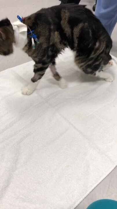
Clinical signs of an aortic thromboembolism in cats include pain and paresis or lower motor neuron paralysis of the hindlimbs. The arterial pulse (either femoral or pedal) is decreased to absent in affected limbs, which are cooler than normal and have firm, swollen gastrocnemius muscle bellies.
Emboli can also infarct other arterial beds in dogs and cats, including the right forelimb, renal, splanchnic, cerebral, or myocardial circulation.
Decompensation of the underlying myocardial disease is not uncommon and can result in congestive heart failure (manifesting as pulmonary edema or pleural effusion).
Ischemia and necrosis of infarcted pelvic limb musculature results in increases in serum CK and AST activities. Subsequent reperfusion of affected muscles can result in life-threatening hyperkalemia and acidosis.
Differential glucose and lactate concentrations help confirm a diagnosis of arterial thromboembolism. In peripheral venous blood samples from affected limbs, glucose concentration is lower and lactate concentration higher, compared with those in unaffected limbs or central veins.
Echocardiography is the imaging modality of choice to assess cardiac structure, function, and presence of an intracardiac thrombus in dogs and cats. Thoracic radiography is used to diagnose left-sided congestive heart failure or to evaluate for noncardiac etiologies such as pulmonary neoplasia.
Although they are rarely performed, nuclear perfusion studies using the unbound radioisotope 99mTc can provide sensitive information regarding the extent of perfusion of the hindlimbs and areas that might require amputation in select cases.
Infrared thermography can provide additional support in diagnosing arterial thromboembolism in cats and other animals.
Systemic hypertension in cats, as well as patent ductus arteriosis, aortic coarctation, degenerative processes, connective tissue disorders (eg, Marfan syndrome), and infections in dogs, have been associated with aortic aneurysms and dissections. Obtaining differential blood pressure measurements in the forelimbs and hindlimbs can be part of the diagnostic workup.
Thrombosis of the cranial vena cava and subsequent chylothorax and acute respiratory distress have been reported in dogs and cats with transvenous pacemakers and other indwelling jugular devices/catheters. Thrombosis of the cranial vena cava has also been associated with caval cannulation for open heart surgeries performed under cardiopulmonary bypass.
Treatment of Thrombosis, Embolism, Aneurysm, and Dissection
Restoration of blood flow and perfusion
Supportive care
Antithrombotics, thrombolytics, and antimicrobials
Surgical intervention
The treatment of thrombosis or embolism is intended to restore blood flow and perfusion, preventing further clot formation while addressing the underlying cause.
Common approaches include supportive care, pharmacological therapies (eg, antithrombotics, thrombolytics, antimicrobials if sepsis is present), as well as additional interventions (eg, angioplasty/venoplasty, endovascular stenting, thrombectomy). Specific therapeutic approaches vary by species, location, severity, chronicity, underlying etiology, and comprehensive clinical picture of the patient.
Treatment of Bacterial Endocarditis
The treatment of bacterial endocarditis includes long-term antimicrobials (several weeks to months) and, in some cases (depending on the species), intermittent administration of antipyretic, anti-inflammatory, or antithrombotic drugs. The choice of antimicrobial should be based on culture and susceptibility results obtained from blood cultures.
The prognosis for recovery from bacterial endocarditis depends on the organism and on the affected heart valve. In general, the prognosis is poor to guarded, and persistent debilitating cardiac disease can be common even if the active infection can be controlled.
Treatment of Venous Thrombosis in Horses and Cattle
The treatment of venous thrombosis in horses and cattle is usually limited to supportive care, including hydrotherapy of accessible veins, anti-inflammatory agents, and systemic antimicrobials to control secondary sepsis.
Surgical removal of thrombosed jugular veins has been performed successfully in horses; however, unless both veins are severely affected, inflammation resolves with medical treatment, and the formation of collateral vessels usually results in sufficient venous circulation.
Thrombosis of the cranial or caudal vena cava has more severe clinical signs and requires more aggressive therapy, which could include thrombolytic drugs and/or intravascular/surgical removal followed by aggressive anticoagulation. Response to anticoagulation therapy alone is generally inadequate.
Measures to minimize trauma to, as well as bacterial contamination of, veins are the best means to prevent venous thrombosis. Extreme care should be taken when placing catheters or administering IV injections.
Intrinsic thrombus has been treated as follows:
Antiplatelet therapy (as needed, depending on clinical response):
aspirin: 5–10 mg/kg, PO, every 24 or 48 hours
clopidogrel: 1–3 mg/kg, PO, every 24 hours
Anticoagulant therapy (as needed):
unfractionated heparin: 40–80 U/kg, SC or IV, every 8–12 hours
low-molecular-weight heparin: enoxaparin (40–80 U/kg, SC, every 24 hours); or dalteparin (50 U/kg, SC, every 12–24 hours for adults, or 100 U/kg, SC, every 24 hours for foals)
The effectiveness of these therapies at facilitating thrombus resolution is unknown; however, they should at least prevent further thrombus formation.
In horses, aneurysms due to Strongylus vulgaris rarely rupture; the chief concern is thromboembolism of intestinal vasculature with subsequent colic. Generally, the arterial wall is involved enough that thrombus removal is impractical. Antimicrobials and anthelmintics to kill the migrating larvae are of considerable value. The most rational approach to cranial mesenteric and aortic-iliac thrombosis in horses is prevention and control of Strongylus vulgaris–associated disease.
Ultrasonographically guided balloon thrombectomy for treatment of aorto-iliac-femoral thrombosis in a horse has been reported (7).
Treatment of Cardiogenic Embolism in Cats
Acute management of aortic emboli in cats can be approached in several ways.
More than 50% of cats that survive a cardioembolic event regain some function of their hindlimbs over 4–6 weeks with conservative medical therapy.
More aggressive treatment aimed at dissolving the thrombus with thrombolytic drugs or rheolytic intervention (using high-pressure saline and aspiration to break up and remove a clot) can result in short-term functional improvement; however, survival time might be similar to that of conservative therapy. In some cases, survival time can be worse because of the risk of reperfusion injury if the treatment is not administered within the initial 4- to 6-hour window preceding the event (8).
Pearls & Pitfalls
|
Conservative treatment of aortic emboli in cats usually consists of initial pain management and anticoagulant therapy as follows:
Pain management options:
hydromorphone: 0.1–0.2 mg/kg, SC, IM, or IV, every 4–6 hours as needed
buprenorphine HCl: 0.01–0.03 mg/kg, SC, IM, IV, or oral transmucosal [buccal], every 6–8 hours as needed
injectable high-concentration buprenorphine (1.8 mg/mL): 0.24 mg/kg, SC, every 24 hours for up to 3 days while hospitalized
Anticoagulant therapy options:
enoxaparin: 0.75–1 mg/kg, SC, every 6–12 hours long-term
dalteparin: 50–200 U/kg, SC, every 6–12 hours as needed
unfractionated heparin: initial dose 350 U/kg, SC, followed by 150–250 U/kg, SC, every 6–8 hours or 150–300 U/kg, SC, every 8 hours for 24–48 hours
The aPTT can be used to monitor treatment with unfractionated heparin in cats, with a goal of 1.5–1.7 times the pretreatment value. aPTT and PT might not be reliable for therapeutic monitoring with low-molecular-weight heparin; thus, anti-Xa assays can be used for monitoring.
To further decrease the thrombotic potential, antiplatelet therapy (clopidogrel at 37.5–75 mg total per cat for loading dose, PO, once on admission, then 18.75 mg, PO, every 24 hours long-term) should be considered; in addition, antiplatelet therapy can have a beneficial effect on collateral circulation.
Some cats have a genetic mutation in the P2RY1 gene associated with clopidogrel resistance, requiring alteration of the antithrombotic drug protocol (9).
Thrombolytic therapy, although not routinely recommended for treating aortic emboli in cats, could include streptokinase (90,000 U/cat, IV, over 20 minutes, followed by 45,000 U as CRI over 2–24 hours); recombinant tissue-type plasminogen activator (r-tPA, 0.25–1 mg/kg, IV, every hour up to a total dose of 1–10 mg/kg); or urokinase (4,400 U/kg, IV, over 10 minutes, then 4,400 U/kg, every hour for 12 hours).
These drugs promote thrombolysis by converting plasminogen to plasmin, which subsequently breaks down fibrin strands. Streptokinase is considered a nonspecific activator of plasminogen because it activates circulating fibrin, as well as fibrin contained within thrombi/emboli, which can lead to a systemic proteolytic state and bleeding. Streptokinase infusion has been associated with life-threatening hyperkalemia (reperfusion injury) and clinical signs of bleeding requiring blood transfusion in cats.
Although urokinase and r-tPA (alteplase) are more fibrin-specific than streptokinase, bleeding can occur also with these agents. Complications such as hyperkalemia (attributable to reperfusion injury) can also occur. Moreover, all of these agents are prohibitively expensive and difficult to obtain. r-tPA is the most commonly used thrombolytic and can be considered in cats if the thromboembolic event occurred < 6 hours earlier.
Treatment with thrombolytics appears to induce the best response in cats that have acute onset of clinical signs (< 6 hours) and incomplete or unilateral infarction. However, these cats can respond similarly well to conservative therapy, with less expense and without the risk of reperfusion injury.
Cats with severe complete occlusion/blockage of the blood vessel from a thrombus are more likely to develop reperfusion injury with thrombolytic treatment, and they are also less likely to recover with conservative therapy alone.
Survival rates for initial aortic thromboembolic events are similar whether conservative or thrombolytic therapy is used. Cats with single limb thromboembolism have a better rate of survival to discharge (approximately 70–80%) than cats with bilateral hindlimb thromboembolism (approximately 30–40%), regardless of the therapy used (8, 10, 11, 12, 13, 14).
Aspirin as needed (25 mg/cat, PO, every 48–72 hours; or 5 mg/kg, PO, every 48–72 hours) has historically been a widely used preventive therapy for cardioembolic disease in cats. The antiplatelet efficacy of aspirin in cats has been called into question, and currently there is no evidence that aspirin prevents first-time or recurrent cardiogenic embolism. Clopidogrel is recommended over aspirin as an antiplatelet drug in cats.
The use of antiplatelet agents such as clopidogrel has been shown to hasten thrombus dissolution and decrease recurrence of acute arterial thrombosis in experimental studies and human clinical trials, respectively. However, an in vitro feline study did not identify a notable difference in thrombolysis rates (15). It is not known whether these results can be applied to the natural disease.
Clopidogrel (18.75 mg/cat, PO, every 24 hours for chronic preventive therapy) inhibits both primary and secondary platelet aggregation. These effects are more potent than those induced by aspirin. Clopidogrel also impairs the platelet release reaction, decreasing the release of pro-aggregating and vasoconstrictive agents. Although rare, adverse effects can include vomiting in up to 10% of cats; this effect appears to be ameliorated by giving the drug with food.
Some cats have a genetic mutation in the P2RY1 gene associated with clopidogrel resistance, requiring alteration of the antithrombotic drug protocol (9).
A combination protocol of aspirin and clopidogrel was used previously but has fallen out of favor. Aspirin as the sole antithrombotic in cats at risk for arterial thromboembolism is not recommended (1).
A multicenter, randomized, prospective study showed that clopidogrel was associated with a significantly longer survival time compared with aspirin in cats with cardiogenic arterial thromboembolism (16). The time to recurrence of arterial thromboembolism or death in the clopidogrel group was > 365 days, versus 192 days in the aspirin group.
Thus, clopidogrel is currently recommended instead of aspirin for cats at risk of arterial thromboembolism.
A double-blind, randomized controlled study found that single-agent antithrombotic therapy with rivaroxaban delayed recurrence of arterial thromboembolism in cats for a similar time period as single-agent therapy with clopidogrel (17).
Warfarin has also been used to prevent primary or secondary cardiac embolism in cats but is no longer recommended for use in cats because of marked interindividual variation coupled with a narrow therapeutic index. Problems with warfarin therapy include large inter- and intraindividual variability, difficulty dosing because of tablet size, and bleeding, including fatal hemorrhage.
Low-molecular-weight heparins are smaller than unfractionated heparin; however, they maintain the ability to inhibit factor Xa, with greatly decreased inhibition of factor IIa. The decreased anti-IIa activity translates into a negligible effect on the aPTT; however, measurement of anti-Xa activity can be used to monitor dosing efficacy.
Enoxaparin (1–1.5 mg/kg, SC, every 8–12 hours, or 0.75–1 mg/kg, SC, every 6 hours, depending on clinical response) and dalteparin (150–170 U/kg, SC, every 8–12 hours, or 75 U/kg, SC, every 6 hours, depending on clinical response) have both been used in cats.
Enoxaparin and dalteparin have been well tolerated in cats, with only rare bleeding reported; however, objective clinical studies evaluating their efficacy have not been performed. These agents have been frequently combined with clopidogrel in an attempt to provide a more complete antithrombotic effect. This protocol appears to be well tolerated; however, some minor bleeding has been observed.
The use of factor Xa inhibitors such as rivaroxaban (0.5–1 mg/kg, PO, every 24 hours, or 1.25–5 mg/cat, PO, every 12–24 hours for chronic preventive therapy) or apixaban (0.625 mg/cat for cats < 5 kg and 1.25 mg/cat for cats ≥ 5 kg, PO, every 12 hours for chronic preventive therapy) has been reported for thromboprophylaxis in cats. These drugs appear to be well tolerated in combination with clopidogrel.
In a retrospective study of 1,008 cats with hypertrophic cardiomyopathy (including obstructive and nonobstructive forms), the reported cumulative risk of arterial thromboembolism at 1, 5, and 10 years after diagnosis was 3.5%, 9.7%, and 11.3%, respectively (18).
In a retrospective study of 44 cats with distal aortic thromboembolism, 17 (39%) survived the episode; in 12 of the survivors, aspirin prophylaxis was instituted, with recurrence in 2 (17%) (19). Recurrence of aortic thromboembolism has been reported in up to 75% of cats receiving aspirin prophylaxis (20). In a study of 18 cats receiving clopidogrel and rivaroxaban for prophylaxis after aortic thromboembolism, 9 (50%) survived longer than 1 year (21).
In a retrospective study of 46 cats with arterial thromboembolism that were treated with streptokinase, the median survival time was 51 days (22).
In a retrospective study of 100 cats with distal aortic thromboembolism, 37% survived the initial thromboembolic episode, with an average survival time of 11.5 months (23).
In a retrospective study of 44 cats with distal aortic thromboembolism, median survival time was 6 months (19).
In a study of 127 cats with arterial thromboembolism, 39 survived to discharge, with a median survival time of 117 days (13).
In a prospective multicenter investigation of 40 cats with bilateral pelvic limb paralysis, 15 (37.5%) survived to discharge (9 out of 20 [45%] treated with tPA and 6 out of 20 [30%] receiving placebo), with some cats experiencing > 1 year survival (8, 14).
In cases of aortic thromboembolism in cats, owners who are willing to pursue treatment should be encouraged to give cats 24–72 hours of supportive care before deciding on euthanasia, unless severe infarction, severe congestive heart failure, severe azotemia, or reperfusion injury is present.
Treatment of Arterial Thrombosis in Dogs
There is increasing experience with the diagnosis and treatment of arterial thromboembolism in dogs; however, thrombolytic therapy using streptokinase, urokinase, and r-tPA (0.5–1 mg/kg, IV, over 60–90 minutes, once) has been reported in isolated cases with variable success.
There are no clinical trials evaluating the efficacy of antithrombotic therapy for prevention of arterial thromboembolism in dogs; however, dosing protocols for have been reported for the following drugs:
aspirin: 0.5–15 mg/kg, PO, every 24 hours long-term
clopidogrel: 1.1–4 mg/kg, PO, every 24 hours long-term
warfarin: 0.1–0.22 mg/kg, PO, every 24 hours (not recommended for routine use)
unfractionated heparin: 150–300 U/kg, SC, every 6 hours as needed, depending on clinical signs
dalteparin: 100–175 U/kg, SC, every 8–12 hours as needed, depending on clinical signs
enoxaparin: 1–1.5 mg/kg, SC, every 8–12 hours; or 0.8 mg/kg, SC, every 6 hours as needed, depending on clinical signs
rivaroxaban: 0.5–2 mg/kg, PO, every 12–24 hours long-term
apixaban: 0.25–0.5 mg/kg, PO, every 8–12 hours long-term
Treatment recommendations for pulmonary embolism in dogs are similar to those for cardioembolic disease in cats, including a combination of antiplatelet and anticoagulant therapy, in addition to treatment of the underlying cause and of pulmonary hypertension, if present. A valuable resource is the CURATIVE consensus statement and updates on the rational use of antithrombotics and thrombolytics in veterinary critical care (1, 2, 24).
Antiplatelet medications used in dogs include aspirin and clopidogrel. Aspirin (0.5 mg/kg, PO, every 24 hours, depending on clinical response) was shown to improve median survival time in dogs with IMHA when added to standard immunosuppressive therapy in retrospective studies (25). Similar results were also shown for monotherapy with clopidogrel (26). One study showed no significant difference between therapy with aspirin and therapy with clopidogrel in dogs that have IMHA (27). The consensus statement for management and treatment of IMHA provides additional therapeutic recommendations (25).
Anticoagulant medications used in dogs include heparin, low-molecular-weight heparin (enoxaparin, dalteparin), and factor Xa inhibitors (rivaroxaban, apixaban). Warfarin is not recommended, because of inconsistent outcomes associated with severe bleeding complications.
Successful treatment of aortic thrombosis in dogs has been described using a combination of anticoagulants (clopidogrel and apixaban), systemic and local infusions of thrombolytics such as r-tPA, balloon angioplasty, and placement of endovascular stents (kissing-stent technique) (28).
Successful treatment of thrombosis of the cranial vena cava in dogs has been described using a combination of anticoagulants (clopidogrel with either apixaban or rivaroxaban), systemic and local infusions of thrombolytics such as r-tPA, and balloon venoplasty with or without placement of an endovascular stent.
Key Points
Thrombosis, embolism, aneurysms, and dissections can occur in any animal species.
Clinical signs, diagnostic testing, and treatment vary by classification, location, severity, chronicity, and underlying etiology, as well as by the species affected.
Treatment can include management of the underlying disease process, administration of antithrombotic or thrombolytic (systemic or local infusion) drugs, and surgical procedures (surgical thrombectomy) or interventional procedures (balloon thrombectomy, balloon angioplasty/venoplasty with or without endovascular stent placement).
For More Information
deLaforcade A, Bacek L, Blais M-C, et al. 2022 update of the Consensus on the Rational Use of Antithrombotics and Thrombolytics in Veterinary Critical Care (CURATIVE): domain 1—defining populations at risk. J Vet Emerg Crit Care (San Antonio). 2022;32(3):289-314. doi:10.1111/vec.13204.
Also see pet owner content regarding blood clots and aneurysms in dogs, cats, and horses.
References
Sharp CR, Goggs R, Blais M-C, et al. Clinical application of the American College of Veterinary Emergency and Critical Care (ACVECC) Consensus on the Rational Use of Antithrombotics in Veterinary Critical Care (CURATIVE) guidelines to small animal cases. J Vet Emerg Crit Care (San Antonio). 2019;29(2):121-131. doi:10.1111/vec.12804
deLaforcade A, Bacek L, Blais M-C, et al. 2022 update of the Consensus on the Rational Use of Antithrombotics and Thrombolytics in Veterinary Critical Care (CURATIVE): domain 1—defining populations at risk. J Vet Emerg Crit Care (San Antonio). 2022;32(3):289-314. doi:10.1111/vec.13204
Colakoglu EC, Sevim K, Kalinbacak A, Sen Y. Spontaneous type A aortic dissection in a dog. J Vet Cardiol. 2023;47:14-18. doi:10.1016/j.jvc.2023.04.002
Schoster A, Anderson ME. Caudal vena cava thrombosis-like syndrome in a horse. Can Vet J. 2010;51(8):891-894.
Sonneveld M. Aortic Rupture in the Friesian Horse: A Search for Genetic Background. Master's thesis. University of Utrecht; 2014.
Ploeg M, Saey V, de Bruijn CM, et al. Aortic rupture and aorto-pulmonary fistulation in the Friesian horse: characterisation of the clinical and gross post mortem findings in 24 cases. Equine Vet J. 2013;45(1):101-106. doi:10.1111/j.2042-3306.2012.00580.x
Hilton H, Aleman M, Textor J, Nieto J, Pevec W. Ultrasound-guided balloon thrombectomy for treatment of aorto-iliac-femoral thrombosis in a horse. J Vet Intern Med. 2008;22(3):679-683. doi:10.1111/j.1939-1676.2008.0095.x
Guillaumin J, DeFrancesco TC, Scansen BA, et al. Bilateral lysis of aortic saddle thrombus with early tissue plasminogen activator (BLASTT): a prospective, randomized, placebo-controlled study in feline acute aortic thromboembolism. J Feline Med Surg. 2022;24(12):e535-e545. doi:10.1177/1098612X221135105
Ueda Y, Li RHL, Nguyen N, et al. A genetic polymorphism in P2RY1 impacts response to clopidogrel in cats with hypertrophic cardiomyopathy. Sci Rep. 2021;11(1):12522. doi:10.1038/s41598-021-91372-3
Luis Fuentes V. Arterial thromboembolism: risks, realities and a rational first-line approach. J Feline Med Surg. 2012;14(7):459-470. doi:10.1177/1098612X12451547
Borgeat K, Wright J, Garrod O, Payne JR, Luis Fuentes V. Arterial thromboembolism in 250 cats in general practice: 2004–2012. J Vet Intern Med. 2014;28(1):102-108. doi:10.1111/jvim.12249
Rush JE, Freeman LM, Fenollosa NK, Brown DJ. Population and survival characteristics of cats with hypertrophic cardiomyopathy: 260 cases (1990–1999). J Am Vet Med Assoc. 2002;220(2):202-207. doi:10.2460/javma.2002.220.202
Smith SA, Tobias AH, Jacob KA, Fine DM, Grumbles PL. Arterial thromboembolism in cats: acute crisis in 127 cases (1992–2001) and long-term management with low-dose aspirin in 24 cases. J Vet Intern Med. 2003;17(1):73-83. doi:10.1111/j.1939-1676.2003.tb01326.x
Guillaumin J. Feline aortic thromboembolism: recent advances and future prospects. J Feline Med Surg. 2024;26(6). doi:10.1177/1098612X241257878
Hogan DF, Ward MP. Effect of clopidogrel on tissue-plasminogen activator-induced in vitro thrombolysis of feline whole blood thrombi. Am J Vet Res. 2004;65(6):715-719. doi:10.2460/ajvr.2004.65.715
Hogan DF, Fox PR, Jacob K, et al. Secondary prevention of cardiogenic arterial thromboembolism in the cat: the double-blind, randomized, positive-controlled feline arterial thromboembolism; clopidogrel vs. aspirin trial (FAT CAT). J Vet Cardiol. 2015;17(suppl 2):S306-S317. doi:10.1016/j.jvc.2015.10.004
Brainard BM, Coleman AE, Kurosawa A, et al. Therapy with clopidogrel or rivaroxaban has equivalent impacts on recurrence of thromboembolism and survival in cats following cardiogenic thromboembolism: the SUPERCAT study. J Am Vet Med Assoc. 2024;263(4):1-10. doi:10.2460/javma.24.09.0584
Fox PR, Keene BW, Lamb K, et al. International collaborative study to assess cardiovascular risk and evaluate long-term health in cats with preclinical hypertrophic cardiomyopathy and apparently healthy cats: the REVEAL Study. J Vet Intern Med. 2018;32(3):930-943. doi:10.1111/jvim.15122
Schoeman JP. Feline distal aortic thromboembolism: a review of 44 cases (1990–1998). J Feline Med Surg. 1999;1(4):221-231. doi:10.1053/jfms.1999.0049
Pion PD, Kittleson MD. Therapy for feline aortic thromboembolism. In: Kirk RW, ed. Current Veterinary Therapy X. Saunders, 1989;295-302.
Lo ST, Walker AL, Georges CJ, Li RH, Stern JA. Dual therapy with clopidogrel and rivaroxaban in cats with thromboembolic disease. J Feline Med Surg. 2022;24(4):277-283. doi:10.1177/1098612X211013736
Moore KE, Morris N, Dhupa N, Murtaugh RJ, Rush JE. Retrospective study of streptokinase administration in 46 cats with arterial thromboembolism. J Vet Emerg Crit Care. 2000;10(4):245-257. doi:10.1111/j.1476-4431.2000.tb00010.x
Laste NJ, Harpster NK. A retrospective study of 100 cases of feline distal aortic thromboembolism: 1977-1993. J Am Anim Hosp Assoc. 1995;31(6):492-500. doi:10.5326/15473317-31-6-492
Goggs R, Blaise M-C, Brainard BM, et al. American College of Veterinary Emergency and Critical Care (ACVECC) Consensus on the Rational Use of Antithrombotics in Veterinary Critical Care (CURATIVE) guidelines: small animal. J Vet Emerg Crit Care (San Antonio). 2019;29(1):12-36. doi:10.1111/vec.12801
Swann JW, Garden OA, Fellman CL, et al. ACVIM consensus statement on the treatment of immune-mediated hemolytic anemia in dogs. J Vet Intern Med. 2019;33(3):1141-1172. doi:10.1111/jvim.15463
Mellett AM, Nakamura RK, Bianco D. A prospective study of clopidogrel therapy in dogs with primary immune-mediated hemolytic anemia. J Vet Intern Med. 2011;25(1):71-75. doi:10.1111/j.1939-1676.2010.0656.x
Griebsch C, Hall E, Barrs VR. Effectiveness of aspirin vs. clopidogrel in dogs with immune mediated haemolytic anaemia evaluated by serial thromboelastography and platelet mapping. Vet J. 2022;287:105882. doi:10.1016/j.tvjl.2022.105882
Gavin KA, Weisse C, Berent AC. Stenting of the caudal aorta and aortic trifurcation for the treatment of thrombosis in 7 dogs. J Vet Intern Med. 2022;36(2):441-450. doi:10.1111/jvim.16359


