The morphology and the dental formulas (see the table ) of mammalian teeth are variable and closely related to the animal’s alimentation.
Dental Formulas of Various Animal Species
Species | Deciduous Teeth | Permanent Teeth |
|---|---|---|
Horse | ||
Cowc Sheep Goat | ||
Pig | ||
Dog | ||
Cat | d | |
a Canine teeth usually appear only in male horses; the canine teeth are usually regressed or absent in mares. | ||
b Small premolars (wolf teeth) are often present, especially in the upper jaw; however, it can be present in the mandible and can be unilateral or bilateral. | ||
c The canine tooth of domestic ruminants has commonly been counted as a fourth incisor. d The maxillary second premolar and maxillary and first molar can be absent in some cats. Dc = Deciduous canine. Di = Deciduous incisor. Dp = Deciduous premolar. C = Canine. I = Incisor. M = Molar. P = premolar. | ||














Most domestic species are either brachydont (short crowned; dogs and cats) or hypsodont (long crowned, continual eruption; equids, ruminants). Pigs are categorized as bunodonts (low cusps of the crowns), with elodont canine teeth in males (long crowns, continual eruption, continual tooth formation).
Each tooth has a crown above the gum line and one to several roots below the gum line. Dental pulp, which contains nerves, blood and lymphatic vessels, connective tissue, and odontoblasts, occupies the central portion of the tooth (pulp cavity). The pulp cavity is surrounded by dentin, a mineralized but porous material. Enamel, the most mineralized tissue, is formed by ameloblasts prior to tooth eruption.
In brachydonts, the enamel is the peripheral layer to the coronal portion of the tooth. Cementum, another mineralized connective tissue formed by cementoblasts, is the peripheral layer of the root in brachydonts and the peripheral layer of the root and crown in hypsodonts. Gingiva attaches to the tooth at the base of the crown in brachydonts or the base of the clinical crown in hypsodonts. Deeper to the gingival attachment, the periodontal ligament anchors the roots to the alveolar bone and holds the tooth in its alveolus (socket).
Most mammals are diphyodont (ie, having two generations of teeth: an initial deciduous set of teeth succeeded by a permanent set of teeth). Elephants, kangaroos, and manatees are polyphyodont, having successive generations of teeth that are continually replaced throughout life.
Teeth can be specifically identified using anatomic nomenclature by their set (deciduous or permanent), side (left or right), arch (maxillary or mandibular), class (incisor, canine, premolar, and molar), and normal anatomic position in the mouth from mesial to distal (first, second, third, or fourth). (See images , , , , , , and .)
A complete description allows specific identification of and communication about a particular tooth, as in deciduous left maxillary third premolar (notationally abbreviated dLMaxP3) or permanent right mandibular canine (notationally abbreviated RMandC). Variations do occur, with extra teeth designated as supernumerary, as in supernumerary right maxillary second premolar (notationally abbreviated sRMaxP2).
A common dental notation system popular in clinical practice is the modified Triadan system, which assigns a 3-digit number to a specific tooth (see and ) (1). Each dental arch is divided into left and right quadrants. The hundreds-place digit denotes the specific quadrant, and the subsequent digits identify the specific tooth number.
In a clockwise direction (looking onto the animal), the right maxillary quadrant is labeled 100, the left maxillary quadrant 200, the left mandibular quadrant 300, and the right mandibular quadrant 400. When referring to the deciduous dentition, these respective quadrants are numbered 500–800.
Each tooth is given a 2-digit number according to its position from midline and anatomical nomenclature, with the central incisor being 01, the canine tooth 04, and the first molar 09. For example, in horses, the left mandibular second premolar is tooth 306, and the last molar on the right mandible is tooth 411.
Missing teeth are skipped in the numbering sequence. For example, in cats, the tooth distal to the maxillary canine is—phylogenetically, developmentally, and anatomically speaking—the second premolar (106 or 206), whereas the first premolar has been lost during evolutionary history (ie, there is no 105 or 205).


Feline skull with visible teeth labeled. Two additional images show occlusal views of the mandibular and maxillary arches. This specimen is missing the left maxillary canine tooth and the distal root of the right and left mandibular fourth premolars.
Cats have only one molar in each quadrant, which is the first molar (109, 209, 309, or 409). The tooth distal to the maxillary canine is the second premolar (106 or 206), whereas the first premolar has been lost during evolutionary history (ie, there is no 105 or 205).
Feline skull with visible teeth labeled. Two additional images show occlusal views of the mandibular and maxillary arch
Photograph and labels courtesy of Dr. Alexandra Wright. Anatomic specimen courtesy of the Cornell Dentistry and Oral Surgery Service.

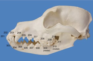
Dog skull with the left maxillary and mandibular permanent teeth numbered according to the modified Triadan system. The teeth visible in the left maxillary quadrant are numbered from 201 to 210. The teeth visible in the left mandibular quadrant are numbered from 303 to 311. Dogs have 42 permanent teeth (12 incisors, 4 canines, 16 premolars, and 10 molars).
Complete Triadan numbering of all quadrants of the dog's dentition is as follows: The incisors (101–103, 201–203, 301–303, 401–403) and canine teeth (104, 204, 304, 404) are single rooted. In the maxillary arch, the first premolars (105, 205) have 1 root, the second and third premolars (106, 107, 206, 207) have 2 roots, and the fourth premolars (108, 208) and first and second molars (109, 110, 209, 210) have 3 roots. In the mandibular arch, the first premolars (305, 405) have 1 root; the second, third, and fourth premolars (306–308, 406–408) and the first and second molars (309, 310, 409, 410) have 2 roots; and the third molars (311, 411) have 1 root.
Dog skull with the left maxillary and mandibular permanent teeth numbered according to the modified Triadan system. The
Courtesy of Dr. Maria Soltero-Rivera

Left side of an equine skull with visible dentition labeled.
Left side of an equine skull with visible dentition labeled.
Photograph and labels courtesy of Dr. Alexandra Wright. Anatomic specimen courtesy of the Cornell Anatomy Laboratory.

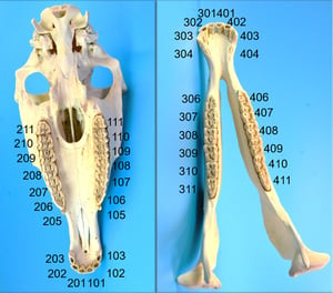
Donkey skull with the maxillary and mandibular permanent teeth numbered according to the modified Triadan system. Each dental arch (maxillary and mandibular) is divided into left and right quadrants. The hundreds-place digit denotes the specific quadrant, and the subsequent digits identify the specific tooth number.
In a clockwise direction (looking onto the animal), the right maxillary quadrant is labeled 100, the left maxillary quadrant 200, the left mandibular quadrant 300, and the right mandibular quadrant 400. Each tooth is given a 2-digit number according to its position from midline and anatomical nomenclature (ie, the central incisor being 01, the canine tooth 04, the first molar 09, etc). For example, the second premolar in the left mandibular quadrant is tooth 306, and the last molar in the right mandibular quadrant is tooth 411. Missing teeth are skipped in the numbering sequence.
This image provides an occlusal view of both dental arches in equids.
Donkey skull with the maxillary and mandibular permanent teeth numbered according to the modified Triadan system. Each
Courtesy of the Archaeozoological Reference Collection of the Institute of Topographic Anatomy, University of Veterinary Medicine, Vienna.

Skull of a small ruminant with visible dentition labeled. When canine teeth are present, they are in line with the incisor teeth in the mandibular arch and grossly appear similar to the incisors. Incisors are single rooted, and the premolars and molars are multirooted. Two additional images show occlusal views of the mandibular and maxillary arches.
Skull of a small ruminant with visible dentition labeled. When canine teeth are present, they are in line with the inci
Photograph courtesy of Dr. Alexandra Wright. Anatomic specimen courtesy of the Cornell Anatomy Laboratory.

Porcine skull with visible dentition labeled. This is a female pig, noted by the smaller canine teeth. Two additional images show occlusal views of the mandibular and maxillary arches.
Porcine skull with visible dentition labeled. This is a female pig, noted by the smaller canine teeth. Two additional i
Photograph courtesy of Dr. Alexandra Wright. Anatomic specimen courtesy of the Cornell Anatomy Laboratory.

Selected premolar and molar anatomic specimens from a horse, a sheep, and a pig. Equine maxillary premolars and molars have three roots, while the mandibular premolars and molars have two roots. Small ruminant premolars have two to four roots, and the molars have two to four roots. Pig premolars have one to two roots, and the molars have two to six roots. Root numbers can vary because of congenital or pathological factors, so advanced imaging is needed to determine root anatomy and pathology in individuals of all species.
Selected premolar and molar anatomic specimens from a horse, a sheep, and a pig. Equine maxillary premolars and molars
Photograph courtesy of Dr. Alexandra Wright. Anatomic specimen courtesy of the Cornell Anatomy Laboratory.
Feline skull with visible teeth labeled. Two additional images show occlusal views of the mandibular and maxillary arches. This specimen is missing the left maxillary canine tooth and the distal root of the right and left mandibular fourth premolars.
Cats have only one molar in each quadrant, which is the first molar (109, 209, 309, or 409). The tooth distal to the maxillary canine is the second premolar (106 or 206), whereas the first premolar has been lost during evolutionary history (ie, there is no 105 or 205).
Feline skull with visible teeth labeled. Two additional images show occlusal views of the mandibular and maxillary arch
Photograph and labels courtesy of Dr. Alexandra Wright. Anatomic specimen courtesy of the Cornell Dentistry and Oral Surgery Service.
Dog skull with the left maxillary and mandibular permanent teeth numbered according to the modified Triadan system. The teeth visible in the left maxillary quadrant are numbered from 201 to 210. The teeth visible in the left mandibular quadrant are numbered from 303 to 311. Dogs have 42 permanent teeth (12 incisors, 4 canines, 16 premolars, and 10 molars).
Complete Triadan numbering of all quadrants of the dog's dentition is as follows: The incisors (101–103, 201–203, 301–303, 401–403) and canine teeth (104, 204, 304, 404) are single rooted. In the maxillary arch, the first premolars (105, 205) have 1 root, the second and third premolars (106, 107, 206, 207) have 2 roots, and the fourth premolars (108, 208) and first and second molars (109, 110, 209, 210) have 3 roots. In the mandibular arch, the first premolars (305, 405) have 1 root; the second, third, and fourth premolars (306–308, 406–408) and the first and second molars (309, 310, 409, 410) have 2 roots; and the third molars (311, 411) have 1 root.
Dog skull with the left maxillary and mandibular permanent teeth numbered according to the modified Triadan system. The
Courtesy of Dr. Maria Soltero-Rivera
Left side of an equine skull with visible dentition labeled.
Left side of an equine skull with visible dentition labeled.
Photograph and labels courtesy of Dr. Alexandra Wright. Anatomic specimen courtesy of the Cornell Anatomy Laboratory.
Donkey skull with the maxillary and mandibular permanent teeth numbered according to the modified Triadan system. Each dental arch (maxillary and mandibular) is divided into left and right quadrants. The hundreds-place digit denotes the specific quadrant, and the subsequent digits identify the specific tooth number.
In a clockwise direction (looking onto the animal), the right maxillary quadrant is labeled 100, the left maxillary quadrant 200, the left mandibular quadrant 300, and the right mandibular quadrant 400. Each tooth is given a 2-digit number according to its position from midline and anatomical nomenclature (ie, the central incisor being 01, the canine tooth 04, the first molar 09, etc). For example, the second premolar in the left mandibular quadrant is tooth 306, and the last molar in the right mandibular quadrant is tooth 411. Missing teeth are skipped in the numbering sequence.
This image provides an occlusal view of both dental arches in equids.
Donkey skull with the maxillary and mandibular permanent teeth numbered according to the modified Triadan system. Each
Courtesy of the Archaeozoological Reference Collection of the Institute of Topographic Anatomy, University of Veterinary Medicine, Vienna.
Skull of a small ruminant with visible dentition labeled. When canine teeth are present, they are in line with the incisor teeth in the mandibular arch and grossly appear similar to the incisors. Incisors are single rooted, and the premolars and molars are multirooted. Two additional images show occlusal views of the mandibular and maxillary arches.
Skull of a small ruminant with visible dentition labeled. When canine teeth are present, they are in line with the inci
Photograph courtesy of Dr. Alexandra Wright. Anatomic specimen courtesy of the Cornell Anatomy Laboratory.
Porcine skull with visible dentition labeled. This is a female pig, noted by the smaller canine teeth. Two additional images show occlusal views of the mandibular and maxillary arches.
Porcine skull with visible dentition labeled. This is a female pig, noted by the smaller canine teeth. Two additional i
Photograph courtesy of Dr. Alexandra Wright. Anatomic specimen courtesy of the Cornell Anatomy Laboratory.
Selected premolar and molar anatomic specimens from a horse, a sheep, and a pig. Equine maxillary premolars and molars have three roots, while the mandibular premolars and molars have two roots. Small ruminant premolars have two to four roots, and the molars have two to four roots. Pig premolars have one to two roots, and the molars have two to six roots. Root numbers can vary because of congenital or pathological factors, so advanced imaging is needed to determine root anatomy and pathology in individuals of all species.
Selected premolar and molar anatomic specimens from a horse, a sheep, and a pig. Equine maxillary premolars and molars
Photograph courtesy of Dr. Alexandra Wright. Anatomic specimen courtesy of the Cornell Anatomy Laboratory.
For More Information
Verstraete FJM, Terpak CH. Anatomical variations in the dentition of the domestic cat. J Vet Dent. 1997; 14(4):137-140.
Staszyk C, Suske A, Pöschke A. Equine dental and periodontal anatomy: a tutorial review. Equine Vet Educ. 2015;27:474-481.
Also see pet owner content regarding dental development ofdogs, cats, and horses.
References
Floyd MR. The modified Triadan system: nomenclature for veterinary dentistry. J Vet Dent. 1991;8(4):18-9. doi:10.1177/089875649100800402