

Endodontic disease (pulpitis) is inflammation or infection of the tooth pulp. Clinical signs include discolored teeth, trauma or fracture of the tooth with or without pulp exposure, reluctance to chew or hold toys in the mouth, tooth sensitivity, hypersalivation, and parulis. Diagnosis is by thorough oral examination and diagnostic imaging of affected teeth. Treatment for irreversible pulpitis is aimed at removing the source of inflammation.
Endodontic disease, also known as pulpitis, occurs when the dental pulp (odontoblasts, fibroblasts, undifferentiated mesenchymal cells, blood vessels, and nerves in the center of the tooth) becomes infected or inflamed.
Etiology and Pathogenesis of Endodontic Disease in Small Animals
Endodontic disease can be categorized as either reversible or irreversible. An inflamed pulp can heal after a minor injury. However, more-severe trauma will cause irreversible pulpitis, eventually leading to pulp necrosis. In dogs and cats, most cases of pulpitis are diagnosed as irreversible.
The neurovascular supply to teeth enters through the apical delta. Because dental pulp has no collateral circulation, injuries are less likely to heal, and extravasated blood remains trapped in the dentinal tubules, where it deteriorates rather than being removed from the tooth.
The pulp is protected from bacteria by the impervious enamel covering the dentin of the crown of the tooth. Damage to the enamel surface, either through trauma or from a developmental abnormality that allows bacteria to reach the pulp, will result in pulpitis and possibly pulp necrosis.
Blunt trauma from a concussive force can also injure the pulp beyond its ability to heal.
Direct exposure of the pulp at a fracture site will lead a tooth to die and therefore necessitates endodontic treatment or surgical extraction. Teeth are fractured from external trauma (eg, catching rocks, automobile impacts, aggressive play) or from biting on inappropriate objects (eg, real bones regardless of the state of processing, hooves, antlers, hard nylon toys, rocks, fences, or cages).
Severe periodontitis may progress apically to reach the apex of the root of a tooth, producing secondary endodontic disease.
In dogs, deep caries may extend into the endodontic system of the tooth (1).

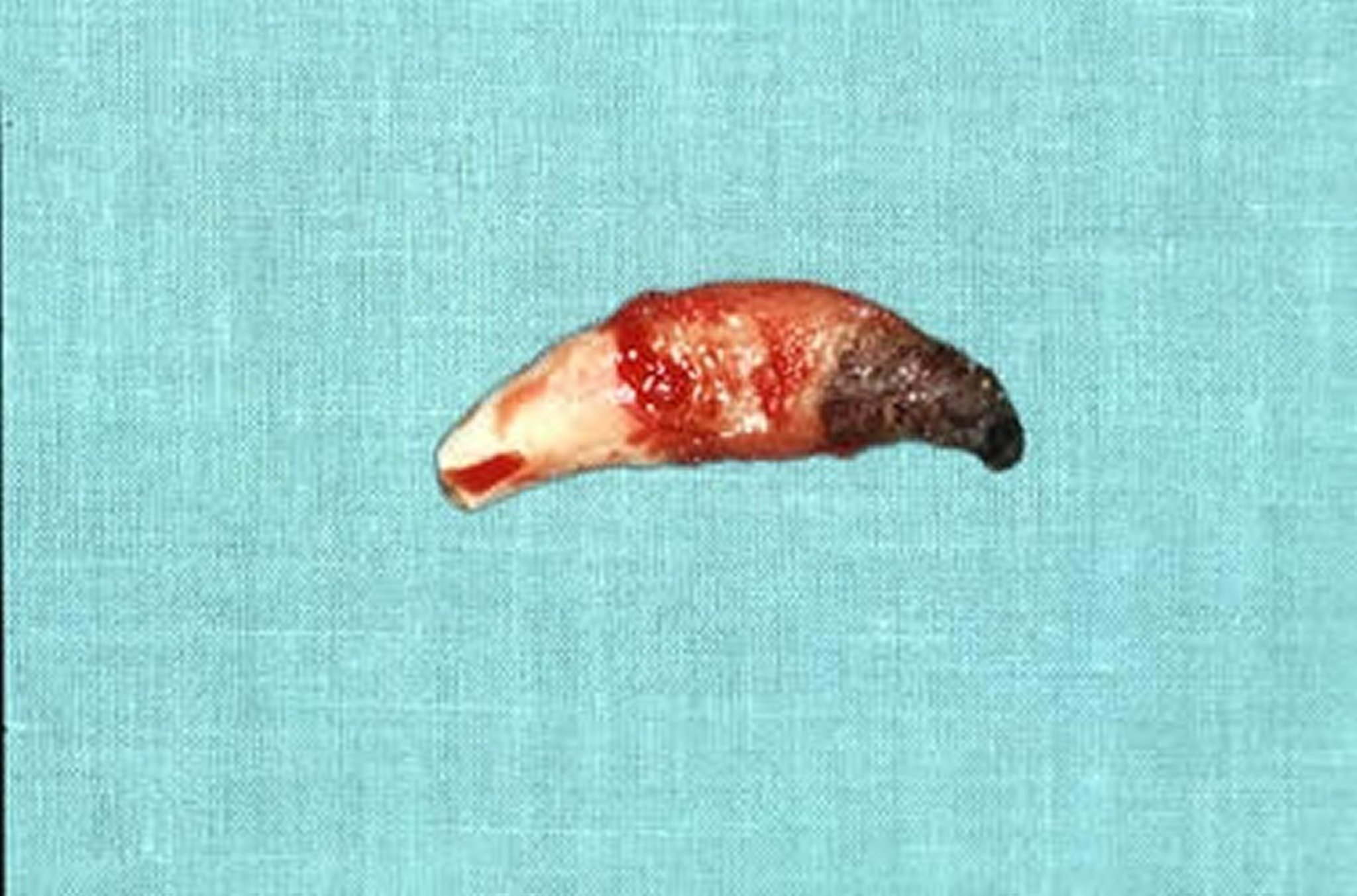
Tooth with a periapical lesion from a dog after surgical extraction. A complicated crown fracture left untreated will result in a periapical abscess. Note the necrotic apex of the tooth.
Courtesy of Dr. Ben Colmery III.
An inflamed or dead pulp releases inflammatory mediators into the periradicular tissues (through furcation canals into the periodontal ligament at the furcation of a multirooted tooth, through lateral canals into the periodontal ligament at the midroot level, and through apical foramina into the periapical tissues). The tissues surrounding the apex of a tooth develop a periapical granuloma, cyst, or abscess (see ).
Clinical Findings and Lesions of Endodontic Disease in Small Animals
Clinical signs of endodontic disease can include signs of pain and a tooth that appears intrinsically stained (discolored).
Teeth with endodontic disease are thought to be painful as long as the neurovascular supply is still alive. It can take weeks, months, or longer for the neurovascular supply to die, depending on the age of the patient and the tooth affected. Once the nerve becomes nonvital, the tooth is thought to enter a quiescent phase in which it is no longer painful. This period may last months to years. Once the tooth begins to exhibit evidence of endodontic infection, the tooth becomes painful once again.
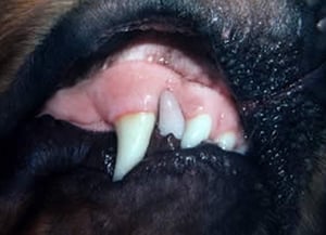
A discolored tooth signifies endodontic disease and is evidence of previous trauma and hemorrhage from the pulp into the dentinal tubules (see ). The discoloration can be any shade or color; however, most frequently pink, purple, gray, or green, or a dull appearance of the affected tooth is observed. It has been reported that 87%–92% of intrinsically stained teeth are nonvital (2, 3).
The most obvious indication of endodontic disease is a fractured tooth with exposure of the pulp chamber or canal. The exposed pulp bleeds for only a short time. After the initial injury, it may appear as a red dot at the site of the exposure if the pulp remains vital, or as a black hole if it becomes necrotic. Treatment is required for a complicated crown fracture (see , , and photographs).

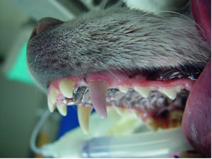
The image depicts an intrinsically stained tooth of a dog. From 87% to 92% of intrinsically stained teeth are thought to be nonvital and therefore warrant treatment with surgical extraction or root canal therapy.
The image depicts an intrinsically stained tooth of a dog. From 87% to 92% of intrinsically stained teeth are thought t
Courtesy of Dr. Brenda Mulherin.
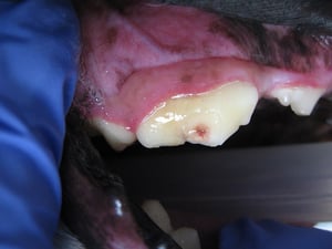
The image depicts a complicated crown fracture of the right maxillary fourth premolar tooth of a dog. This can also be referred to as a “slab” fracture. The red area within the center of the tooth depicts pulp exposure. While the tooth is not currently bleeding, the pulp is exposed and will become nonvital without immediate treatment.
The image depicts a complicated crown fracture of the right maxillary fourth premolar tooth of a dog. This can also be
Courtesy of Dr. Brenda Mulherin.

Pulpitis in a dog. A complicated crown fracture exposing the pulp of a tooth will lead to pulpitis. Teeth with pulp exposure should be treated with vital pulp therapy, root canal, or extraction, depending on the timing of the injury and the expectations of the client.
Pulpitis in a dog. A complicated crown fracture exposing the pulp of a tooth will lead to pulpitis. Teeth with pulp exp
Courtesy of Dr. Ben Colmery III.

Pulpal hemorrhage in a dog. Concussive trauma to a tooth without loss of crown structure can result in tooth nonvitality. The tooth may become discolored in various shades of pink, purple, or gray, or have a dull appearance. If this is observed, the main differential diagnosis would be a nonvital tooth.
Pulpal hemorrhage in a dog. Concussive trauma to a tooth without loss of crown structure can result in tooth nonvitalit
Courtesy of Dr. Ben Colmery III.

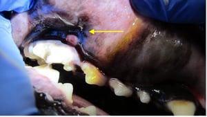
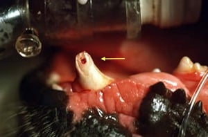
The image depicts a parulis (yellow arrow), or draining tract, associated with an endodontically compromised right maxillary fourth premolar tooth. Parulides frequently occurs at the mucogingival line.
The image depicts a parulis (yellow arrow), or draining tract, associated with an endodontically compromised right maxi
Courtesy of Dr. Brenda Mulherin.

A swelling or draining tract that is observed ventral to the medial canthus of the eye may be an indication of an endodontically compromised tooth. This is most commonly associated with the maxillary fourth premolar or first or second molar teeth. A thorough oral examination and radiographic imaging of the oral cavity should be considered.
A swelling or draining tract that is observed ventral to the medial canthus of the eye may be an indication of an endod
Courtesy of Dr. Brenda Mulherin.
The image depicts an intrinsically stained tooth of a dog. From 87% to 92% of intrinsically stained teeth are thought to be nonvital and therefore warrant treatment with surgical extraction or root canal therapy.
The image depicts an intrinsically stained tooth of a dog. From 87% to 92% of intrinsically stained teeth are thought t
Courtesy of Dr. Brenda Mulherin.
The image depicts a complicated crown fracture of the right maxillary fourth premolar tooth of a dog. This can also be referred to as a “slab” fracture. The red area within the center of the tooth depicts pulp exposure. While the tooth is not currently bleeding, the pulp is exposed and will become nonvital without immediate treatment.
The image depicts a complicated crown fracture of the right maxillary fourth premolar tooth of a dog. This can also be
Courtesy of Dr. Brenda Mulherin.
Pulpitis in a dog. A complicated crown fracture exposing the pulp of a tooth will lead to pulpitis. Teeth with pulp exposure should be treated with vital pulp therapy, root canal, or extraction, depending on the timing of the injury and the expectations of the client.
Pulpitis in a dog. A complicated crown fracture exposing the pulp of a tooth will lead to pulpitis. Teeth with pulp exp
Courtesy of Dr. Ben Colmery III.
Pulpal hemorrhage in a dog. Concussive trauma to a tooth without loss of crown structure can result in tooth nonvitality. The tooth may become discolored in various shades of pink, purple, or gray, or have a dull appearance. If this is observed, the main differential diagnosis would be a nonvital tooth.
Pulpal hemorrhage in a dog. Concussive trauma to a tooth without loss of crown structure can result in tooth nonvitalit
Courtesy of Dr. Ben Colmery III.
The image depicts a parulis (yellow arrow), or draining tract, associated with an endodontically compromised right maxillary fourth premolar tooth. Parulides frequently occurs at the mucogingival line.
The image depicts a parulis (yellow arrow), or draining tract, associated with an endodontically compromised right maxi
Courtesy of Dr. Brenda Mulherin.
A swelling or draining tract that is observed ventral to the medial canthus of the eye may be an indication of an endodontically compromised tooth. This is most commonly associated with the maxillary fourth premolar or first or second molar teeth. A thorough oral examination and radiographic imaging of the oral cavity should be considered.
A swelling or draining tract that is observed ventral to the medial canthus of the eye may be an indication of an endod
Courtesy of Dr. Brenda Mulherin.
Drainage of infected pulp material most commonly occurs through the fracture site. However, a periapical abscess can occur if the site becomes occluded. The skin ventral to the medial canthus of the eye is a common site for swelling and purulent drainage from a fistula due to an endodontically diseased maxillary fourth premolar. This can also cause a parulis, an intraoral red draining fistula near the mucogingival junction adjacent to the tooth (see ).
An abscessed maxillary canine tooth in dogs can cause swelling along the side of the nose; in cats, the swelling is often immediately rostral to the eye (see ). Veterinary patients often do not give an indication of discomfort, even for conditions that cause severe orofacial pain in humans.
Diagnosis of Endodontic Disease in Small Animals
Diagnosis of endodontic disease is by thorough oral examination and diagnostic imaging of affected teeth. A tooth with a periapical granuloma or cyst will typically demonstrate radiographic evidence of a periapical lucency, ie, an irregular circular lesion with decreased radiopacity around a root tip (see ).

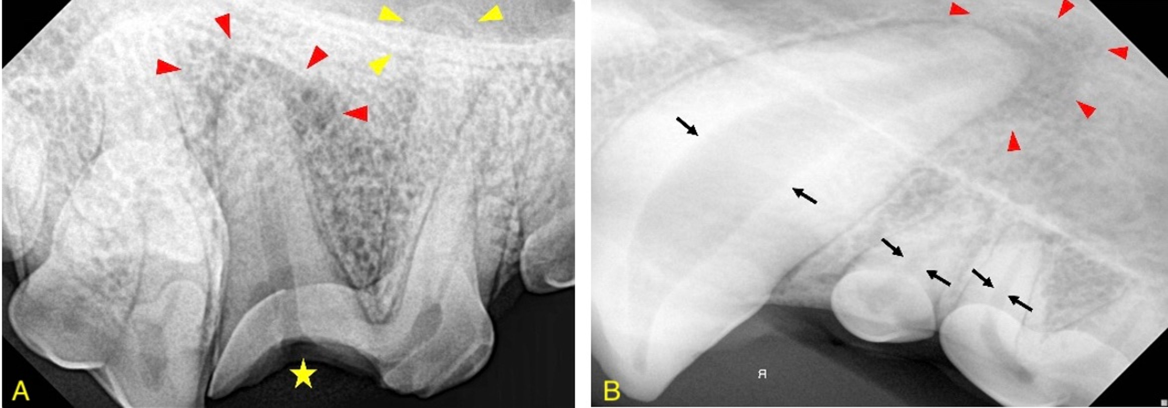
The image depicts radiographic evidence of a periapical lucency associated with a right maxillary fourth premolar tooth (A) and left maxillary canine tooth (B). (A) Note the radiolucent halo around the distal root identified by the red arrowheads. In addition, note the radiolucency associated with one of the mesial roots (yellow arrowheads). The visible loss of crown structure, identified by the yellow star, is likely contributing to the endodontic compromise of this tooth. Extraction or root canal therapy may be considered treatment options for this tooth. (B) The periapical lucency is identified by the red arrowheads. Note the wide pulp chamber of the left maxillary canine tooth compared to the maxillary first and second premolar teeth.
Courtesy of Dr. Brenda Mulherin.
A tooth with an acute periapical abscess (painful accumulation of pus around the apex of a nonvital tooth) may not show distinct radiographic signs.
Throughout life, the pulp produces dentin on the inside surface of the pulp cavity, resulting in a constantly decreasing cross-sectional width of the pulp chamber in the crown and root canal in the root of the tooth. A necrotic pulp discontinues its normal dentin production, and thus it falls behind that of a normally maturing tooth adjacent to it or on the contralateral side. Conversely, an inflamed pulp may produce dentin at an accelerated rate.
If there is generalized pulpitis, the effect can be an apparent accelerated aging of the entire tooth with an abnormally narrow root canal space and pulp chamber. Generally, when evaluating a tooth with endodontic or periapical disease, the focus should be on the gross structural defects at the crown of the tooth, the radiographic appearance of the root apex, the width of its pulp cavity, and the appearance of the periapical tissues.
Treatment of Endodontic Disease in Small Animals
Root canal therapy
Vital pulp therapy
Dental extraction
In teeth with a mature apex (closed apex), extraction or root canal therapy is indicated for every tooth in which a fracture has exposed the pulp chamber. Treatment for a tooth that is suspected to be nonvital usually involves either endodontic therapy or extraction of the affected tooth. Depending on how long the pulp has been compromised and the stage of pulpitis present, vital pulp therapy, root canal therapy, and extraction of the affected tooth are the most common recommended treatment options.
Canine teeth in dogs and cats and carnassial teeth (maxillary fourth premolars and mandibular first molars) in dogs are considered strategic teeth. Root canal therapy of these teeth is more frequently recommended over extraction when these teeth are in need of treatment (see ).

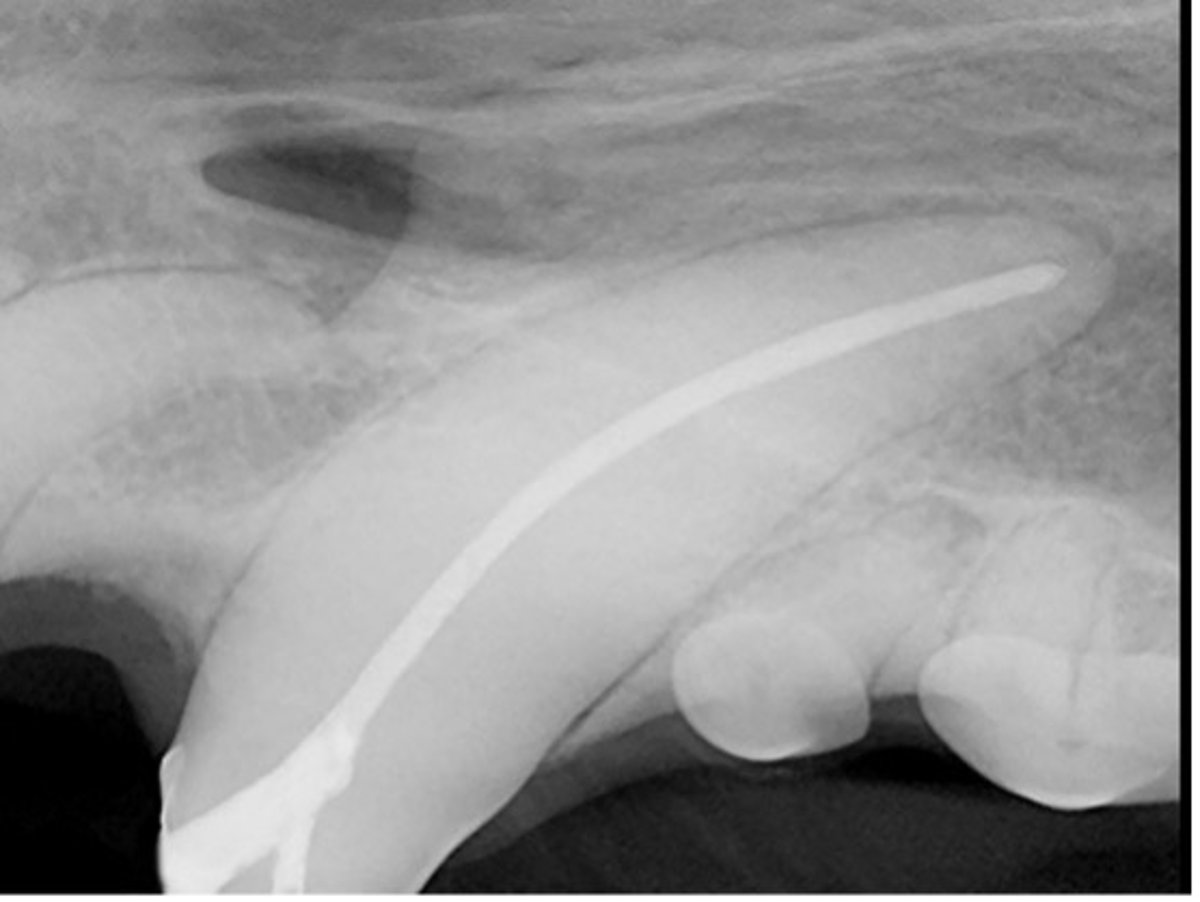
Root canal therapy of the left maxillary canine tooth of a dog. This image depicts the radiographic appearance of root canal therapy. During root canal therapy, the pulp is removed, and the canal is antiseptically treated, dried, and obturated. A radiopaque restoration is then placed to seal the canal from the external environment.
Courtesy of Dr. Brenda Mulherin.
Root canal therapy involves removing the entire pulp contents, antiseptically preparing the inside of the tooth, and filling it with a radiographically detectable cement to seal the canal. Finally, a restoration is placed to seal the inside of the tooth from the external environment.
All openings of the tooth need to be sealed, including the instrumentation site and the fracture site. Furthermore, all root canals need to be cleaned and sealed. If they are unable to be sealed, they should be extracted, and the communication of the extracted root with other roots should be restored to limit exposure to the external environment.
Surgical extraction of a tooth with pulp exposure frequently involves making a surgical flap, removing alveolar bone, and elevating the tooth out of the socket. After the removal of the tooth, bone and soft tissue management to create an environment for healing is performed by curettage of the alveolus, smoothing the bone, and freshening tissue edges. The extraction site is then closed with monofilament absorbable suture material.
The benefit of a root canal over dental extraction includes the ability to keep the form and function of the tooth, improved cosmetic appearance of the tooth, and the less invasive nature of the treatment. Disadvantages of root canal therapy include increased costs, expertise required, possibility of failure, potential loss of restorations, and increased follow-up.
Military, police, assistance, and working dogs frequently have root canal therapy performed to assist them in their duties. These animals may also require fabrication and placement of a full or partial prosthodontic crown to help maintain the remaining tooth structure and prevent restorations from falling out.
Young animals (< 11 months old) with trauma to the permanent dentition that has led to exposure of the pulp are not candidates for root canal therapy. These teeth are candidates for extraction. Alternatively, depending on when the fracture occurred and how rapidly the patients are presented for assessment and treatment, vital pulp therapy, regenerative, or revascularization treatments can be performed in efforts to save the tooth.
Vital pulp therapy involves removing a portion of the diseased pulp and placing a restoration to seal the pulp from the external environment. This procedure removes the diseased portion of the pulp, but not the entire pulp, in efforts to keep the tooth alive. This treatment is most successful when it is performed as soon as possible after a trauma, ideally within 24–48 hours following the initial injury (4).
Key Points
Trauma to a tooth frequently ends in endodontic compromise of the pulp, leading to irreversible pulpitis. Clinical signs may include discoloration of the tooth or visible evidence of loss of the crown structure, frequently exposing the pulp of the tooth.
Intrinsic staining of a tooth is an indication that the tooth is no longer vital.
Treatment for endodontically compromised mature permanent teeth includes extraction or root canal therapy. Root canal therapy is frequently considered for strategic teeth, including the maxillary and mandibular canine teeth and the carnassial teeth (maxillary fourth premolar tooth and mandibular first molar tooth).
Treatment for endodontically compromised immature permanent teeth may be extraction or vital pulp therapy, depending on how soon the patient presents after the injury.
References
Hale FA. Dental caries in the dog. Can Vet J. 2009;50(12):1301-4. doi:10.1177/089875649801500203
Feigin K, Bell C, Shope B, Henzel S, Snyder C. Analysis and assessment of pulp vitality of 102 intrinsically stained teeth in dogs. J Vet Dent. 2022;39(1):21-33. doi:10.1177/08987564211060387.
Hale FA. Localized intrinsic staining of teeth due to pulpitis and pulp necrosis in dogs. J Vet Dent. 2001;18(1):14-20. doi:10.1177/089875640101800102
Clarke DE. Vital pulp therapy for complicated crown fracture of permanent canine teeth in dogs: a three-year retrospective study. J Vet Dent. 2001;18(3):117-21. doi:10.1177/089875640101800301
For More Information
Lee DB, Arzi B, Kass PH, Verstraete FJM. Radiographic outcome of root canal treatment in dogs: 281 teeth in 204 dogs (2001-2018). J Am Vet Med Assoc. 2022;260(5):535-542. doi:10.2460/javma.21.03.0127
Girard N, Southerden P, Hennet P. Root canal treatment in dogs and cats. J Vet Dent. 2006;23(3):148-60. doi:10.1177/089875640602300304
Also see pet health content regarding dental disorders of dogs and dental disorders of cats.


