

Benign Fibroblastic Tumors
Collagenous nevi are benign, focal, developmental defects associated with increased deposition of dermal collagen. They are common in dogs, uncommon in cats, and rare in large animals. They generally are found in middle-aged or older animals, most frequently on the proximal and distal extremities, head, neck, and areas prone to trauma. They are sessile to raised, dermal nodules, often with a papillated surface. Two forms are seen: one develops in the interfollicular dermis or subcutaneous fat that is not accompanied by adnexal involvement, and one incorporates adnexa and induces enlarged, often malformed follicles, sebaceous glands, and apocrine glands. This latter form has been called focal adnexal dysplasia. Excision of both forms is generally curative, although, infrequently, expansive forms have been identified that may grow too large to be surgically removed.
Generalized nodular dermatofibrosis (dermatofibromas), recognized rarely in German Shepherd Dogs (believed to be an inherited, autosomal dominant trait) and even less commonly in other canine breeds, is a syndrome in which multiple collagenous nevi are associated with renal cystadenocarcinomas and, in females, multiple uterine leiomyomas. Skin lesions, first recognized when animals are 3–5 years old, are characterized by the development of multiple collagenous nevi varying from barely palpable to large and nodular, generally on the limbs, feet, head, and trunk. They may be symmetrically distributed. Renal disease develops ~3–5 years after the skin lesions are recognized. No known therapy can prevent development of the renal and uterine neoplasms.
Acrochordons (cutaneous tags, soft fibromas, fibrovascular papillomas) are distinctive, benign, cutaneous lesions of older dogs. These lesions are common, may be single or multiple, and can develop in any breed, although large breeds may be at increased risk. Most commonly, they appear as pedunculated exophytic growths, often covered by a verrucous epidermal surface. Treatment is optional, but a biopsy is recommended to confirm the diagnosis. Acrochordons are amenable to excision, electrosurgery, and cryosurgery, but dogs that develop one are prone to develop others over time.
Fibromas are discrete, generally cellular proliferations of dermal fibroblasts. Histologically, they resemble collagenous nevi or cutaneous tags. Fibromas occur in all domestic species but are primarily a tumor of senior dogs. Doberman Pinschers, Boxers (predisposed to developing multiple tumors), and Golden Retrievers are most at risk. The head and extremities are the most likely sites. Clinically, the lesions appear as discrete, generally raised, often hairless nodules originating in the dermis or subcutaneous fat. They palpate as either firm and rubbery (fibroma durum) or soft and fluctuant (fibroma molle). These lesions are benign, but complete excision is recommended if they change appearance or grow large. These lesions may be confused with low-grade fibrosarcoma on the muzzle of dogs, which have a malignant, infiltrative biological behavior.
Soft tissue Sarcomas
This group of malignancies includes equine sarcoids, fibromatoses, fibrosarcomas, malignant fibrous histiocytomas, neurofibrosarcomas, leiomyosarcomas, rhabdomyosarcomas, and variants of liposarcomas, angiosarcomas, synovial cell sarcomas, mesotheliomas, and meningiomas. As a group, sarcomas are widely recognized, yet poorly characterized neoplasms with up to 78% diagnostic disagreement. High-grade soft tissue sarcomas have a 30–40% metastatic rate, which suggests that most are incorrectly classified.
Spindle-cell sarcomas demonstrate much greater morphologic heterogeneity than carcinomas; often, features of one sarcoma are intermixed with features of another. Consequently, it is widely accepted that the cell of origin of all soft tissue sarcomas is a primitive mesenchymal cell that can differentiate in many different directions. This makes it difficult to define histopathologic criteria necessary to make an unequivocal diagnosis of specific spindle-cell sarcomas. In addition, comparing neoplastic mesenchymal cells with the normal cell they most closely resemble does not imply origin from those cells. Immunohistochemistry and molecular diagnostics can identify biomarkers to differentiate cell of origin and help guide treatment.

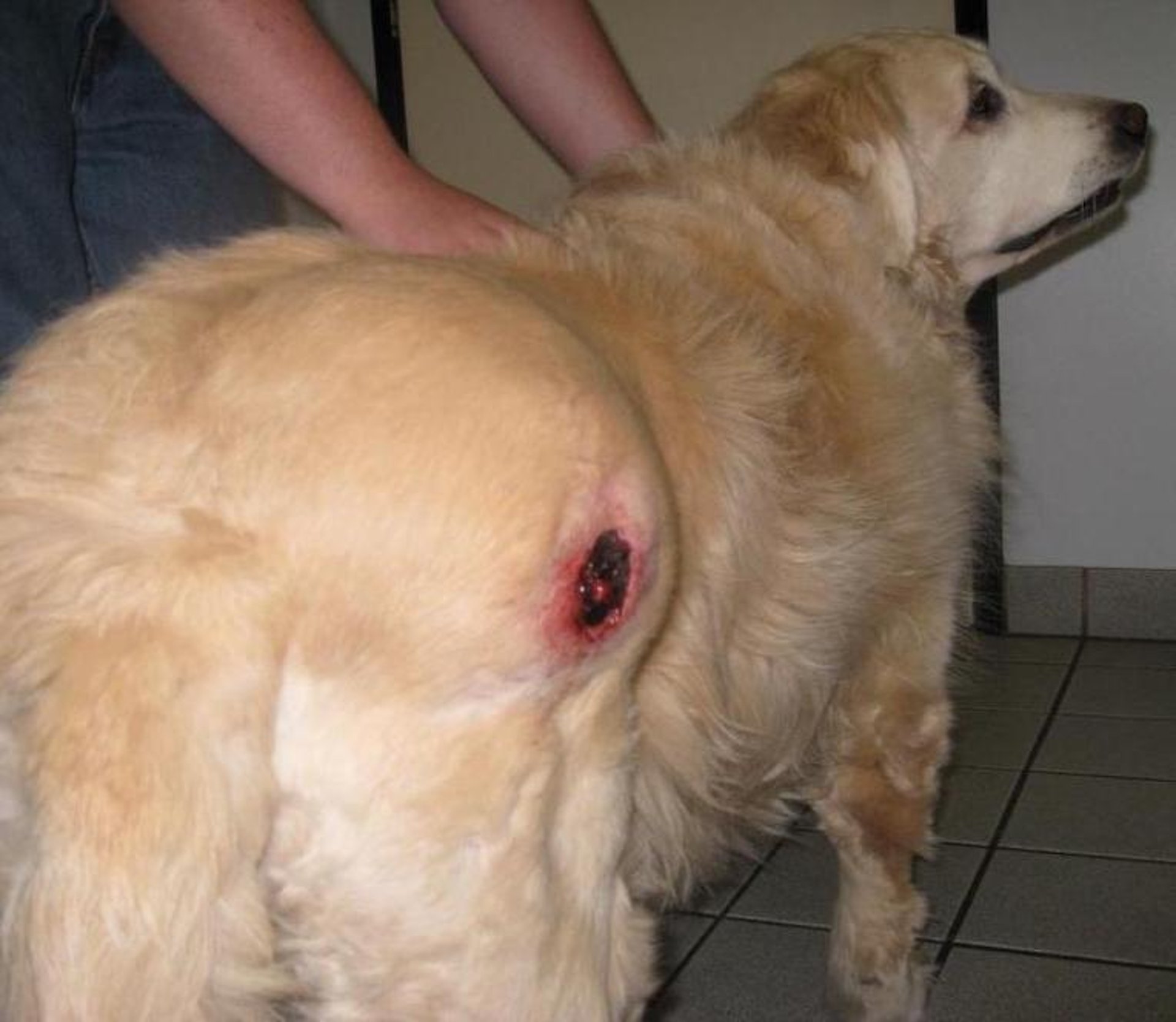
Clinical photograph of an ulcerated mass causing signs of pain and lameness in a 15-year-old Golden Retriever mix.
Courtesy of Dr. Alice Villalobos.

Post-treatment photograph of a soft tissue sarcoma in a 15-year-old Golden Retriever mix after palliative intralesional chemotherapy. The tumor reduced to 15% of its original size, and the lameness resolved.
Courtesy of Dr. Alice Villalobos.
Further confusion stems from the difficulty in determining whether these tumors are benign or malignant or what their biological behavior will be in certain locations or breeds. Most spindle-cell sarcomas of domestic animals are locally infiltrative, difficult to excise, and yet seldom metastasize. Because, by definition, only malignant tumors have metastatic potential, these tumors should be considered benign; however, again by definition, benign neoplasms are not infiltrative, and those tumors should be considered malignant and treated aggressively from the start. In human pathology, infiltrative but nonmetastasizing mesenchymal spindle-cell tumors have been defined as sarcomas of intermediate malignancy, a concept used below.
Clinically, four general principles relate to spindle-cell sarcomas and soft tissue sarcomas: The more superficial the location, the more likely the tumor is to be benign (deep tumors tend to be malignant). The larger the tumor, the more likely it is to be malignant. A rapidly growing tumor is more likely to be malignant than one that develops slowly. Benign tumors are relatively avascular, whereas most malignancies are hypervascular. Knowing the type of sarcoma, its size, location, stage, and histologic grade (low, intermediate, high [or I, II, III]), which depends on the degree of differentiation, the number of mitotic figures per 10 high-power fields, percent necrosis, and Ki-67 (a proliferation marker) level, will help guide treatment planning.
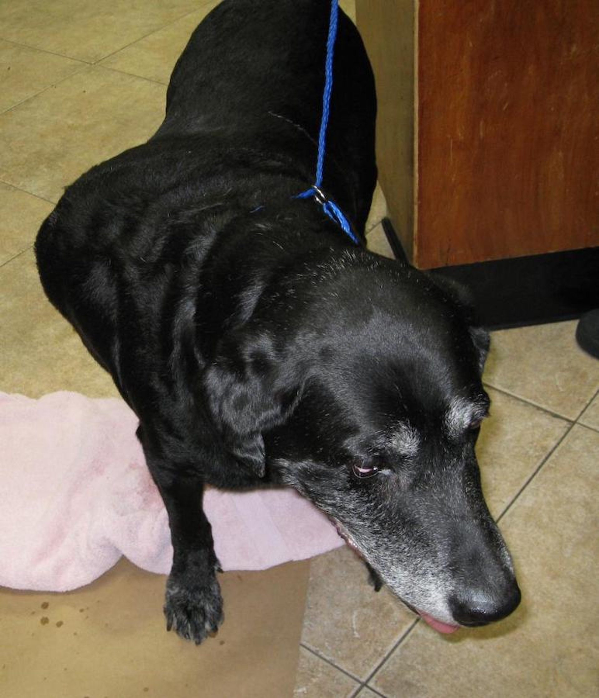
Clinical photograph of a fibrosarcoma of the rostral maxilla in an senior shepherd mix causing dysphagia, displacement of teeth, and foul breath.
Courtesy of Dr. Alice Villalobos.
Surgical excision is the treatment of choice. Wide excision or amputation should be performed when anatomically feasible because spindle-cell sarcomas often infiltrate along fascial planes, making it difficult to determine from gross examination the peripheral margins of the tumor. The best, if not only, opportunity to completely remove a spindle-cell sarcoma is during the first surgical attempt. A presurgical biopsy should be performed along with, if possible, tumor imaging with CT or MRI or ultrasound to provide a clear surgical plan that includes the intention of complete removal, with biopsy samples submitted for margin determinations.
Those sarcomas that recur have a greater potential for metastasis, and the time to recurrence often shortens with each subsequent attempt at excision. In addition, many soft tissue tumors have a pseudocapsule, which on gross examination gives the impression of complete encapsulation; these tumors should not be shelled out, because neoplastic cells are usually present in the pericapsular connective tissues.
Many sarcomas are shaped like an octopus, with tentacles that extend deeply into the tumor bed. Except for equine sarcoids, cryosurgery is usually not used for these tumors because some types, most notably fibrosarcomas, are resistant to freezing.
Spindle-cell sarcomas generally do not respond well to conventional doses of radiation; however, higher doses and stereotactic radiation have reportedly controlled ~50% of them for 1 year. Surgical debulking followed by options to enhance local control such as electroporation, intraoperative placement of carboplatin-containing biodegradable beads, intracavitary chemotherapy with follow-up intratumor bed chemotherapy, localized hyperthermia therapy, or follow-up radiation therapy may be helpful.
Chemotherapeutic protocols for sarcomas that include targeted therapy agents such as tyrosine kinase (T-K) inhibitors may improve treatment efficacy. Most protocols involve the use of doxorubicin often in combination with other agents, including carboplatin, cyclophosphamide, vincristine, dacarbazine, and methotrexate. Some clinicians prefer to use a combination of carboplatin, T-K inhibitors, and metronomic chemotherapy, whereas others rotate carboplatin with doxorubicin. Although chemotherapy may cause temporary adverse events, it can improve the length and overall quality of life; it is seldom curative.
Fibromatosis (aggressive fibromatosis, extra-abdominal desmoids, desmoid tumors, low-grade fibrosarcomas, nodular fasciitis) is a sclerosing and infiltrative proliferation of well-differentiated fibroblasts derived from aponeuroses and tendon sheaths. They are generally seen on the heads of dogs, especially Doberman Pinschers and Golden Retrievers, where they are commonly diagnosed as nodular fasciitis or fibroma. In veterinary medicine, the term nodular fasciitis is applied to two different diseases: one that behaves as a fibromatosis and one that commonly affects the periocular tissues (known as canine fibrous histiocytoma.
Fibromatoses are infrequently diagnosed in cats and horses. Grossly, fibromatoses are generally indistinguishable from infiltrative fibrosarcomas; however, they can be differentiated on histologic examination. Focal lymphoid nodules are scattered throughout the tissues. The fibromatoses are locally infiltrative, with essentially no metastatic potential. If feasible, wide, complete excision with local control techniques as described above is the treatment of choice at diagnosis. Recurrence is common, and radiation therapy may be of value for local control.
Clinical photograph of a low-grade "fibroma" fibrosarcoma in the muzzle of a Golden Retriever. These classic tumors on the muzzle are confusing in that in the biopsy report "nodular fasciitis," or fibroma, seems benign and does not indicate its slowly expanding, infiltrative biological behavior. This tumor's locally infiltrative nature caused severe disfigurement and discomfort for this Golden Retriever as it continued to enlarge at the end of the patient's life. These confusing tumors should be treated aggressively from the start.
Courtesy of Dr. Alice Villalobos.

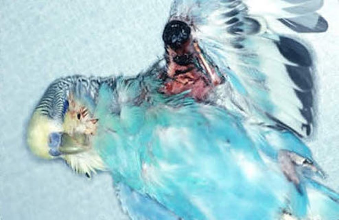
Photograph of a fibrosarcoma in a parakeet showing the typical ulcerated surface.
Courtesy of Dr. Louise Bauck.
Fibrosarcomas are aggressive mesenchymal tumors in which fibroblasts are the predominant cell type. They are the most common soft tissue tumors in cats and are also common in dogs but rare in other domestic animals. In dogs, these tumors are most common on the trunk and extremities. Gordon Setters, Irish Wolfhounds, Brittany Spaniels, Golden Retrievers, and Doberman Pinschers may be predisposed.
Fibrosarcomas vary markedly in their appearance and size. Neoplasms arising in the dermis may appear nodular. Those arising in the subcutaneous fat or subjacent soft tissues may require palpation to identify. They appear as firm, fleshy lesions involving the dermis and subcutaneous fat and often invade musculature along fascial planes. When tumors are multiple, they are usually found within the same anatomic region. Fibrosarcomas with abundant interstitial proteoglycans (connective tissue mucins) are called myxosarcomas or myxofibrosarcomas. Myxosarcomas remain poorly defined in veterinary medicine, and many of them could be characterized as variants of liposarcomas or malignant fibrous histiocytomas. Fibrosarcomas in dogs are invasive tumors; ~10% metastasize. Factors that affect whether a fibrosarcoma can be completely excised include the skill of the surgeon; rate of growth (defined by mitotic index, quantity of necrosis, and Ki-67 level); degree of cellular atypia; and the tumor’s infiltrative nature, size, and location (which may require imaging to define properly).

Clinical photograph of a cat with an injection site sarcoma in a typical intrascapular location. These tumors develop in some cats (1–16 cases/10,000 vaccinations) due to chronic inflammation following administration of adjuvanted vaccines, inactivated rabies vaccines, long-acting corticosteroid injections, or other injections. The chronic inflammation causes neoplastic transformation of fibroblasts.
Courtesy of Dr. Alice Villalobos.

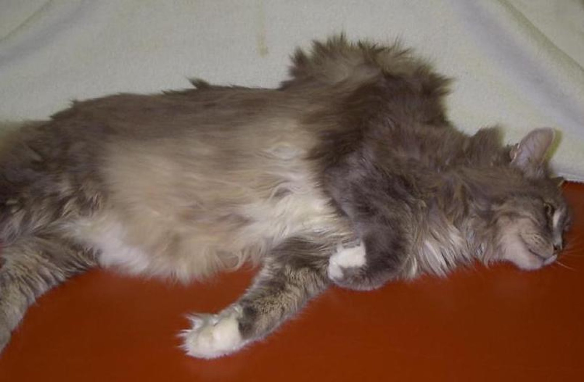
Clinical photograph of a fibrosarcoma on the right metatarsal area of a geriatric Maine Coon cat.
Courtesy of Dr. Alice Villalobos.
Three forms of fibrosarcoma are recognized in cats: a multicentric form in the young (generally < 4 years old) caused by the feline sarcoma virus (FSV); a solitary form in the young or old, in which FSV has not been implicated; and a fibrosarcoma that develops in the soft tissues where cats are commonly vaccinated ( see Preventative Health Care for Small Animals).
An association with adjuvanted, inactivated rabies and feline leukemia virus vaccinations and long-acting corticosteroid injections is better defined than with vaccinations for other viral or bacterial diseases. Aluminum hydroxide (commonly used in adjuvants) was suspected as the main causal agent; however, three large epidemiologic studies did not provide evidence that it posed a greater risk. Prolonged inflammation of fibroblasts, induced by adjuvants and injectable medications, may predispose these cells to undergo neoplastic transformation resulting in uncontrolled proliferation of fibroblasts and myofibroblasts. These tumors appear as nodules or plaques between the shoulder blades, in the soft tissues of the proximal hind limbs, or less commonly, over the lumbar areas. Although commonly classified as fibrosarcomas, injection-site sarcomas (ISSs) are extremely heterogeneous and may be appropriately called malignant fibrous histiocytomas (giant cell tumors), liposarcomas, osteosarcomas, or chondrosarcomas. Immunohistochemical staining has identified growth factors and proto-oncogenes that may be therapeutic targets. Feline leukemia virus and FSV are not involved in the pathogenesis of ISS.
Wide and deep surgical excision, with 5-cm lateral margins and two facial layers, is the treatment of choice for ISSs. Because the necessary margin extent is commonly underestimated and not achieved, recurrence is common (>70% within 1 year following the initial surgery). The rate of recurrence is >90% for ISSs. Even when surgical excision is clinically and histologically complete, recurrence is still the rule when margins are < 5 cm or two facial layers are not removed. Therefore, multimodal therapy combining presurgical 3D imaging, aggressive surgery, electroporation, intracavitary chemotherapy with carboplatin, followed by radiation therapy and IV carboplatin every 21 days for 4–6 treatments, along with tyrosine kinase (T-K) inhibitors as adjunctive therapy, may yield improved results.
Chemotherapy with carboplatin, doxorubicin and cyclophosphamide, or dacarbazine, along with T-K inhibitors and targeted therapies, are recommended for nonresectable tumors. Immunotherapy using biologic response modifiers from vaccinia used after surgery or canarypox virus vector (used intratumorally before excision and followed by radiation therapy) reduced relapse. Immunotherapy, as an adjunct to surgery and radiation, may increase tumor-free intervals up to 20% compared with historical controls.
Preventive measures include use of nonadjuvanted or intranasal vaccines and avoiding polyvalent vaccines. Vaccine schedules should be customized for individual cats based on risk assessment depending on the patient’s age, environment, and lifestyle so that cats are vaccinated only as frequently as necessary according to the principles of current vaccination guidelines in combination with local laws. To facilitate amputation with clean surgical margins in the event of an ISS, published guidelines recommend injection of vaccines subcutaneously in the most distal aspects of the limbs or tail when possible.
Fibrohistiocytic Tumors
Fibrohistiocytic tumors are pleomorphic, mesenchymal tumors composed of fibroblasts and histiocytic cells (often present as multinucleated giant cells) and remain poorly defined in veterinary medicine. A lesion called canine fibrous histiocytoma (nodular granulomatous episclerokeratitis, nodular fasciitis, proliferative keratoconjunctivitis, conjunctival granuloma, Collie granuloma) is recognized at the episcleral junction and cornea primarily in young to middle-aged (2–4 years old) Collies; however, the histologic features are more suggestive of a granulomatous inflammatory response than a neoplasm. As might be expected for a noninfectious inflammatory process, these are generally responsive to sublesional injections of 10–40 mg of methylprednisolone.
Malignant fibrous histiocytomas (extraskeletal giant cell tumors, giant cell tumors of soft parts, dermatofibrosarcomas) are most frequently found in the skin and soft tissues of cats, occasionally found in horses and mules, and rarely in the skin of other domestic species, including dogs. In cats, malignant fibrous histiocytomas are most common on the distal aspects of extremities or ventral cervical regions of senior cats; however, they may also be diagnosed at vaccination sites. In horses and mules, these have been described as giant cell tumors of soft parts. Seen in young adult to middle-aged equids, they are firm, nodular to diffuse swellings that are white on cut surface, with variable hemorrhage. Malignant fibrous histiocytomas are sarcomas of intermediate malignancy. They are locally invasive and tend to recur after attempts at complete excision but seldom metastasize. Radical excision is recommended. Local control may improve with adjuvant electroporation or radiation therapy.
Peripheral Nerve Sheath Tumors
Amputation neuromas (traumatic neuromas) are non-neoplastic, disorganized proliferations of peripheral nerve parenchyma and stroma that form in response to amputation or traumatic injury. They are most commonly identified after tail docking in dogs or neurectomy in the distal extremities of horses. The most common clinical presentation is a young dog that continuously traumatizes its docked tail. In horses, such a lesion appears as a firm, often painful swelling at a neurectomy surgery site. Excision is curative.
Neurofibromas and neurofibrosarcomas (perineuromas, neurilemmomas, nerve sheath tumors, hemangiopericytomas, neurothekeomas, schwannomas) are spindle-cell tumors that arise from the connective tissue components of the peripheral nerve. They are believed to arise from Schwann cells, but they could also arise from mesenchymal cells, which produce the nonmyelinated connective tissues that surround the myelinated nerve fiber. In dogs, forms of this tumor can be virtually indistinguishable from hemangiopericytomas and may be the same tumor.

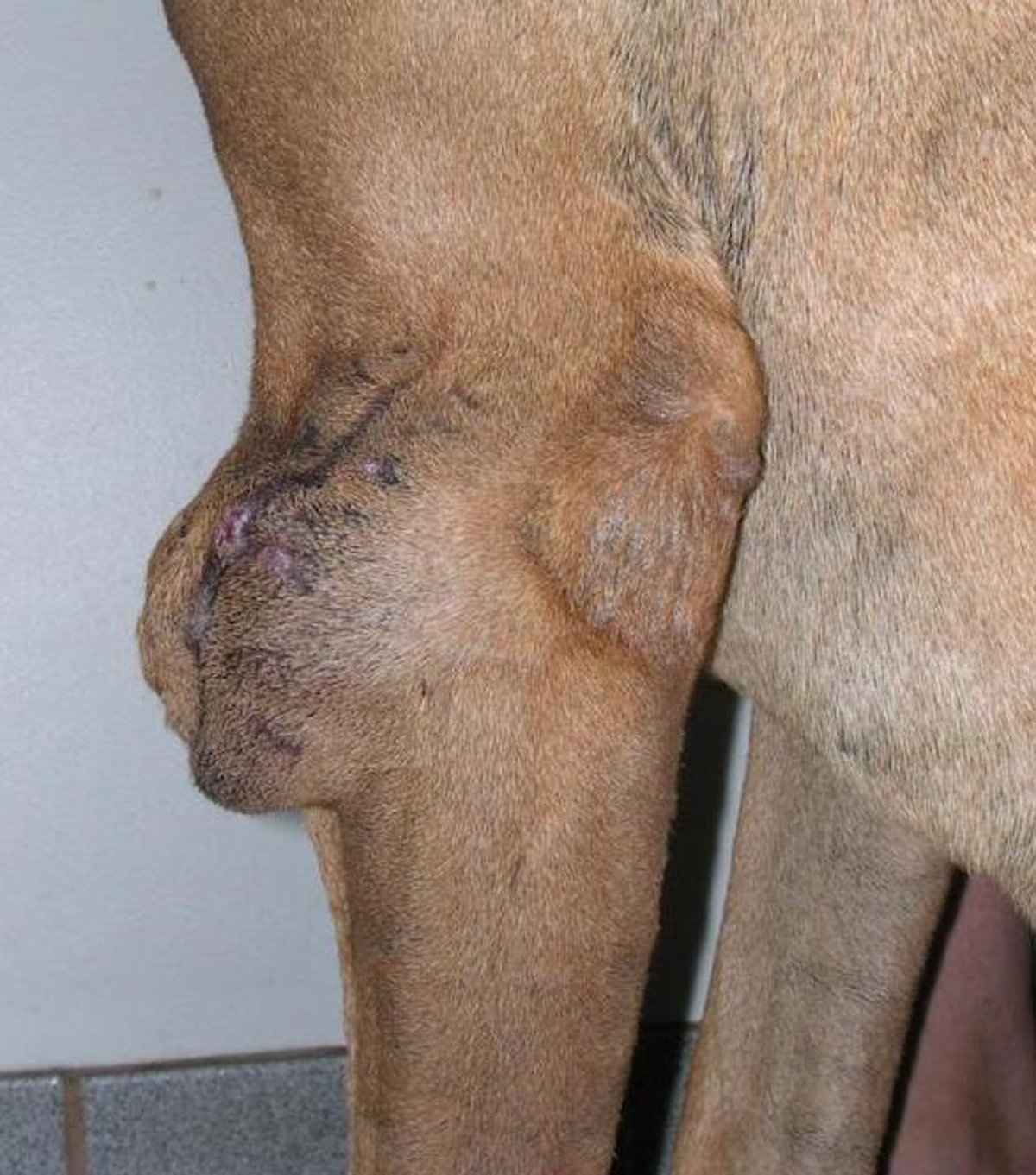
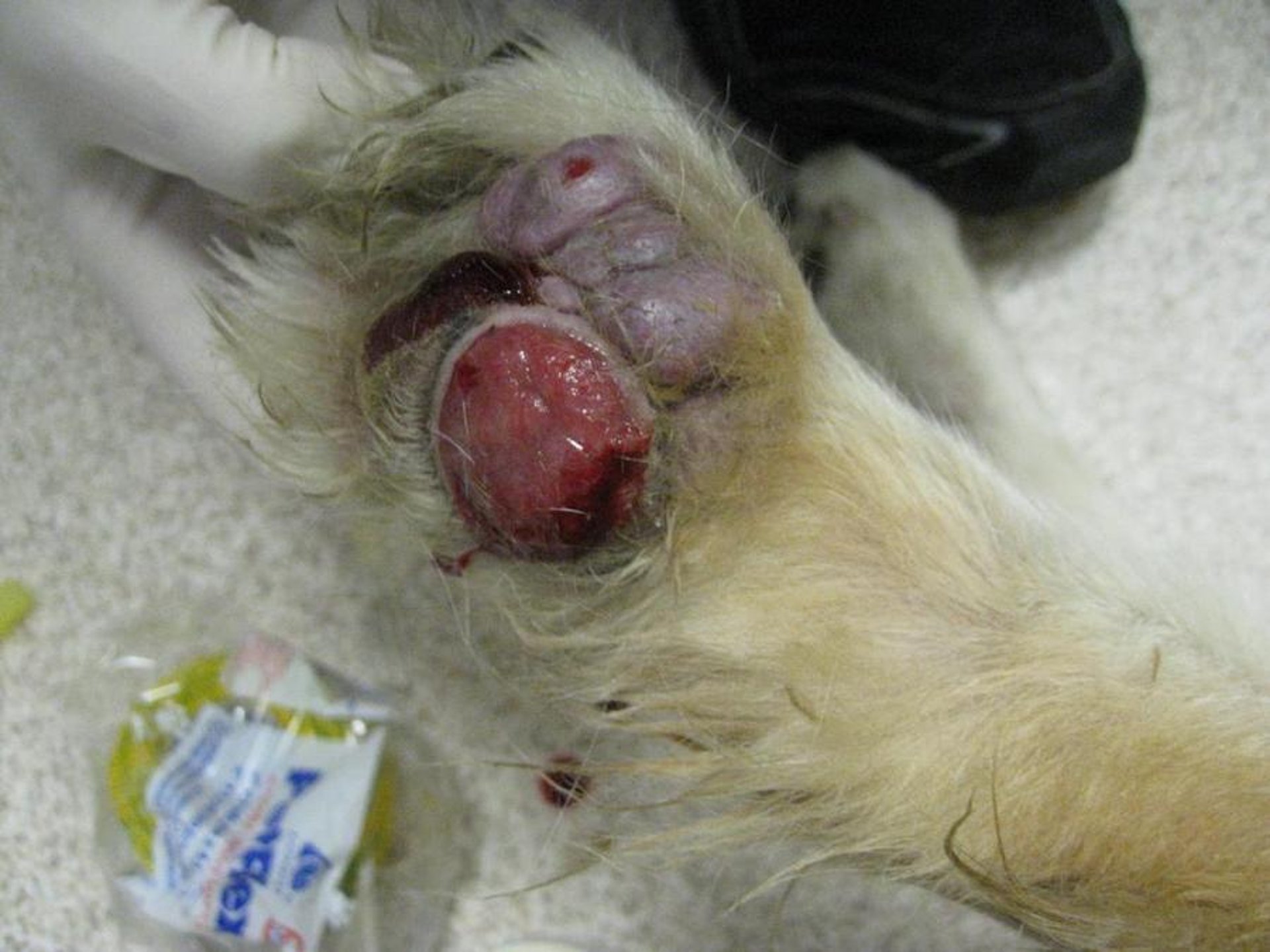
Clinical photograph of the area of the left elbow of a Great Dane mix with a recurrence of a peripheral nerve sheath tumor. These tumors are locally infiltrative; therefore, if adequate excision is not achieved, follow-up adjunctive treatment to gain local control is required to increase the tumor-free interval.
Courtesy of Dr. Alice Villalobos.
In dogs and cats, peripheral nerve sheath tumors of the skin are found in older animals. In cattle, they have a suspected genetic basis, may be multiple, can develop in both the young and old, and are generally an incidental finding at slaughter; they arise from the deep nerves of the thoracic wall and viscera, and cutaneous involvement is rare. Regardless of species, these tumors appear as white, firm, nodules. Attachment to a peripheral nerve may occasionally be noted.
Both benign and intermediate-grade malignant variants are recognized. Benign tumors are most common in cattle in which, because of their indolent nature, treatment is optional; also, additional tumors often develop spontaneously at other sites over time. In dogs, cats, and horses, most are locally infiltrative but do not metastasize. Complete excision is the treatment of choice. When margins are narrow or insufficient, follow-up radiation therapy, postoperative intralesional tumor bed chemotherapy (using the patient's serum mixed with the chemotherapy agent), electrochemotherapy, or systemic chemotherapy with carboplatin or metronomic chemotherapy may increase the tumor-free interval.
Adipose Tissue Tumors
Clinical photograph of a 12-year-old spayed female Aussie-Labrador mix with a lipoma. This mass developed over the left hip area and was diagnosed as a lipoma on the basis of fine-needle aspiration and cytologic evaluation.
Courtesy of Dr. Margaret Norton.
Lipomas are benign tumors of adipose tissue, perhaps more accurately characterized as hamartomas. They are common in dogs, occasionally identified in cats and horses, and rare in other domestic species. In dogs, they generally develop in older, obese females, most commonly on the trunk and proximal limbs. The breeds most at risk are Doberman Pinschers, Labrador Retrievers, Miniature Schnauzers, and mixed-breed dogs. Older neutered male Siamese cats are predisposed, and tumors are most commonly found on the ventral abdomen. Obesity does not appear to be a factor in the development of lipomas in cats. Affected horses are generally < 2 years old. Lipomas typically appear as soft, occasionally pedunculated, discrete nodular masses, and most are freely movable. In dogs and cats, >5% are multiple. In general, these tumors float when placed in formalin.
A rare variant of this tumor, diffuse lipomatosis, has been identified in Dachshunds. Virtually the entire skin is affected, resulting in prominent folds on the neck and truncal skin. Many lipomas merge imperceptibly with the adjacent non-neoplastic adipose tissue, making it difficult to determine when the entire lesion is excised. Lipomas with an abundant connective tissue stroma (fibrolipomas), cartilaginous stroma (chondrolipomas), or a prominent vascular component (angiolipomas) are also recognized. Despite their benign nature, lipomas should not be ignored because they tend to enlarge over time, and their gross presentation may be indistinguishable from that of infiltrative lipomas or liposarcomas. Excision is curative. In dogs, dietary restriction to 70% of normal intake for several weeks before surgery may allow for better definition of the surgical margins of the tumor.
Infiltrative lipomas (intra- and intermuscular lipomas) are rare in dogs and even less common in cats and horses. In dogs, they are most common in middle-aged females, usually on the thorax and limbs. The breeds of dog most at risk are the same as those for lipomas. These tumors are poorly confined, soft, nodular to diffuse swellings that typically involve the subcutaneous fat and underlying muscle and connective tissue stroma. Infiltrative lipomas, which dissect along fascial planes and between skeletal muscle bundles, are considered sarcomas of intermediate malignancy. They rarely metastasize. Aggressive excision is recommended, and amputation may be necessary.
Liposarcomas are rare neoplasms in all domestic animals. Most are recognized in older male dogs, in which they usually develop on the trunk and extremities; Shetland Sheepdogs and Beagles may be predisposed. In cats, feline leukemia virus infection has been infrequently associated with their development; whether this is a coincidence or such infections play a causative role remains unclear. Liposarcomas are nodular and soft to firm. They may exude a mucinous fluid when sectioned. Many have palpable, partially encapsulated areas; however, these zones should not be construed as evidence of a benign tumor. Liposarcomas are malignant neoplasms that have a low metastatic potential but are frequently pseudoencapsulated. Wide excision is recommended. Recurrence is common, so follow-up radiation therapy is indicated in cases with insufficient margins.
Vascular Tumors
Hemangiomas of the skin and soft tissues are benign proliferations that closely resemble blood vessels. Whether these are neoplasms, hamartomas, or vascular malformations remains undefined, and no clear criteria exist that allow for their separation. They are most commonly identified in dogs, occasionally in cats and horses, and rarely in cattle and pigs; they are an exceptional finding in other domestic animals.
In dogs, they are tumors of adult dogs and most commonly develop on the trunk and extremities. Many canine breeds (including Gordon Setters, Boxers, Airedale Terriers, Scottish Terriers, and Kerry Blue Terriers) are considered to be at risk.
Cats most frequently develop hemangiomas when they are adults. Lesions are most common on the head, extremities, and abdomen.
In horses, they are most common on the distal extremities of young (< 1-year-old) animals.
In cattle, they may be seen as congenital lesions or in older animals. Dairy cattle are predisposed to developing disseminated hemangiomas (angiomatosis) in the skin and internal organs.
In pigs, these lesions generally develop in the scrotal or perineal skin of Yorkshire, Berkshire, and less commonly Chester White boars. In the first two breeds, the disease is believed to be genetically transmitted.

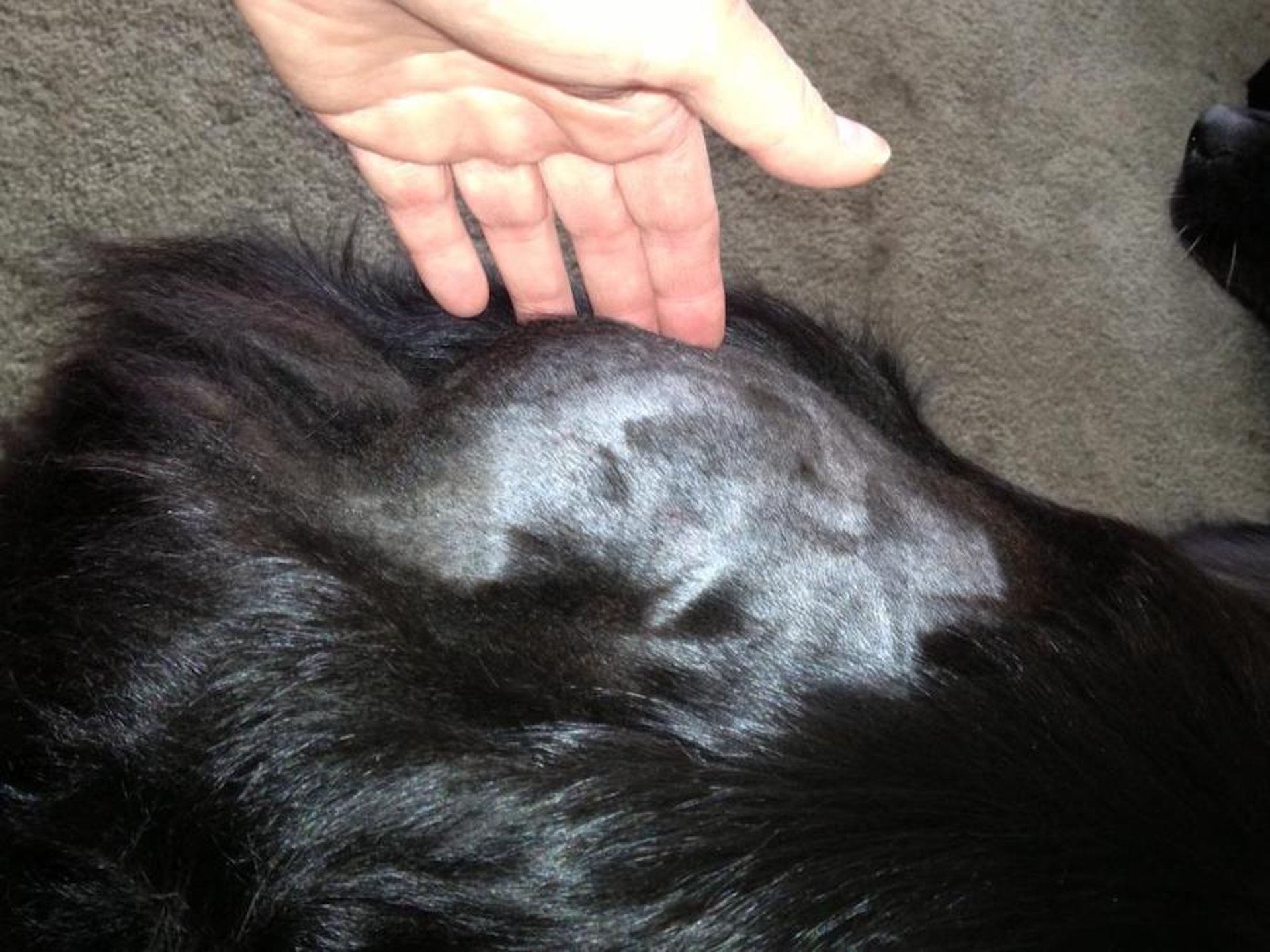
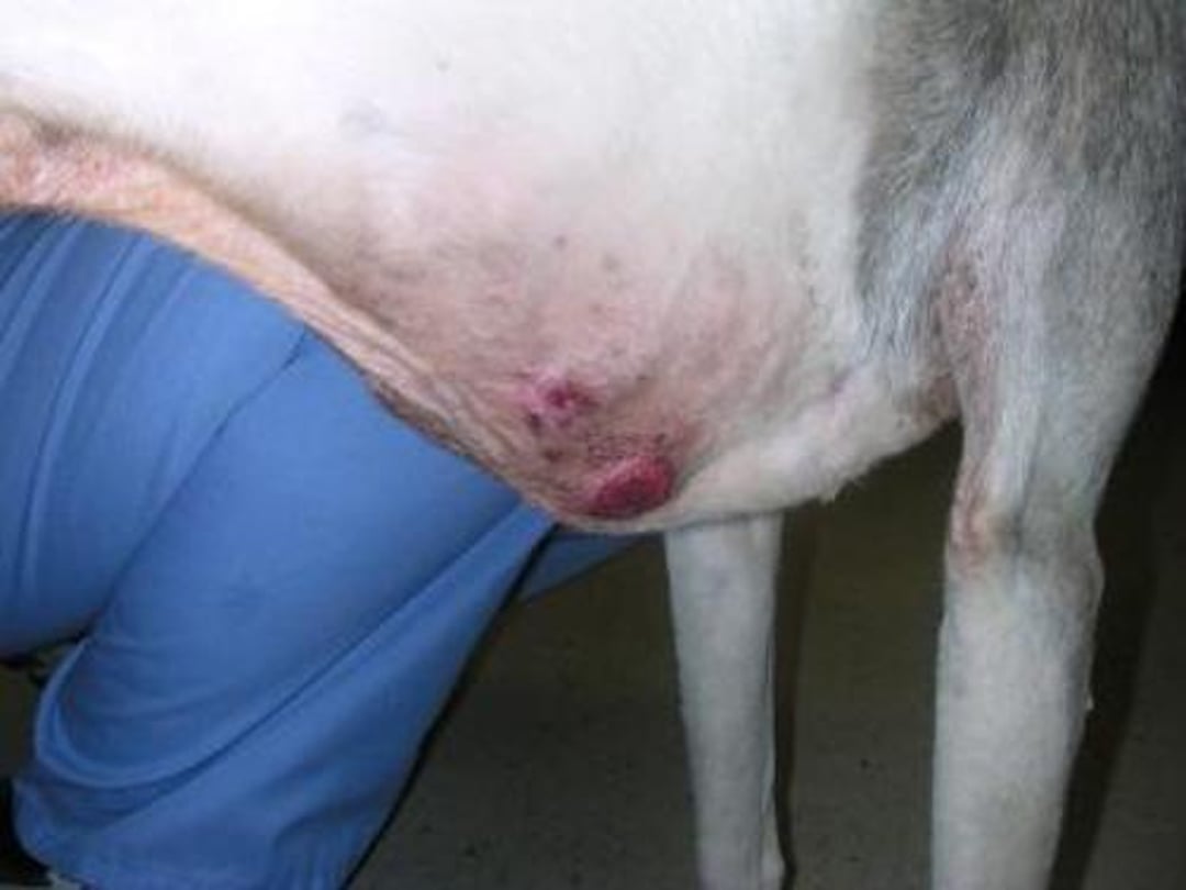
Clinical photograph of a older Black Labrador with a hemangiopericytoma over the lateral right thorax. Hemangiopericytomas can become very large. They recur after incomplete excision or complete excision with narrow, inadequate margins.
Courtesy of Dr. Alice Villalobos.

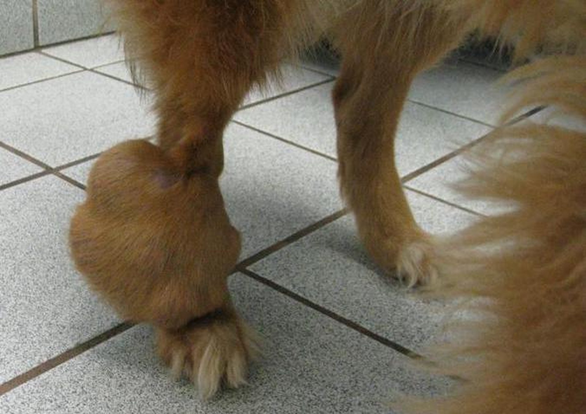
Clinical photograph of a mixed-breed dog with a hemangiopericytoma of the right hind leg. This multilobulated mass was not painful, but its weight made it more difficult for this dog to walk normally.
Courtesy of Dr. Alice Villalobos.
Hemangiomas are single to multiple, circumscribed, often compressible, red to black nodules. The lining epidermis may be unaffected or ulcerated or papillated. Small, superficial hemangiomas that often appear as a blood blister are known as angiokeratomas. When erythrocytes are sparse or absent within vascular lumens, the term lymphangioma is applied. Hemangiomas are benign but tend to ulcerate and grow quite large. Excision is the treatment of choice; however, in large animals in which the lesions may be large and involve the distal extremities, this may be difficult. In these cases, cryosurgery or radiation therapy may be necessary. Except in dairy cattle with angiomatosis, development of additional tumors at new sites after complete excision is uncommon.
Hemangiopericytomas (canine spindle-cell sarcoma, canine malignant fibrous histiocytoma, canine neurofibrosarcoma, canine perineuroma) are common in dogs and rare in cats (if they occur at all). This tumor was initially named because it was thought to derive from fibroblastic cells that surround small vessels; however, the appropriateness of the name remains a topic of debate. These tumors develop most commonly on the distal aspect of the extremities and thorax of older dogs. Females appear to be predisposed, and Siberian Huskies, mixed-breed dogs, Irish Setters, and German Shepherd Dogs are most at risk.
Hemangiopericytomas typically present as firm, multilobulated, solitary lesions with irregular borders, most commonly in the subcutaneous fat but sometimes in the dermis. They are of intermediate malignancy and have limited metastatic potential. Complete excision with adequate margins is the treatment of choice; however, because of the infiltrative nature of these tumors, excisions are incomplete or margins are narrow in most surgeries; therefore, ~30% recur. If the first excision of any sarcoma does not have adequate margins (ie, clean, wide surgical margins, whereas complete but narrow margins are not considered adequate for local tumor control), follow-up surgery to remove the tumor bed and increase the margins is indicated. At surgery, intracavitary chemotherapy with carboplatin or 5-fluorouracil mixed with the patient's serum, or intraoperative radiation therapy with follow-up intratumor bed chemotherapy, electroporation, or follow-up radiation therapy, may increase the tumor-free interval.
Angiosarcomas, arguably the most aggressive of all soft tissue tumors, are composed of cells that have many functional and morphologic features of normal endothelium. Although these tumors are often divided into hemangiosarcomas (of purported blood vessel origin) and lymphangiosarcomas (of lymphatic vessel origin), such a distinction is arbitrary. The term angioendothelioma is also used.
These tumors generally arise spontaneously. In the nonpigmented skin of dogs with short, often white coats, chronic solar injury may induce transformation in the superficial vascular plexus, which initially appears as a hemangioma and then progresses to a malignant vascular tumor. The light-skinned breeds prone to actinically induced dermal angiosarcomas are Whippets, Italian Greyhounds, white Boxers, and pit bull–type dogs. Pathologists often diagnose these lesions as cutaneous hemangiosarcomas. Because histogenesis of dermal hemangiosarcoma is often related to that of chronic solar irritation, predisposed breeds should be protected from sun exposure. Dermal hemangiosarcomas have a moderately high potential for malignancy; therefore, wide surgical excision is the treatment of choice for tumors >0.5 cm in diameter. Cryosurgery or laser surgery can effectively control multifocal, small, early lesions. When metastasis occurs, it is by the hematogenous route to the lungs and spleen. Up to one-third of splenic hemangiosarcomas arise in the skin or peripheral tissue and end up in the spleen because of splenic filtering of cells in the bloodstream.

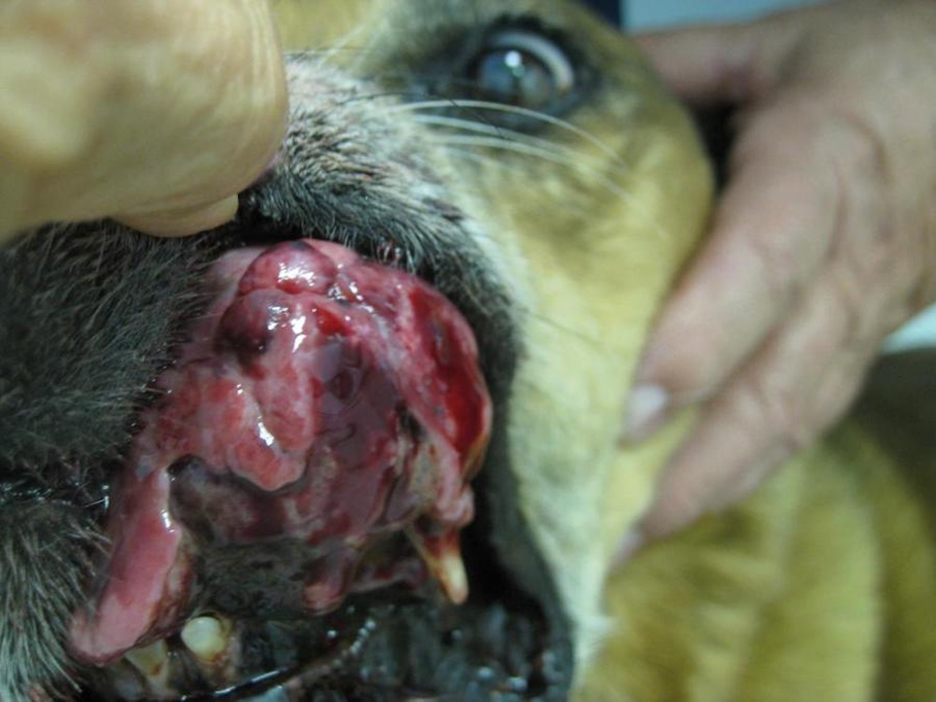
Photograph of a 13-year-old Golden Retriever with angiosarcoma of the hip. This tumor grew rapidly and developed necrosis and ulceration. Distant metastasis, especially to the lungs and liver, is common.
Courtesy of Dr. Alice Villalobos.

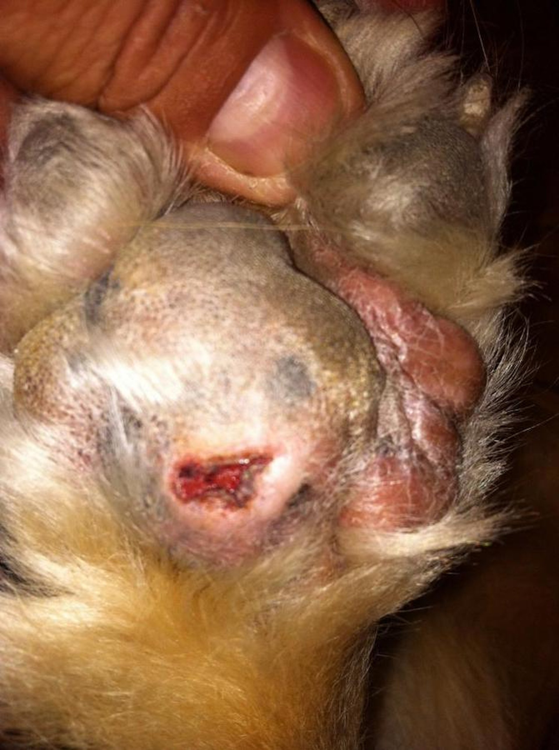
Clinical photograph of a 10-year-old Whippet with cutaneous hemangiosarcoma.
Courtesy of Dr. Alice Villalobos.

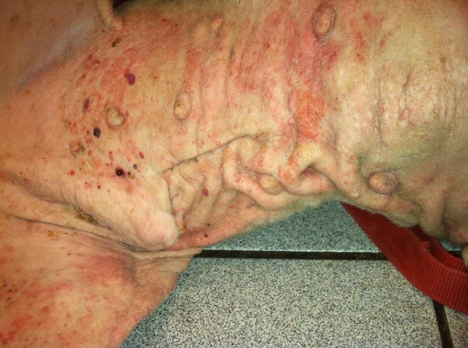
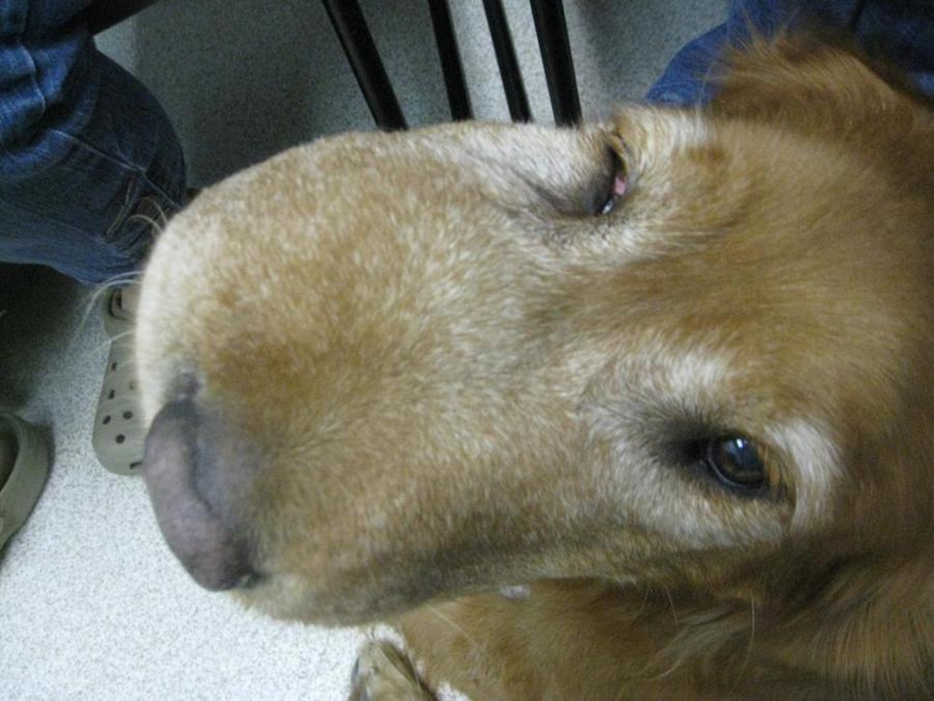
Clinical photograph of solar-induced cutaneous hemangiosarcoma in an older Bulldog. Lesions appear as multiple, red, flat or blood-filled cysts in sun-exposed nonpigmented skin.
Courtesy of Dr. Alice Villalobos.

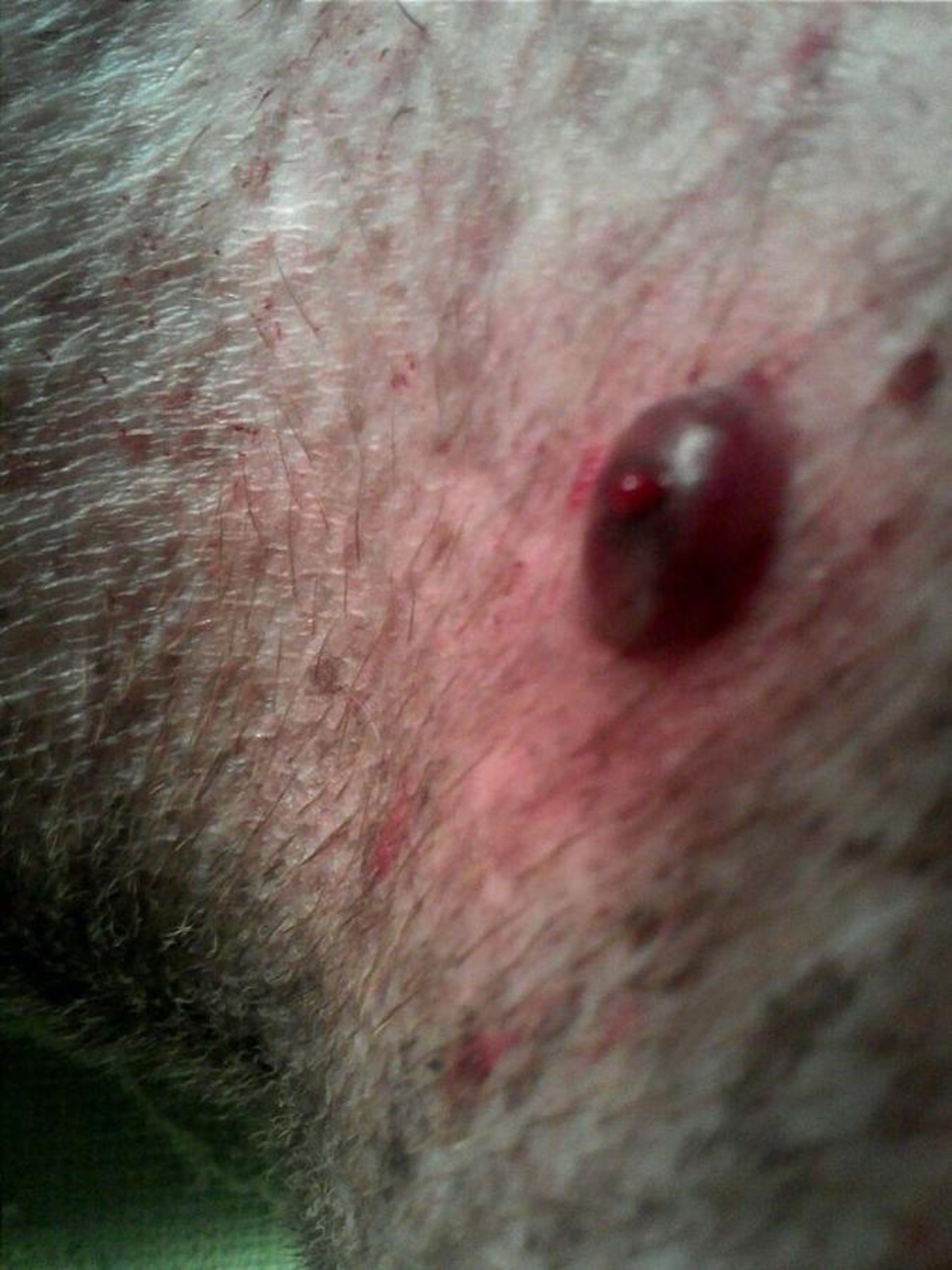
Clinical photograph of an Italian Greyhound with solar-induced cutaneous hemangiosarcoma demonstrating the typical appearance of a blood-filled cystic lesion.
Courtesy of Dr. Alice Villalobos.
Angiosarcomas of the skin and soft tissues are seen in all domestic animals; however, they are most common in dogs, generally in adult or senior animals. In dogs, they most frequently develop on the trunk, hip, thigh, and distal extremities. In addition to the breeds prone to actinically induced angiosarcomas, Irish Wolfhounds, Vizslas, Golden Retrievers, and German Shepherd Dogs are also at risk.
In cats, angiosarcomas are seen most commonly in older, castrated males, on the extremities and trunk. Cats with skin, subcutaneous, or visceral involvement develop distant metastasis.
Angiosarcomas can vary markedly in appearance. Most commonly, they appear as one or more erythematous nodules present anywhere in the skin or underlying soft tissues. Less frequently, they appear as a poorly defined bruise. All grow rapidly, often are associated with large zones of necrosis and thrombosis, and typically are red to black on cut section. Tumors often diagnosed as lymphangiosarcomas may have much less lumenal blood, and the vascular spaces are typically filled with serum. Characteristically, angiosarcomas create their own vascular space by dissecting through soft tissues. Distant metastasis, especially to the lungs and liver, is common. In other domestic animals, these tumors do not appear to behave as aggressively, and postexcisional recurrence rather than metastasis is more common.
For all species, because of the aggressiveness of these nodules, wide excision with intraoperative placement of carboplatin beads or intracavitary carboplatin mixed with the patient's serum may be the treatment of choice. Solar-induced canine cutaneous hemangiosarcomas generally do not have an aggressive biological behavior, although numerous lesions may continue to appear over several years. Superficial lesions are easily destroyed with topical cryotherapy, which may control the disease for years; however, if lesions become large (>1 cm in diameter) or cystic, they are best controlled with excision. If multiple cystic lesions appear, the animal may benefit from a combination of local control and antiangiogenic therapy.
Avoidance of further sun irradiation injury may reduce the development of new lesions; however, previously exposed skin may continue to develop lesions as a result of field cancerization. Adjuvant chemotherapy consisting of administration of vincristine, doxorubicin, and cyclophosphamide (the VAC protocol) has been reported to shrink angiosarcomas; carboplatin is also helpful. However, the effects of chemotherapy for systemic control or radiation therapy for local control and long-term survival time remain to be defined. Electrochemotherapy with bleomycin or cisplatin has shown efficacy. The antiangiogenic role of NSAIDs such as piroxicam, meloxicam, etc, is still not completely understood and may vary from drug to drug. Antiangiogenic or angiostatic compounds that attack the blood supply of tumors may control and prevent metastases; however, results of clinical trials remain elusive.
Cutaneous Smooth Muscle Tumors
Because they either are not recognized or do not develop with any regularity in domestic animals, cutaneous smooth muscle tumors (leiomyomas or leiomyosarcomas) are diagnosed rarely. Those reported generally have been malignant and found in dogs and cats. Usually, they are firm, cutaneous masses. Leiomyomas are small and tend to be limited to the dermis, whereas leiomyosarcomas are larger and most arise from (or extend into) the subcutaneous fat. The behavior of malignant smooth muscle tumors remains poorly defined. Complete excision is the treatment of choice for both leiomyomas and leiomyosarcomas. They have been responsive to vincristine chemotherapy administered intravenously weekly for 6 weeks then tapered; they may also respond to administration of oral tyrosine-kinase inhibitors.
For More Information


