

Epidemics of bacterial diseases are common in dense populations of cultured food or aquarium fish. Predisposition to such outbreaks frequently is associated with poor water quality, organic loading of the aquatic environment, handling and transport of fish, marked temperature changes, hypoxia, or other stressful conditions. Most bacterial pathogens of fish are aerobic, gram-negative rods. Diagnosis is by isolation of the organism in pure culture from infected tissues and identification of the bacterial agent. Sensitivity testing before antimicrobial use is recommended.
Aeromonas, Pseudomonas, and Vibriosis in Fish
A number of bacteria produce a similar syndrome, generically referred to as hemorrhagic septicemia and characterized by external reddening and hemorrhage in the peritoneum, body wall, and viscera. Morbidity and mortality are highly variable, depending on predisposing conditions such as low dissolved oxygen, other water quality problems, handling stress, or trauma. Ulcerative lesions are common as disease progresses, and mortality can be significant if stress is not controlled. Antimicrobial therapy is recommended if fish are dying. Common bacterial isolates from affected fish include Aeromonas and Pseudomonas spp, which are more common in freshwater animals, and Vibrio spp, more commonly isolated from marine fish. Control is based on removal of predisposing factors. If antimicrobial therapy is warranted, drug selection should be based on sensitivity testing when possible.

Courtesy of Dr. Ruth Francis-Floyd.
Aeromonas salmonicida, a gram-negative, nonmotile rod, is the causative agent of goldfish ulcer disease and furunculosis in salmonids and is a very important disease of koi and goldfish. The disease also occurs in other freshwater and marine species. In the acute form, hemorrhages are found in the fins, tail, muscles, gills, and internal organs. In more chronic forms, focal areas of swelling, hemorrhage, and tissue necrosis develop in the muscles. These lesions progress to deep crateriform abscesses that discharge from the skin surface. Liquefactive necrosis develops in the spleen and kidney. Diagnosis is made by isolating and identifying a pure culture of the organism from infected tissue. Avoidance through use of good quarantine practices, and vaccination when appropriate, is preferable to treatment. Successful treatment is possible, based on appropriate antimicrobial therapy. Bacteriologic culture of blood is an effective and nonlethal method for effective identification and sensitivity testing of A salmonicida isolates from valuable koi. Commercial vaccines are available for prevention of A salmonicida in salmonids and koi, but information on efficacy in koi is limited.
Vibriosis is a potentially serious, common systemic disease of many cultured, aquarium, and wild marine and estuarine fish; it is less common in freshwater fish. Three genera of the family Vibrionaceae are frequently associated with infection in fish: Vibrio, Listonella, and Photobacterium. These genera can result in hemorrhages and ulcerations of the skin, fin, and tail; hemorrhagic and degenerative changes of internal organs; and other systemic changes. Diagnosis requires identification of pure isolates from infected tissues. Isolation of V cholerae from fish is not uncommon and should not cause alarm as long as the isolate is the non-O type. Preventive measures include minimizing stress and crowding. Coldwater vibriosis (Hitra disease), a serious problem in sea farming of salmonids, is characterized by high mortality, resistance to drug therapy, and stress mediation. The etiologic agent is Aliivibrio salmonicida. Because members of this family are ubiquitous in marine environments, avoidance is difficult. Preventive vaccination with formalin-killed Vibrio is used in the salmonid industry. Antimicrobial therapy should be based on results of antimicrobial susceptibility testing.
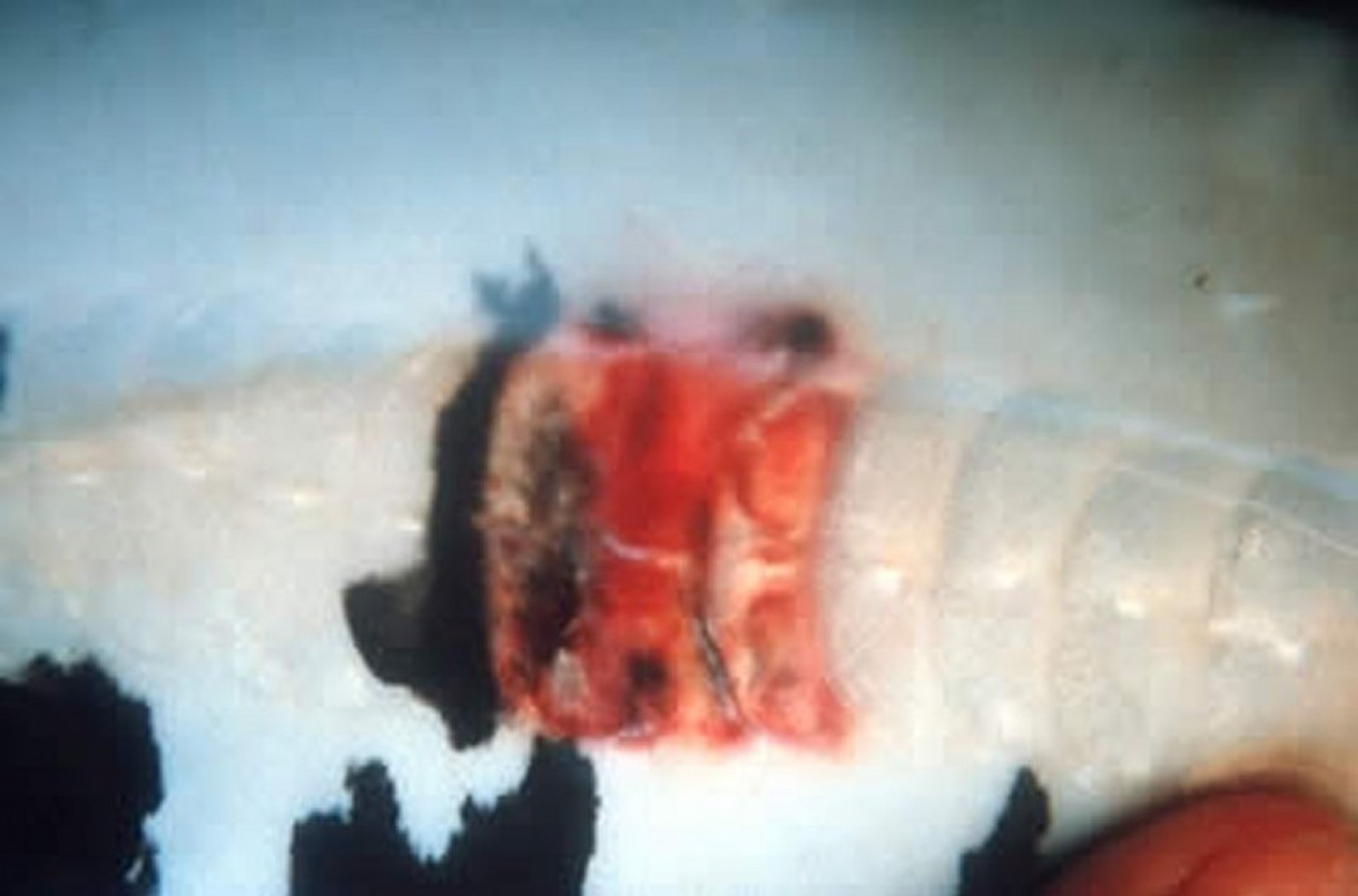
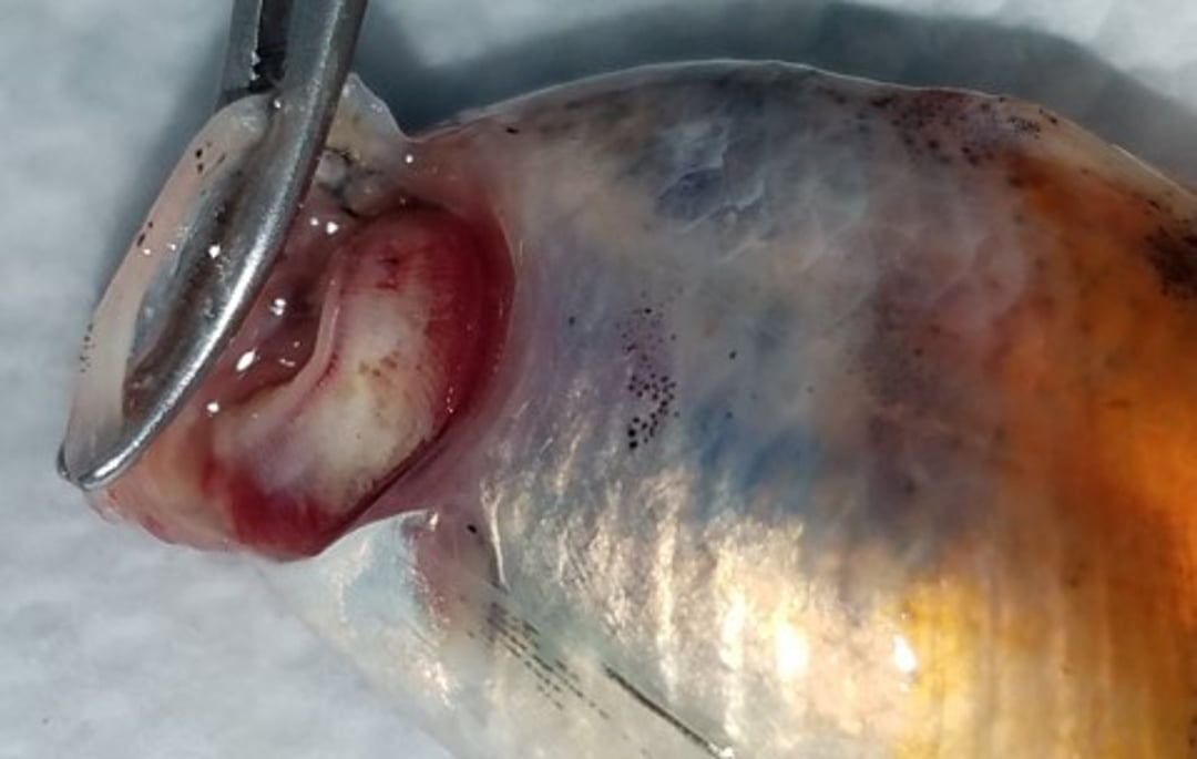
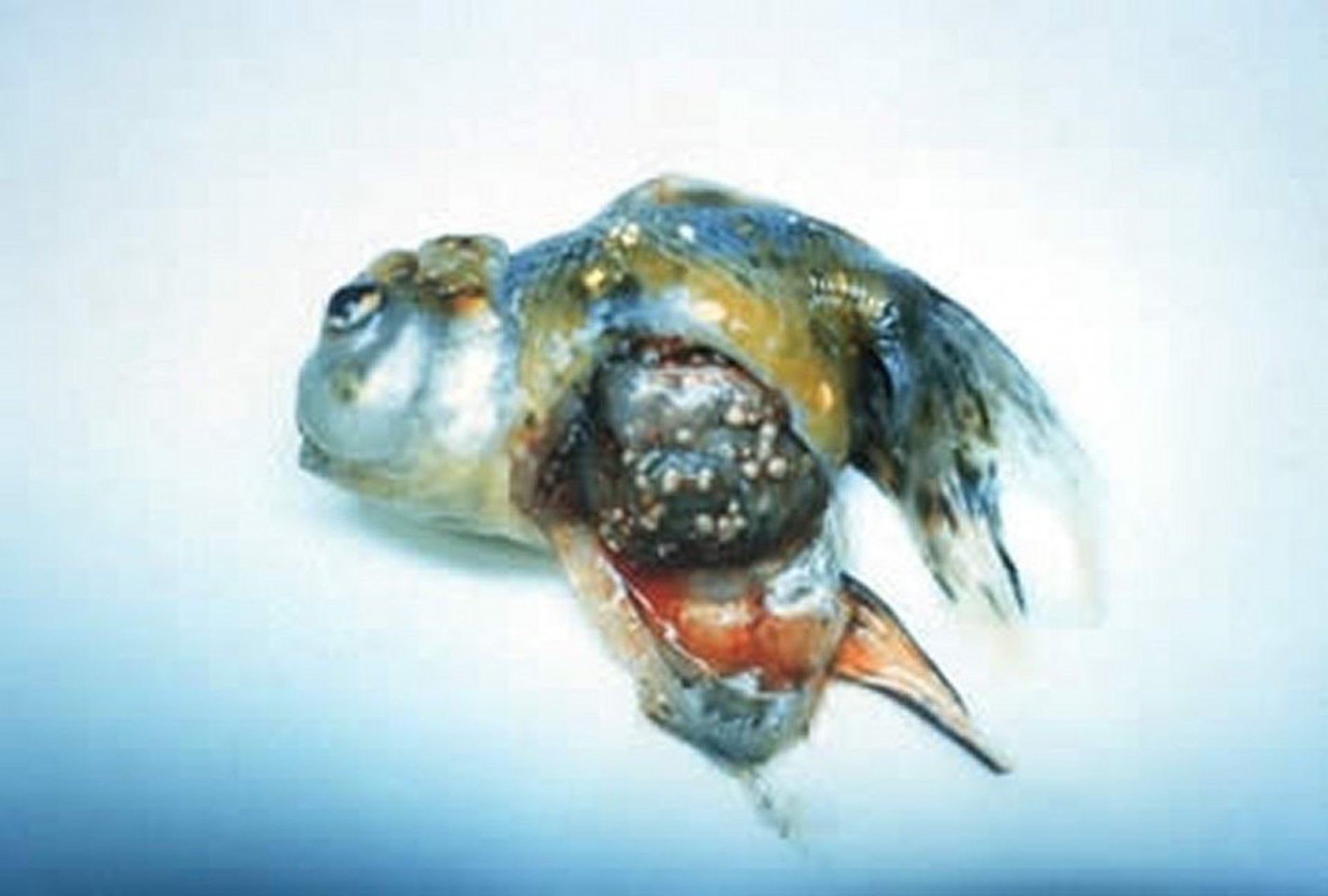
Edwardsiella in Fish

Courtesy of Dr. Denise Petty.
Edwardsiella ictaluri is commonly associated with disease in channel catfish; however, it is also responsible for high mortality in zebrafish, both in research laboratories and aquarium fish. It is an obligate pathogen and can be transmitted by direct contact with infected fish, water, and feces. Clinical signs of disease in infected zebrafish include hemorrhaging in the skin, pale gills, lethargy, and splenomegaly. On histologic evaluation, bacteria frequently can be found in high numbers in spleen, anterior and posterior kidneys, nares, and forebrain. It can be isolated on standard culture media, but it is slow-growing. Coinfections with Aeromonas spp are common; Aeromonas spp are fast growers and can easily overgrow E ictaluri.
Edwardsiella piscicida is an emerging disease and has been reported in many fish species, both freshwater and marine, and in ornamental, game, and food fish. When phenotypic identification of isolates are performed, it cannot be distinguished from E tarda; consequently it is recommended to submit bacterial isolates identified as E tarda for complete genome sequencing. Some fish infected with E piscicida often have a granulomatous response. Gram-negative bacteria may be seen in the granulomas.
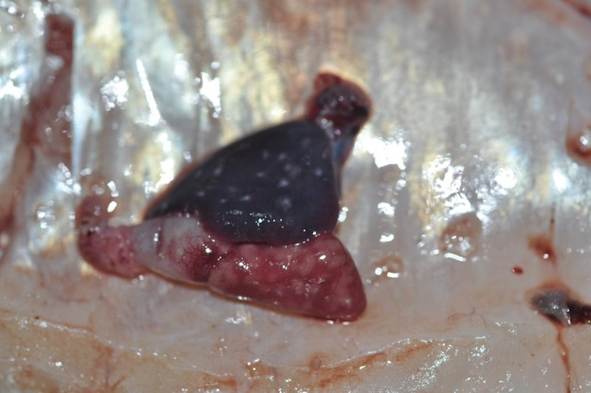
Epitheliocystis in Fish

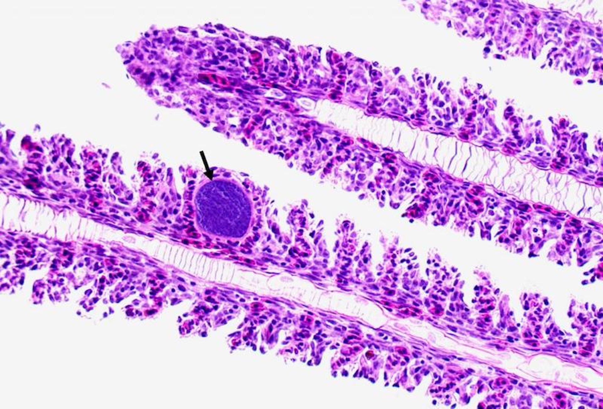
Courtesy of Dr. Denise Petty.
Although more commonly reported in aquacultured fish species, epitheliocystis also occurs in aquarium fish and is frequently seen in discus (Symphysodon spp), oscar (Astronotus ocellatus), and occasionally other pet fish. Epitheliocystis is caused by an obligate intracellular bacteria in the order Chlamydiales. Infection most frequently involves the gills and occasionally the skin. Infections are more common in freshwater-reared fish species. Epitheliocystis should be suspected if colorless spherical structures (infected, hypertrophied cells) that have granular contents are seen on gill biopsy or skin scraping. Histopathologic findings typically include basophilic hypertrophied cells. Clinical signs are variable and depend on severity of infection. Heavily infected fish may be lethargic and dyspneic. Orally administered oxytetracycline has been used to successfully control infections.
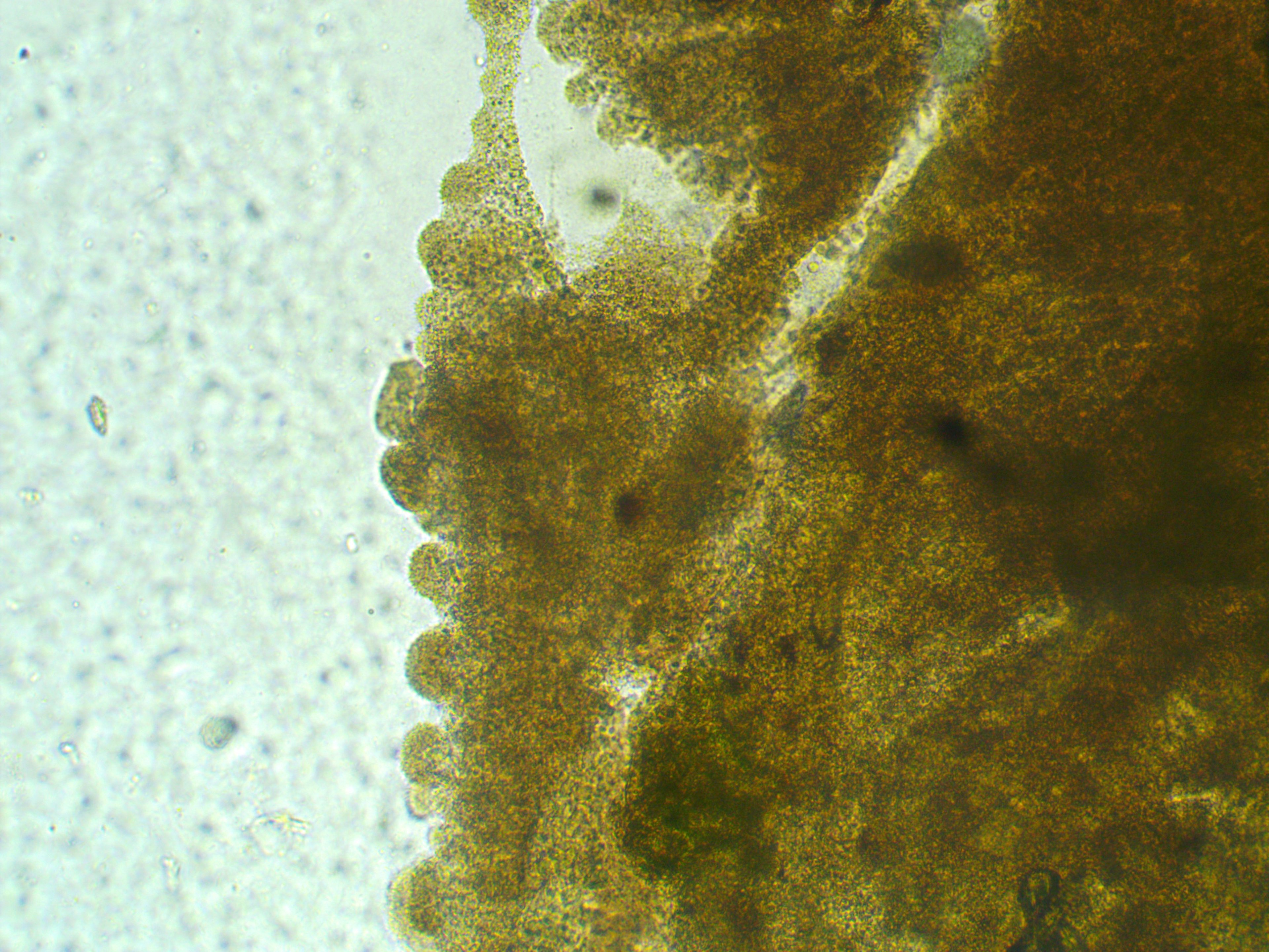
Flavobacterium in Fish

Courtesy of Dr. Denise Petty.

Courtesy of Dr. Denise Petty.
Flavobacterium columnare, the member of this group responsible for columnaris disease, is most common in warmwater species of fish. A presumptive diagnosis can be made from visualization of typical organisms on wet mounts of infected skin or gill tissue. There are several strains of this bacterium, which range from low to high virulence. As yet unidentified water quality conditions predispose susceptible fish to infection with the virulent strain. Mortality can be acute, and by the time the condition is seen it may be difficult to control.
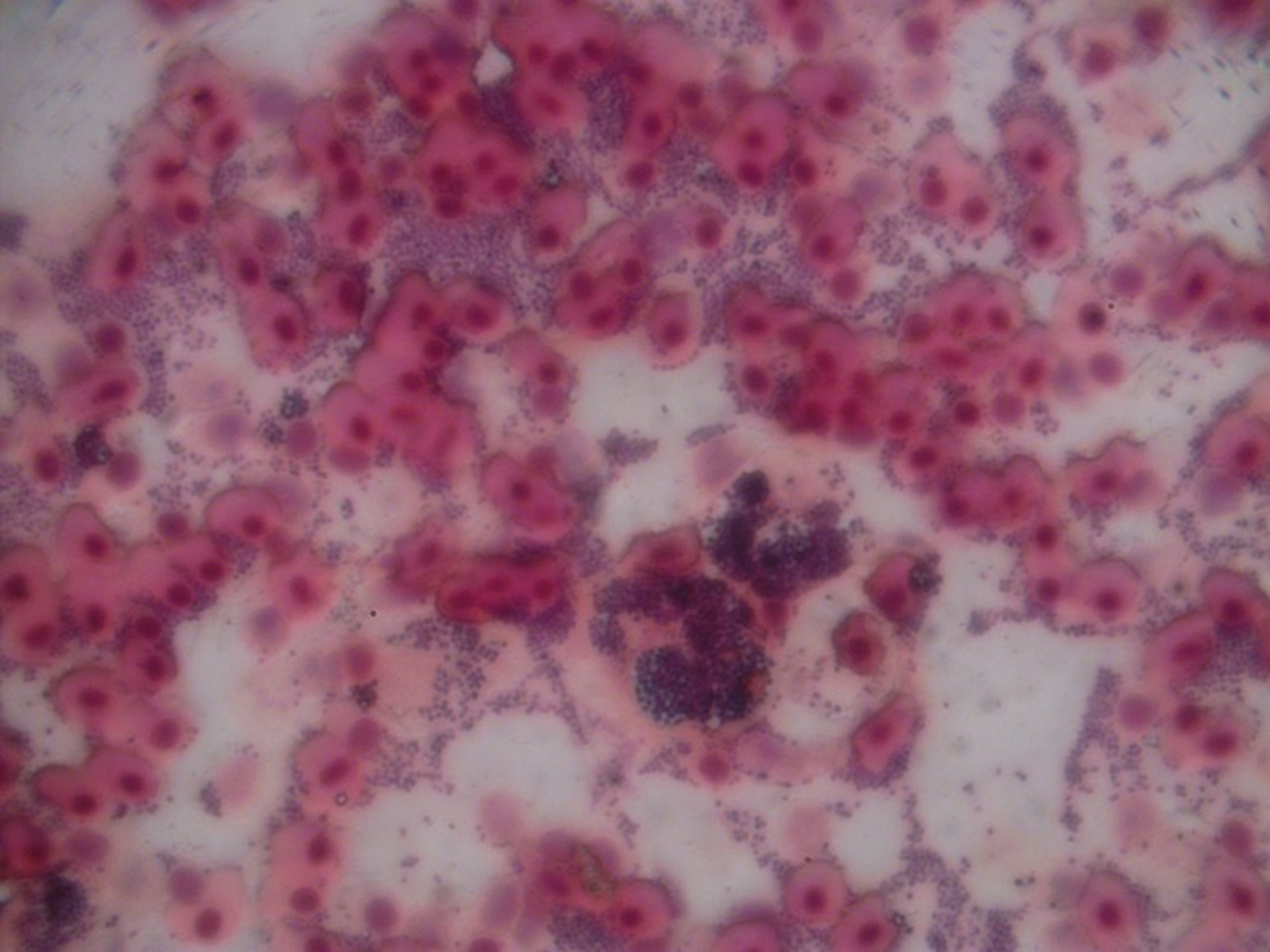
Gram-positive Bacterial Infections of Fish
These infections of concern to fish culturists and aquarists may be caused by Streptococcus and related genera Lactococcus, Enterococcus, and Vagococcus. Infections are uncommon but can cause high mortality (>50%) when they do occur. Chronic infections may continue for weeks, with only a few fish dying each day.

Courtesy of Dr. Denise Petty.
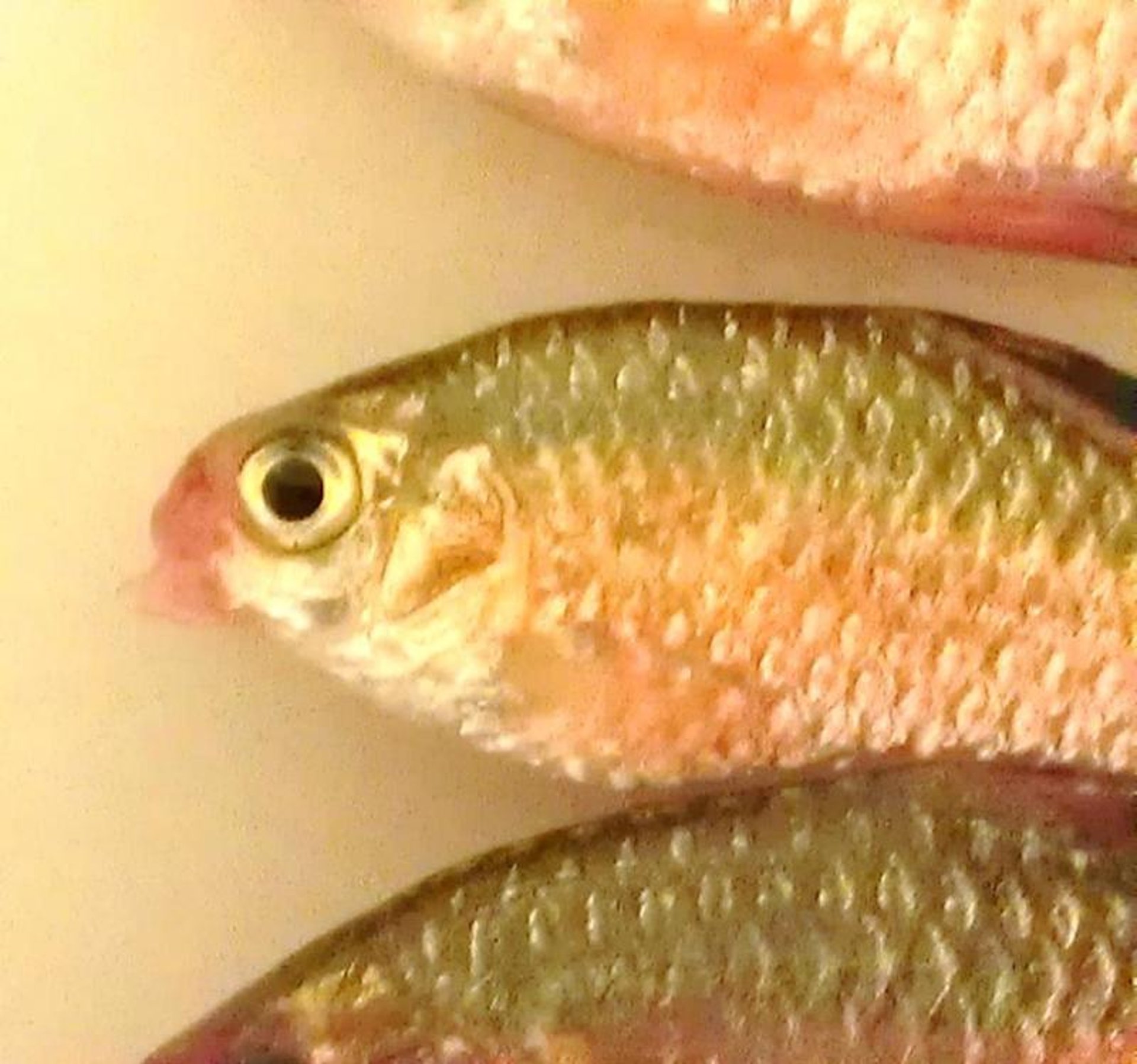
Species known to be susceptible include salmonids, assorted marine fish (eg, mullet and sea bass), tilapia, sturgeon, and striped bass. Susceptible aquarium species include rainbow sharks, red-tailed black sharks, rosy barbs, danios, and some tetras and cichlids. In general, all fish should be considered susceptible. A characteristic manifestation of Streptococcus infection is neurologic disease, often manifested by spinning or spiraling in the water column.

Courtesy of Dr. Denise Petty.
Brain and kidney cultures from suspect fish should be incubated on blood agar at 25°C for 24–48 hours. Gram stains of pinpoint bacterial colonies reveal typical chains of gram-positive cocci, which allow a presumptive diagnosis. Confirmation requires definitive identification of the organism.
Antimicrobial therapy should be based on antimicrobial susceptibility testing. Erythromycin is often the drug of choice, but it is not FDA approved for this use.
Sources of infection may be environmental or include live foods, such as tubeficid worms, amphibians, or previously infected fish. Future epizootics can be prevented if the source of infection is identified and eliminated. Streptococcus iniae has been isolated from cichlids and other aquarium fish and has zoonotic potential. Autogenous vaccines are available for use in aquaculture facilities.
The gram-positive bacterium Erysiopelothrix sp. is frequently associated with rostral ulceration and stomatitis in cyprinids such as tiger barbs (Puntigrus tetrazona), rosy barbs (Pethia conchonius), and tetras. Infected fish often live for days before succumbing to infection. Fish that exhibit minimal damage due to infection often respond well to treatment with erythromycin. This species is not a zoonotic concern.
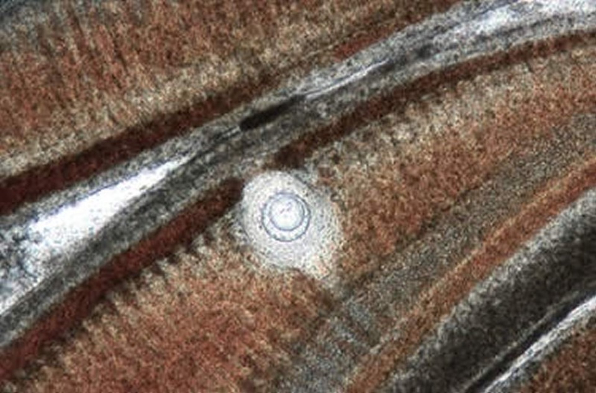
Mycobacterial Infections in Aquarium Fish

Courtesy of Dr. Ruth Francis-Floyd.

Courtesy of Dr. Ruth Francis-Floyd.
Mycobacteriosis is a chronic or acute, systemic, granulomatous disease of aquarium fish and cultured food fish, particularly those reared under intensive conditions. Predisposing environmental factors include low dissolved oxygen, low pH, and high organic load, all found in recirculating aquaculture systems. Correct use of ultraviolet light as a way to disinfect system water reduces bacterial counts and can be a useful tool to control infection in exhibit animals.
The causative bacteria can be any number of species of Mycobacterium, including M chelonae, M marinum, and M fortuitum. Syngnathids (sea horses) are particularly susceptible, but the disease can be seen in any fish. Signs are variable and nonspecific; they can include emaciation, ascites, skin ulceration and hemorrhages, exophthalmos, paleness, and skeletal deformities.
On necropsy, gross lesions of viscera consist of grayish white, necrotic foci that sometimes coalesce to form tumor-like masses. Granulomas may not be grossly visible and are often found first on wet mounts, especially of anterior kidney and spleen, or other viscera from infected fish.
A presumptive diagnosis is based on visualization of acid-fast rods in granulomatous material from suspect lesions. Definitive diagnosis requires isolating and identifying the bacteria. These weakly gram-positive, acid-fast, nonmotile bacteria are difficult to grow but can be isolated using Lowenstein-Jensen or Middlebrook media after incubation at 25°C for 3–4 weeks.
There are no effective treatments that eliminate mycobacteria in fish. Mycobacteria can cause zoonotic infections, and aquarists should be informed of potential risks if handling or cleaning contaminated fish or exhibits. An infected aquarium should be disinfected before other fish are added. Bleach is not an effective disinfectant against mycobacteria but should be used to eliminate organic material. It should then be followed by disinfection with 70% ethanol or phenolic compounds.
For More Information
Also see pet health content on routine health care for fish and emergencies of fish.


