Four-toed hedgehogs can develop a variety of illnesses, including neoplasia, acariasis, oral cavity and dental disorders, cardiovascular disease, and pneumonia. Clinical signs are usually nonspecific (eg, lethargy, weakness, anorexia), which emphasizes the importance of diagnostic testing, despite the need for chemical restraint to perform diagnostic tests.
Cardiovascular and Hematologic Diseases of Four-Toed Hedgehogs
Cardiomyopathy is a common postmortem finding in captive four-toed hedgehogs. The etiology of the disease is not known; however, genetic and nutritional causes have been suggested (1). Affected hedgehogs are typically ≥ 3 years old; however, cardiomyopathy can occur in hedgehogs as young as 1 year old.
Clinical signs of cardiomyopathy in hedgehogs include the following:
dyspnea
decreased activity
weight loss
an auscultable murmur
ascites
acute death
Radiographic analysis can demonstrate varying degrees of cardiac enlargement, pulmonary edema, pleural effusion, hepatic congestion, and ascites.
Common gross necropsy findings associated with cardiovascular disease in hedgehogs include cardiomegaly, hepatomegaly, pulmonary edema and/or congestion, hydrothorax, ascites, and pulmonary or renal infarcts.
Suspected cardiac disease indicates a need for full-body imaging and a full cardiovascular examination, including an ECG and echocardiogram. Normal echocardiographic measurements have been published for four-toed hedgehogs (2).
Hematologic and biochemical testing can be done to screen for concurrent problems and monitor drug effects.
Cardiovascular drugs commonly used to treat congestive heart failure (CHF) in animals (eg, diuretics, ACE inhibitors, pimobendan) might be helpful initially; however, the long-term prognosis for hedgehogs with CHF is poor.
Gastrointestinal and Hepatic Diseases of Four-Toed Hedgehogs
GI obstructions in four-toed hedgehogs are often caused by ingestion of rubber, hair, or carpet fibers. Clinical signs of obstruction include the following:
acute anorexia
lethargy
collapse
vomiting (possibly)
Marked gaseous dilatation of the GI tract can be a nonspecific finding in ill hedgehogs, complicating the diagnosis of GI obstruction.
In addition to GI obstruction, alimentary inflammation, including esophagitis, gastritis, enteritis, colitis, and gastric ulceration, has been reported in hedgehogs (3, 4, 5, 6).
Enteritis in hedgehogs can be caused by Salmonella or other bacteria. Hedgehogs with salmonellosis might be subclinical, or they might present with one or more of the following clinical signs:
diarrhea (can be bright green)
weight loss
decreased appetite
dehydration
lethargy
death
Diagnosis of salmonellosis should be confirmed with fecal culture testing, using a Salmonella-enriching medium. Although treatment is indicated in hedgehogs with clinical signs of salmonellosis, owners should be advised of the zoonotic potential and the risks of creating antimicrobial resistance.
Alimentary candidiasis (Candida albicans) and cryptosporidiosis are other infectious diseases that can cause enteritis in hedgehogs. Many species of nematodes, cestodes, and protozoa have been identified in wild hedgehogs; however, they seem to be far less prevalent in pet hedgehogs.
Some commercial diets cause diarrhea, as do foods inappropriate for hedgehogs (eg, milk).
GI neoplasia, particularly lymphosarcoma, is relatively common in hedgehogs.
Other possible reasons for GI signs include diet change, toxins, hepatic disease, and malnutrition. Hedgehog digestion does not rely on bacterial fermentation, and there is no evidence of antimicrobial sensitivity as occurs in hindgut-fermenting rodents and rabbits.
Hepatic lipidosis is somewhat common in hedgehogs and can be a sequela of numerous disease processes. Clinical signs include the following:
lethargy
inappetence
icterus
diarrhea
clinical signs of hepatic encephalopathy
Obese hedgehogs might have subclinical hepatic lipidosis noted on ultrasonographic or CT imaging. Diagnosis of hepatic lipidosis is supported by testing for hepatic enzyme activities, plasma bilirubin concentration, and bile acid concentration, as well as by diagnostic imaging and ultrasonography-guided liver aspiration. Treatment for hepatic lipidosis, with assisted nutrition, subcutaneous fluids, and administration of liver-supportive supplements (such as silymarin), is similar to that in other species.
Integumentary Diseases of Four-Toed Hedgehogs
Acariasis caused by Caparinia tripilis (a psoroptic mite) is very common in four-toed hedgehogs. Both male and female C tripilis mites have three long setae on the third pair of legs (see ). The mnemonic “three-on-three” may help with recollection. Infestation with Notoedres spp (sarcoptic mites) has also been reported in hedgehogs (7).

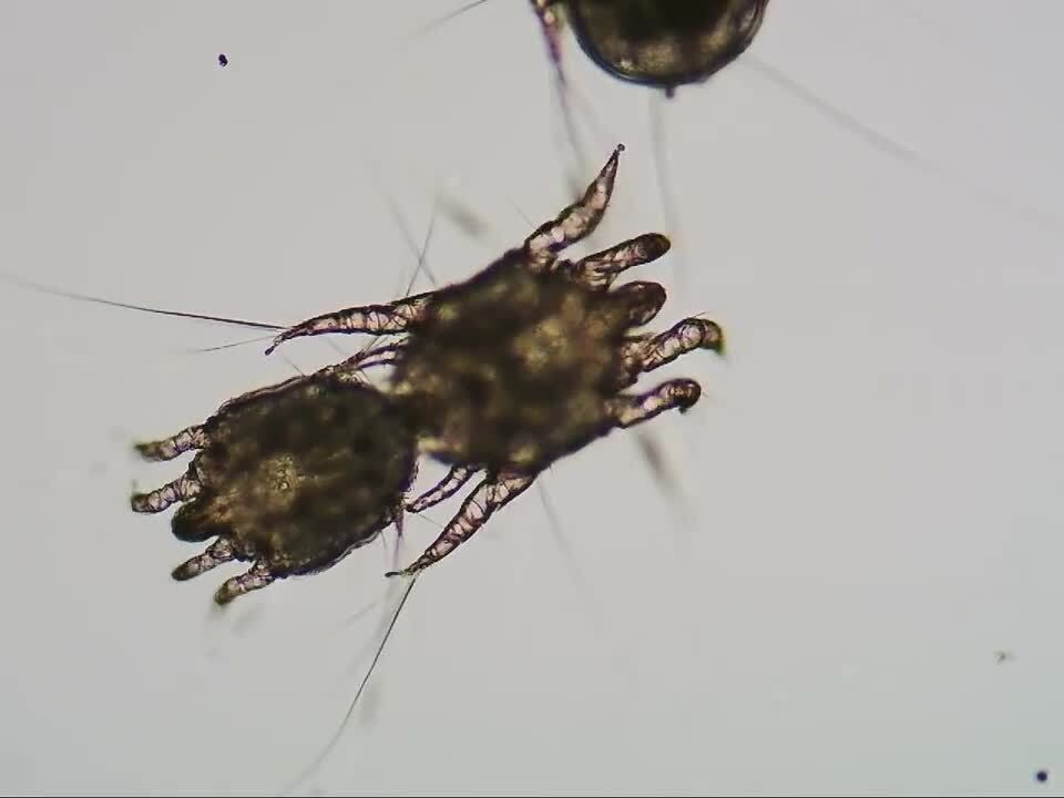
Clinical signs of acariasis include the following:
scaling (see )
loss of spines
white or brownish crusts (mite droppings) at the base of the spines and around the eyes
white powdery deposits around the nose or eyes
nonspecific signs (eg, lethargy, anorexia)
pruritus


Marked scaling of the skin in a hedgehog infested with Caparinia tripilis.
Courtesy of Dr. Grayson Doss.
Hedgehogs with acariasis might scratch or rub themselves; however, many are not pruritic. Some hedgehogs can harbor subclinical mite infestations.
Diagnosis of acariasis in hedgehogs is confirmed by identifying mites and eggs (nits) in a skin sample obtained from a pruritic hedgehog for cytological evaluation. Mites and eggs can also be identified on a tape impression. Traditional skin scrape methods can damage the thin epidermis of the mantle.
Selamectin (20–30 mg/kg, topically, every 21–28 days for 2–3 treatments) or ivermectin (0.3–0.4 mg/kg; topically, SC, or PO; every 10–14 days for 3–5 treatments) can be used to treat acariasis (8). In some cases, ivermectin alone might not be effective. Bedding must be removed, and any items in the cage must be disinfected or discarded. During treatment, the cage should be lined with paper that is changed daily. All hedgehogs in the home should be treated concurrently.
Dermatophytosis is a common clinical disease in four-toed hedgehogs; however, infection without notable clinical signs is also possible. Dermatophytes (Trichophyton erinacei, T mentagrophytes, Microsporum spp, and Arthroderma benhamiae) cause crusting dermatitis, especially around the face and pinnae. Spine loss might also be noted. Although some hedgehogs might scratch with the hindlimbs or rub against stationary objects, many do not display clinical signs of pruritus. Many dermatophyte infections are likely secondary to other dermatoses, such as acariasis or skin trauma.
Diagnosis of dermatophytosis in hedgehogs is confirmed by culturing spines in dermatophyte test medium or by testing spines for the presence of dermatophytes via PCR assay. Treatment consists of administration of oral antifungal agents (eg, terbinafine, itraconazole) and/or topical antifungals (eg, enilconazole, clotrimazole) in spray or shampoo formulations. Lime-sulfur dips may also be used. Other hedgehogs in the home can be subclinically infected; therefore, treatment of all hedgehogs in the home is recommended.
Dermatophytosis in hedgehogs is frequently zoonotic (see Zoonoses of Four-Toed Hedgehogs for details).
Skin neoplasia is common in hedgehogs. The following (among others) have been reported in hedgehogs (8):
mammary tumors
squamous cell carcinomas
epitheliotropic T-cell lymphomas
papillomas
sebaceous gland carcinomas
Pinnal dermatitis is also common in hedgehogs. Clinical signs include skin crusts, accumulated secretions, and a ragged pinnal margin. Dermatophyte infection and acariasis are important differential diagnoses for pinnal dermatitis; other possibilities include nutritional deficiencies, dry skin from inappropriate husbandry, or nonspecific seborrhea. Ear mites (Notoedres cati) are occasionally present; clinical signs, diagnosis, and treatment are as for cats. Otitis externa due to bacterial or yeast infection is often secondary to acariasis or another cause of chronic inflammation. Clinical signs of otitis externa include purulent discharge, odor, and sensitivity of the face and ear. Otic cytological evaluation, skin cytological evaluation, cleansing, and topical antimicrobial and/or anti-inflammatory medications are used as for other species. Otitis media and interna also occur in hedgehogs.
Musculoskeletal Diseases of Four-Toed Hedgehogs
Fractures in four-toed hedgehogs can occur when a limb becomes trapped in a cage wire or exercise wheel. Distal limb fractures can be splinted. Surgical correction might be indicated; however, any fixation device must be able to withstand the hedgehog's strong balling-up mechanism.
Lameness can occur in hedgehogs. Causes include the following:
ingrown toenails
arthritis
pododermatitis
constriction of a foot or digit by fibrous material
A thorough physical examination and diagnostic imaging (eg, radiographs) are excellent initial diagnostic measures when evaluating hedgehogs for lameness.
Neoplasia of Four-Toed Hedgehogs
Neoplasia is extremely common and a major cause of death in four-toed hedgehogs. A wide variety of tumor types can affect virtually every body system. Major body systems affected are the integumentary, reproductive, hemolymphatic, and alimentary systems. Commonly reported tumors in hedgehogs include the following (3):
mammary gland tumors
lymphosarcomas
oral squamous cell carcinomas
proliferative uterine tumors or polyps
Proliferative uterine tumors or polyps are associated with vaginal bleeding, hematuria, and weight loss; ovariohysterectomy is typically curative.
Although neoplasia is most commonly diagnosed in hedgehogs > 3 years old, tumors can occur in patients as young as 1 year old. In one survey, > 80% of the tumors were reported to be malignant (9), and it is not rare for hedgehogs to have more than one type of neoplasia concurrently.
Oral squamous cell carcinomas are extremely common in four-toed hedgehogs and often present initially as gingival swellings. These tumors are locally invasive, and affected patients might have facial asymmetry, loose or missing teeth, and gingivitis.
Eosinophilic leukemia has been reported in hedgehogs (3); clinical signs were nonspecific. A peripheral eosinophilia is not always present. Postmortem findings in hedgehogs with eosinophilic leukemia can include gross splenic pathology and infiltration of multiple organs (including bone marrow) with neoplastic eosinophils. The prognosis for patients with eosinophilic leukemia appears poor: in most reports, the disease course was rapid, and patients died soon after diagnosis (3).
Clinical signs of neoplastic disease in four-toed hedgehogs depend on the location and severity of disease and can include the following:
palpable masses
marked lethargy
neurological signs
weight loss
anorexia
diarrhea
dyspnea
abdominal enlargement
ascites
Diagnosis of neoplastic disease is based on cytological or histological evaluation. Diagnostic imaging to evaluate for evidence of local invasion or metastasis, as well as a CBC and biochemical panel, can be helpful when attempting to determine the prognosis for a hedgehog with neoplastic disease.
For many types of neoplasia, surgical excision and supportive care are attempted treatments; however, other treatment modalities can be used.
Although malignant neoplastic disease is very common in hedgehogs, other causes of masses have been reported in this species, including abscesses, bone cysts, papillomas, and uterine polyps (3).
Neurological Diseases of Four-Toed Hedgehogs
Four-toed hedgehogs with severe, systemic disease without CNS involvement also commonly present with apparent neurological abnormalities from weakness, confounding the diagnosis.
Neurological signs (particularly ataxia) can be caused by the following:
torpor
neoplasia (with or without CNS involvement)
white matter demyelination (“wobbly hedgehog syndrome”)
intervertebral disk disease
postpartum eclampsia
malnutrition
trauma
infectious disease (eg, parasitic migration)
polioencephalomalacia
Wobbly hedgehog syndrome (WHS) has been observed since the mid-1990s in captive four-toed hedgehogs (10). The onset of WHS can occur at any age.
A typical early indication of WHS is ataxia, particularly in the pelvic limbs. Hindlimb paraparesis or monoparesis also commonly develop early. Clinical signs gradually increase in severity over several weeks and can include falling to one side, the inability to right the body or ambulate, and marked weight loss resulting from an inability to acquire food. Other neurological signs occur less often. The neurological deficits often ascend from the pelvic limbs to the thoracic limbs and can lead to complete paralysis; however, most hedgehogs are euthanized before this point because of poor quality of life.
In one study, the median time from onset of clinical signs of WHS to euthanasia was 51 days (11). Appetite is usually normal until the terminal stages, at which point most hedgehogs become dysphagic.
Diagnosis of WHS is confirmed via histological evaluation. WHS has been described as a spongiform myelinopathy, with vacuolar lesions in the white matter of multiple CNS sites, including the cerebellum, medulla oblongata, and spinal cord. The etiology of WHS remains unknown, but a hereditary basis is suspected.
Numerous treatments for WHS have been attempted without success. Although supportive care and hand-feeding can be implemented, euthanasia is warranted when quality of life is compromised.
Torpor in hedgehogs can be caused by cold temperatures (< 20°C [68°F]) or sometimes by very high temperatures. A hedgehog in torpor has a greatly diminished response to stimulation, decreased heart and respiratory rates, and possibly increased susceptibility to infection. Dormancy can last for several weeks, during which time the hedgehog might have periods of activity with ataxia. If a hedgehog housed at inappropriate temperatures is nonresponsive, a diagnosis of torpor should be considered. The prognosis for hedgehogs with torpor is good once a quiet, warm environment is provided and fluid therapy administered. The patient should be monitored for greater alertness over the next several hours.
Pearls & Pitfalls
|
Hypocalcemia in hedgehogs, manifesting as muscle tremors, twitching, and muscle cramping, can result from postpartum eclampsia, malnutrition, or for unknown reasons and usually responds to calcium supplementation.
Intervertebral disk disease (IVDD) has been reported in hedgehogs (12). Clinical signs are similar to those of WHS and include progressive pelvic limb ataxia, urinary stasis, loss of proprioception, and lameness. Cervical and lumbar lesions have been identified in hedgehogs with IVDD; multiple disks can be affected. Radiographic findings include spondylosis, disk-space narrowing, and disk mineralization. Necropsy findings in hedgehogs with IVDD can include degeneration of the nucleus pulposus and annulus fibrosus, dorsal extrusion of disk material, and mineralization of the nucleus pulposus. Evidence of fibrocartilaginous embolism was noted in one hedgehog (12). Temporary improvement with corticosteroids has been described in two cases of IVDD (12).
As in other species, head tilt or circling in hedgehogs can be caused by otitis media and interna or primary neurological disease.
Nutritional Disorders of Four-Toed Hedgehogs
Obesity is very common in four-toed hedgehogs kept as pets. Obese hedgehogs are often unable to completely roll up into a ball because of large subcutaneous fat deposits. These deposits are commonly located in the axillary and rump areas. Obesity management includes decreasing the hedgehog's intake of high-fat foods (eg, high-fat larval insects, pinky mice, egg), avoiding free-choice feeding, and increasing exercise. Exercise wheels should always be provided. Hiding food in the enclosure substrate or distributing it around the enclosure increases exercise through foraging activity.
Weight reduction should be gradual to prevent clinical signs associated with hepatic lipidosis, a common finding in four-toed hedgehogs. Nutritional excess or deficiency can occur with unbalanced diets; for example, calcium deficiency can result from a diet consisting mainly of non–gut-loaded invertebrates.
Ocular Diseases of Four-Toed Hedgehogs
Four-toed hedgehogs are susceptible to corneal ulcers and other ocular injuries. Diagnosis and treatment are similar to those for other species; however, administration of topical medication can be difficult. Blindness does not seem to appreciably decrease the quality of life of pet hedgehogs or impair their ability to move around their cages.
Ocular proptosis can occur; the prognosis for viability of the affected eye(s) is poor. Hedgehogs' shallow orbits might predispose them to proptosis, especially with excessive fat accumulation or orbital inflammation. Concurrent neurological disease might result in ocular trauma.
Oral and Dental Diseases of Four-Toed Hedgehogs
Oral neoplasia, particularly squamous cell carcinoma, is very common in four-toed hedgehogs. Dental disease, including calculus, gingivitis, and periodontitis, is also common. Gingival hyperplasia is often encountered in hedgehogs and can cause dysphagia.
Etiology of oral and dental disease in hedgehogs is poorly understood, as are treatment options. Periodontal disease is often associated with a bacterial component. Intraoral radiographic images (13) or skull CT scans are helpful for identifying periapical infections. Treatment is with antimicrobials. Mixed aerobic and anaerobic infections should be considered in cases of dental abscesses in hedgehogs.
Preventive measures include adding abrasive items to the diet (eg, hard kibble), brushing the teeth with an enzymatic toothpaste, and regular dental prophylaxis. Tooth extraction is sometimes necessary to treat severe dental disease. In cases of advanced periodontal disease requiring extraction of all teeth, hedgehogs can be maintained on a diet of soft food.
Hedgehog teeth do not grow continuously and should not be trimmed.
Reproductive Diseases of Four-Toed Hedgehogs
Hemorrhagic vulvar discharge and hematuria are common in female four-toed hedgehogs and are often caused by uterine neoplasia or endometrial polyps; however, less common differential diagnoses, such as cystitis or lower urinary tract infections, should be considered. Unlike the urethral opening in rodents, which opens into an external urinary papilla, the urethral opening of hedgehogs is located in the distal vagina, which makes ascertaining the source of vaginal hemorrhage on examination difficult. Looking for ultrasonographic abnormalities of the uterus and bladder can help identify the source of hemorrhage.
Pyometra and metritis also occur in hedgehogs. Dystocia is treated as in other small mammals. Premature births occasionally occur; the prognosis for young without a suckling reflex is poor. Agalactia may be suspected if neonates lose condition within 72 hours after birth. Diagnosis of agalactia can be confirmed by attempting to express the dam's mammary glands; however, this usually requires anesthesia and might cause the dam to abandon or cannibalize its young.
Respiratory Diseases of Four-Toed Hedgehogs
Predisposing factors for upper and lower respiratory tract infection in four-toed hedgehogs include the following:
suboptimal environmental temperature
aromatic, dusty, or unsanitary bedding
concurrent disease causing immunocompromise
aspiration of material from an oral infection
Clinical signs of upper and lower respiratory tract infection in hedgehogs include the following:
dyspnea
increased respiratory noise
nasal discharge
lethargy
inappetence
sudden death
Radiographic imaging, CT imaging, hematologic testing, and culture of respiratory secretions are useful for diagnosis. The patient's spines must be elevated away from the thorax when taking lateral radiographs because they create a summation artifact, mimicking disease.
Pearls & Pitfalls
|
Treatment of respiratory disease in hedgehogs includes systemic antimicrobials, nebulization, supportive care, and correction of underlying husbandry problems.
Major differential diagnoses for dyspnea in hedgehogs are pulmonary neoplasia and cardiac disease.
Urinary Diseases of Four-Toed Hedgehogs
Cystitis and urolithiasis in four-toed hedgehogs cause changes in urine color, stranguria, pollakiuria, inappetence, and lethargy. Calcium phosphate uroliths are the most commonly reported uroliths in hedgehogs (14). Urinalysis with culture should be obtained, and diagnostic imaging performed.
Kidney disease is also common in hedgehogs, and in many cases can be secondary to systemic disease. Genetic or dietary factors might contribute to the high prevalence of kidney disease among hedgehogs. Types of kidney disease observed in hedgehogs include the following:
glomerulonephropathy
glomerulosclerosis
infarcts
neoplasia
nephritis
polycystic kidneys
tubular nephrosis
Clinical signs associated with kidney disease tend to be nonspecific; however, polyuria and/or polydipsia can occur.
Diagnosis of kidney disease in hedgehogs should be based on urinalysis and serum chemistry panels. Treatment consists of correcting the underlying cause (if possible), administration of fluid therapy, and supportive care.
Zoonoses of Four-Toed Hedgehogs
Several strains of Salmonella commonly infect four-toed hedgehogs, particularly S enterica serotype Tilene, S enterica serotype Typhimurium, and S enterica serotype Enteritidis (15). In many cases, hedgehogs are subclinical carriers. Numerous cases of transmission to humans have been documented, particularly in young children, even without direct contact with affected hedgehogs (16).
Hedgehogs can have soft feces and tend to walk through their droppings, facilitating the transmission of Salmonella. Anyone handling hedgehogs should assume that the hedgehog carries and transmits Salmonella and should take appropriate measures, including the following:
washing hands immediately after handling
not allowing hedgehogs or fomites to contact food or food preparation areas
keeping cages clean
Because infected hedgehogs can shed Salmonella intermittently, a negative culture does not rule out carrier status. Treatment aimed at eliminating the carrier state is unlikely to be successful and might lead to antimicrobial resistance.
Pearls & Pitfalls
|
Lancefield group A Streptococcus spp, which are documented human pathogens, have been reported in both four-toed hedgehogs and the wild European hedgehog (Erinaceus europaeus) (15, 17). There are no documented reports of zoonotic transmission of Streptococcus infection to humans.
Dermatophytosis in humans, transmitted from pet hedgehogs, is also well documented (15). Four-toed hedgehogs can be subclinical carriers of Trichophyton mentagrophytes varerinacei, Microsporum spp, and Arthroderma benhamiae. In addition, some humans are extremely sensitive to contact with hedgehog spines and develop transient, markedly pruritic urticaria within minutes of handling hedgehogs.
Wild four-toed hedgehogs are susceptible to foot-and-mouth disease. Importation of four-toed hedgehogs into the US, therefore, was banned by the USDA in 1991 to prevent introduction of this disease.
Rabies has not been reported in wild or captive four-toed hedgehogs; however, the salivation that occurs during self-anointing behavior can be mistaken as a sign of rabies.
Key Points
Dermatological diseases, particularly acariasis and dermatophytosis, are common in four-toed hedgehogs kept as pets.
Neoplasia, including oral squamous cell carcinoma and uterine neoplasia, is a common cause of morbidity and mortality in four-toed hedgehogs.
Transmission of infectious dermatophytes and Salmonella spp from pet hedgehogs to humans is well-documented in the medical literature.
Generalized weakness from severe illness often causes ataxia or abnormal gait in hedgehogs, which can be mistaken for neurological disease; however, other major differentials for neurological signs include torpor and wobbly hedgehog syndrome.
Obesity is common and often secondary to overfeeding, inappropriate foods, lack of an exercise wheel, or a combination thereof.
For More Information
Doss G, Carpenter JW. African pygmy hedgehogs. In: Quesenberry KE, Orcutt CJ, Mans C, Carpenter JW, eds. Ferrets, Rabbits, and Rodents: Clinical Medicine and Surgery. 4th ed. Saunders; 2021:401-415.
References
Raymond JT, Garner MM. Cardiomyopathy in captive African hedgehogs (Atelerix albiventris). J Vet Diagn Invest. 2000;12(5):468-472. doi: 10.1177/104063870001200516
Black PA, Marshall C, Seyfried AW, Bartin AM. Cardiac assessment of African hedgehogs (Atelerix albiventris). J Zoo Wildl Med. 2011;42(1):49-53. doi:10.1638/2010-0012.1
Doss G, Carpenter JW. African pygmy hedgehogs. In: Quesenberry KE, Orcutt CJ, Mans C, Carpenter JW, eds. Ferrets, Rabbits, and Rodents: Clinical Medicine and Surgery. 4th ed. Saunders; 2021:401-415.
Raymond JT, White MR. Necropsy and histopathologic findings in 14 African hedgehogs (Atelerix albiventris): a retrospective study. J Zoo Wildl Med. 1999:30(2)273-277.
Pei-Chi H, Jane-Fang Y, Lih-Chiann W. A retrospective study of the medical status on 63 African hedgehogs (Atelerix albiventris) at the Taipei Zoo from 2003 to 2011. J Exot Pet Med. 2015;24(1):105-111. doi:10.1053/j.jepm.2014.11.003
Hrysyzen TM, Malmberg JL, Johnston MS. Diagnosis and clinical management of eosinophilic gastroenteritis in an African pygmy hedgehog (Atelerix albiventris). J Exot Pet Med. 2019;30:88-91. doi:10.1053/j.jepm.2019.02.001
Pantchev N, Hofmann T. Notoedric mange caused by Notoedres cati in a pet African pygmy hedgehog (Atelerix albiven tris). Vet Rec. 2006;158(2):59-60. doi:10.1136/vr.158.2.59
Doss GA. Dermatologic diseases of four-toed hedgehogs. Vet Clin North Am Exot Anim Pract. 2023;26(2):443-453. doi:10.1016/j.cvex.2022.12.007
Raymond JT, Garner MM. Spontaneous tumours in captive African hedgehogs (Atelerix albiventris): a retrospective study. J Comp Pathol. 2001;124(2-3):128-133. doi:10.1053/jcpa.2000.0441
Graesser D, Spraker TR, Dressen P, et al. Wobbly hedgehog syndrome in African pygmy hedgehogs (Atelerix spp.). J Exot Pet Med. 2006;15(1):59-65. doi:10.1053/j.jepm.2005.11.010
Gonzalez GA, Balko JA, Sadar MJ, et al. Retrospective evaluation of wobbly hedgehog syndrome in 49 African pygmy hedgehogs (Atelerix albiventris): 2000–2020. J Am Vet Med Assoc. 2023;261(9):1-6. doi:10.2460/javma.23.03.0167
Raymond JT, Aguilar R, Dunker F, et al. Intervertebral disc disease in African hedgehogs (Atelerix albiventris): four cases. J Exot Pet Med. 2009;18(3):220-223. doi:10.1053/j.jepm.2009.06.007
Doss GA, Jeon YN, Snyder C. Feasibility of dental and oral cavity examination and dental radiography in sedated four-toed hedgehogs (Atelerix albiventris). J Exot Pet Med. 2025;56:1-7. doi:10.1053/j.jepm.2025.11.001
Frerichs K, Lennox AM. Urolith analysis in five African pygmy hedgehogs (Atelerix albiventris). J Exot Pet Med. 2022;40(1):40. doi:10.1053/j.jepm.2021.11.005
Ruszkowski JJ, Hetman M, Turlewicz-Podbielska H, Pomorska-Mól M. Hedgehogs as a potential source of zoonotic pathogens—a review and an update of knowledge. Animals (Basel). 2021;11(6):1754.
Riley PY, Chomel BB. Hedgehog zoonoses. Emerg Infect Dis. 2005;11(1):1. doi:10.3201/eid1101.040752
Rodenbaugh C, Ramachandran A, Brandão J. Lancefield group A Streptococcus-associated dermatitis in an African pygmy hedgehog (Atelerix albiventris). J Exot Ped Med. 2020;33:27-30. doi:10.1053/j.jepm.2020.02.002