


Ocular Diseases of Marine Mammals
Ocular disease is common in captive pinnipeds and cetaceans and is often associated with environmental factors. Overuse of oxidative disinfectants and high bacterial loads have been associated with disease in both indoor and outdoor pools. Excessive bright light, including reflection from light-colored paint and shallow pools, as well as lack of shade, also have been implicated in ocular disease. Pinnipeds housed in freshwater are also more prone to developing ocular disease. Corneal edema is often the first indication of an environmental problem, but keratopathies and uveitis are not uncommon and may be due to trauma or other underlying disease. Corneal defects may be painful and difficult to see because animals may tightly clamp their eyelids shut.
Diagnosis of ocular disease in marine mammals is similar to diagnosis in other species, including ophthalmic examination, fluorescein stain, intraocular pressure measurements, and other testing. Bacterial or fungal culture of the lesion prior to therapy can help direct treatment.
Treatment includes topical or systemic treatments or both, depending often on the temperament of the animal. Doxycycline administered at 5–10 mg/kg, PO, every 12 hours can result in therapeutic concentrations in the tear film of pinnipeds. Subconjunctival injections of therapeutic agents, particularly when combined with a sustained release poloxamer gel, can provide effective treatment for approximately a week. Consultation with an ophthalmologist is recommended for treatment of persistent and severe lesions and advanced procedures. Oral administration of antioxidants and carotenoids (lutein, grapeseed, vitamin C, etc) may help support ocular health; however, successful resolution and prevention of recurrence depend on removal of the underlying cause.
Gastric Foreign Bodies of Marine Mammals

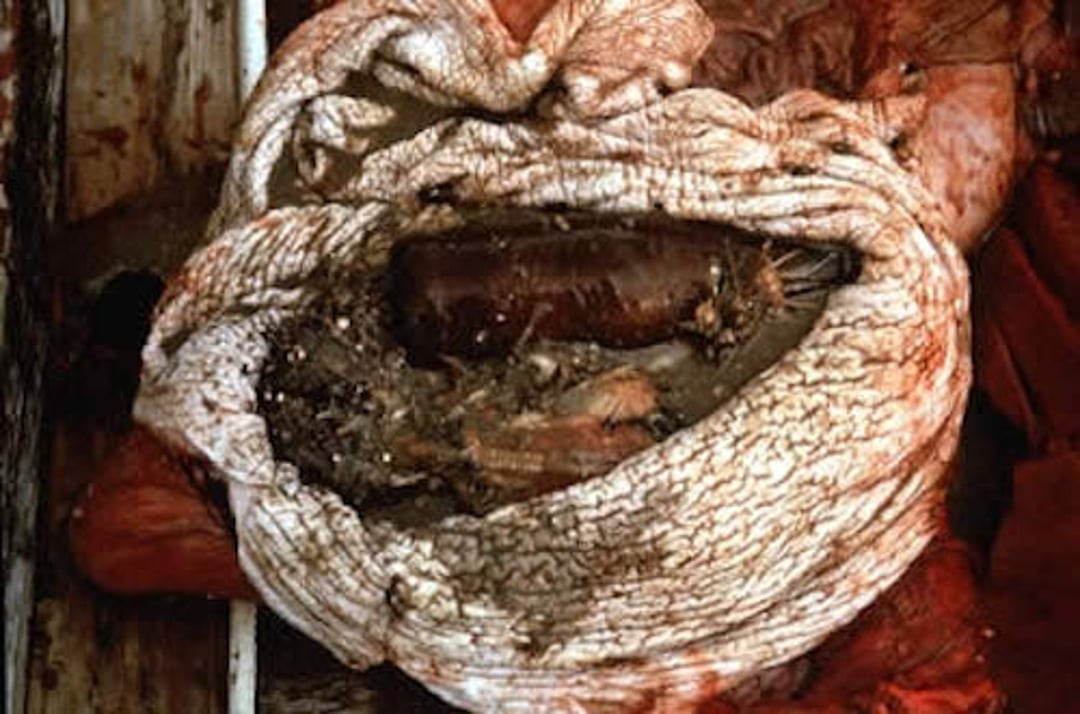
Foreign bodies, including a beer bottle, in a cetacean stomach.
Courtesy of Dr. James McBain.
Many captive marine mammals develop the habit of swallowing objects dropped into their pools. In cetaceans, the opening to the second compartment of the stomach is small, and foreign objects remain in the first compartment. In pinnipeds, the small pylorus prevents passage of most foreign bodies. Frequently, no clinical signs are evident. On occasion, anorexia, regurgitation, or lethargy may be seen. Diagnosis is often made by having witnessed the animal swallow an object. Smaller animals can be radiographed; in small cetaceans, the esophagus can be palpated to establish the presence of foreign bodies.
Animals occasionally regurgitate foreign bodies; however, assisted removal is usually indicated. Removal is usually best performed by gastroscopy, which is also used as a method of diagnostic confirmation. All efforts should be made to prevent ingestion of foreign bodies. Training animals to retrieve for reward as a displacement to swallowing foreign objects is thought to be beneficial. Foreign bodies are also found in free-ranging marine mammals. Some items, such as small rocks, may be incidental and not problematic; however, ingestion of marine debris such as plastic bags can cause severe gastric impaction and death.
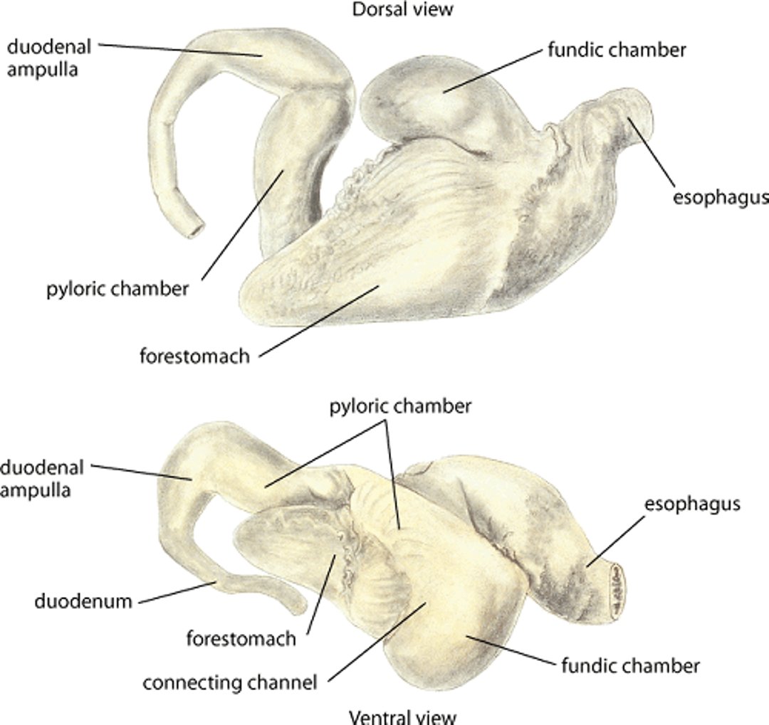
Stomach, dolphin
Diagram of stomach, dolphin. Top) Dorsal view. Bottom) Ventral view. Illustration by Dr. Gheorghe Constantinescu. Drawn, with permission, from slides courtesy of Dr. Raymond Tarpley, Texas A & M University. |

Gastrointestinal Ulcers of Marine Mammals
Gastrointestinal ulcers are a notable problem in both captive and free-ranging marine mammals. Ulcers of the esophagus and first compartment of the cetacean stomach are a common finding and pose less severe clinical problems than do ulcers of the pyloric region or proximal duodenum. Gastric and proximal duodenal ulcers in pinnipeds frequently progress to perforation, which results in peritonitis and subsequent death. Gastric ulcers also are found in sirenians, sea otters, and polar bears. Although ulcers in cetaceans perforate less frequently than in pinnipeds, they should be treated as a serious problem. Various causes, including parasitic damage and increased histamine content of spoiled fish, may be involved in the cause of a GI ulcer, but the disease in captive animals should be considered associated with environmental or stress-related conditions. Dramatic environmental changes, including changes of personnel or companion animals, can precipitate serious GI ulceration in cetaceans, pinnipeds, and sea otters.
Clinical signs include lethargy, partial anorexia, abdominal splinting, pallor, and occasionally regurgitation. Animals with bleeding ulcers often develop anemia and possibly leukocytosis (or leukopenia with sepsis). Diagnosis generally is based on identification of mammalian RBCs and abnormal mucosal cells, such as basal cells, in gastric washes; confirmation is by endoscopic visualization of the lesions.
Palliative treatment of nonperforating ulcers usually consists of administration of histamine blockers and alumina gel–based antacids with or without sucralfate and simethicone, along with frequent small meals. The underlying cause must be identified and corrected for successful resolution. Management of perforating ulcers with resulting peritonitis includes intensive broad-spectrum antimicrobial and fluid therapy. Stress-induced GI ulcers are more likely to develop in marine mammals that have previously had an ulcer.
Trauma of Marine Mammals

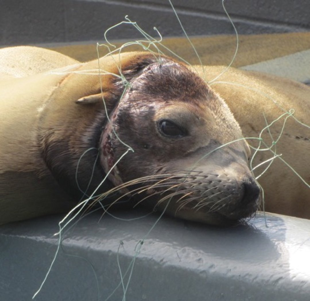
Sea lion with trauma due to fishing line entanglement.
Courtesy of Dr. Cara Field.

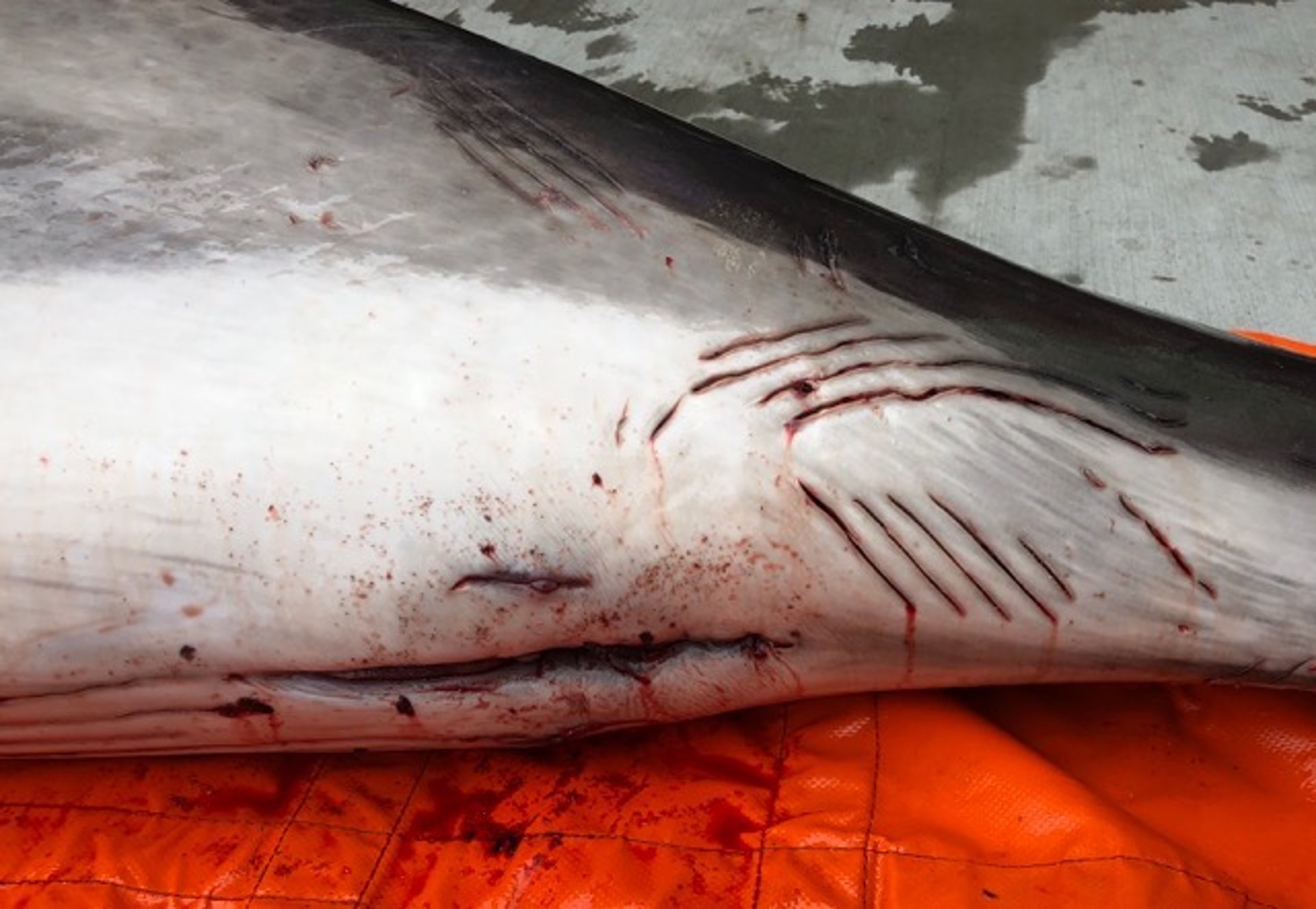
Female porpoise with numerous rake marks around the anogenital region.
Courtesy of Dr. Cara Field.
Traumatic lesions (eg, cuts, wounds from gunshots or propeller blades, and entanglements) are common in marine mammals. Propeller injuries and blunt force trauma are a major problem in manatees, which commonly enter heavily navigated recreational waters in Florida. Conspecific and interspecific trauma are particularly common in free-ranging animals, including bites and blunt force, and may result in myopathy and infection. Odontocete bites generally appear as parallel linear lacerations (rake marks) and are usually more superficial, whereas pinniped and sea otter bite wounds can yield more severe trauma and abscess formation following deep puncture. Male sea otters can cause substantial nasal trauma to females during mating.
Traumatic wounds should be cleaned, debrided, and generally allowed to heal as open wounds unless body cavities are breached. Antimicrobials often should be administered during convalescence to prevent gross infection. Maintenance of good water quality and a high plane of nutrition are beneficial to the healing process. Large wounds frequently heal uneventfully. More severe trauma, such as fractures, may require surgical intervention.
Oil Exposure of Marine Mammals
Exposure of marine mammals to spills of petroleum hydrocarbons is a major concern. Sea otters are particularly susceptible to such exposure because of their natural grooming habits and their lack of an insulating blubber layer. Fur seals are similarly vulnerable but also have blubber to protect against severe hypothermia, and young animals of any species are also highly vulnerable. Hepatotoxicity; renal toxicity; GI, mucosal and ocular damage; and loss of homeothermic ability are important effects of exposure to petroleum hydrocarbons. However, the most devastating effects are usually due to direct pulmonary damage from inhalation of volatile hydrocarbons.
Despite experimental evidence suggesting cetaceans, pinnipeds, and polar bears— unlike sea otters—avoid petroleum spills if possible, these species can be greatly affected. Ingestion of large quantities of oil by these species is less likely except through affected prey, and in pinnipeds and polar bears, through grooming. Baleen fouling occurs in mysticete whales but usually resolves within 24–36 hours. Petroleum ingestion in sirenians has not been reported but could result in dysbiosis as they are hindgut fermenters with a long GI transit time. Moderate to severe pulmonary damage has been documented in free-ranging cetaceans for years after a massive oil spill, in additional to chronic adrenal insufficiency and increased perinatal mortality. Efforts to reduce human exposure to hydrocarbons while dealing with oil-contaminated animals is a top priority, and proper training prior to handling oiled marine mammals is strongly recommended.
Capture, transport, and holding stresses lower the threshold of hydrocarbon toxicity in these animals. Priority treatment of exposed animals is stabilization prior to washing, including hydration, correction of life-threatening electrolyte imbalances including hypoglycemia, correction of hypothermia or hyperthermia, and potential nutritional compromise and other emergency care. Further treatment generally includes removal of oil from both the hair and skin using mild detergents (eg, 2% liquid dishwashing detergent) and the GI system if possible. Activated charcoal is not effective for oil itself but may be effective in binding other potentially dangerous chemicals, such as heavy metals, bound to petroleum products.
Washing oiled marine mammals requires large volumes of warm, soft (3–5 grains of hardness) water, especially for animals with a dense fur coat (eg, sea otters and fur seals). Weathered oil and tar may be pretreated with warmed methyl soyate or vegetable oil worked vigorously into fur, followed by one or more washes with detergent and thorough rinsing and drying. Full recovery and waterproofing will occur as the animal resumes normal grooming behavior, and animals should be gradually reintroduced to water with regular monitoring of recovery. Temporary housing in soft water after washing can reduce recovery time.
Pneumonia of Marine Mammals
Pneumonia is a common cause of morbidity and death in both captive and free-ranging marine mammals. Most cases of marine mammal pneumonia have substantial bacterial, fungal, or viral involvement, and most organisms cultured from terrestrial species have been identified in marine mammals. Pneumonia often can be the result of errors in management, although pneumonia-associated death is common even in carefully managed captive animals. Marine mammals require good air quality, including high rates of air exchange at the water surface in indoor facilities. Tempered air or acclimation to cold temperatures is also important to prevent lung disease, even in polar species. Animals acclimated to cold temperatures are usually quite hardy; however, sudden transition from warm environments to cold air, even with warmer water, can precipitate fulminating pneumonias, particularly in nutritionally or otherwise compromised animals.
Clinical signs include lethargy, anorexia, severe halitosis, dyspnea, pyrexia, and possibly marked leukocytosis. Subcutaneous emphysema has been noted in pinnipeds with pneumonia from different causes. The disease can progress rapidly.
Preliminary diagnosis is usually based on clinical signs and auscultation and confirmed by diagnostic techniques such as thoracic radiography and ultrasonography, in addition to response to therapy. Advanced imaging, bronchoscopy, and fine-needle aspirates can help evaluate the extent and establish the cause of disease.
Treatment consists of correction of environmental factors and appropriate intensive antimicrobial therapy and supportive care.


