Common Disorders
Generally, zoo animals are prone to similar suites of infectious and noninfectious diseases as found in domestic animals, and diagnostic and treatment plans are often translated from domestic animal and human medicine. There are certain diseases, however, that are either specific to zoo-housed species or that warrant additional consideration when managing the health of zoological collections.
Amphibian Chytrid Disease in Zoo Animals
Batrachochytrium dendrobatidis, amphibian chytrid disease or Bd, is a keratinophilic fungus that has been associated with the decrease in global amphibian populations to levels consistent with extinction (1). All amphibians are considered susceptible to Bd, with the ventral area being the most frequent entry point, as it is the area most often in contact with contaminated surfaces.
The affected skin in cases of Bd becomes hyperkeratotic, thus losing its osmoregulatory function, and eventually the animal dies from hyperkalemia. Bd can be successfully eradicated by treating positive individuals with itraconazole or chloramphenicol (2). An increase in ambient temperature helps disinfect contaminated enclosures, although caution should be exercised with heat-sensitive species.
Screening for Bd is recommended as part of pretransport testing and quarantine for amphibians and is performed using quantitative PCR assay on swabs taken from the ventral body and the plantar and palmar surfaces of the feet and toes. Positive samples should be retested and treated, and cases should be reported to the relevant authorities.
Batrachochytrium salamandrivorans, or Bsal, is an emerging infectious disease that has led to population declines of some wild urodeles (salamanders) in Europe. It causes erosion and ulceration of the affected skin. A PCR assay is available. Although, as of mid 2026, the disease has not been identified outside of Europe, it is recommended that zoo veterinarians begin screening for it during animal quarantine.
As of mid 2026, only tailed amphibians (newts and salamanders) have been affected by Bsal, and it is believed that Asian species can be nonclinically affected carriers of the fungus. Bd, Bsal, and ranavirus are diseases notifiable to WOAH (formerly OIE) under the Aquatic Animal Health Code.
Amyloidosis in Zoo Animals
Amyloidosis is disease caused by the deposition of amyloid in tissues. It can occur secondary to chronic inflammation (eg, in cheetahs) (3) or for reasons unknown; however, there is a suspected genetic component (eg, in black-footed cats) (4). It has also been associated with chronic pododermatitis in some bird species (5).
Avian Aspergillosis in Zoo Animals
Avian aspergillosis generally results in chronic respiratory tract disease. Affected birds can exhibit weight loss, marked leukocytosis, and, in later stages, dyspnea. Death can occur peracutely if there is a localized aspergilloma that occludes the trachea or invades a major blood vessel. Antemortem diagnostics, including serological testing, have poor sensitivity and specificity.
Diagnostic imaging (ie, radiographic and CT evaluation) can be used to identify lesions, even if treatment can prove frustrating.
Treatment is with systemic antifungals, such as itraconazole, voriconazole, and/or terbinafine, administered orally, parenterally, and by nebulization. Dose-dependent voriconazole toxicity has been documented in some penguin species (6). Risk factors include enclosures with insufficient ventilation and high humidity, which allow high fungal spore burdens, and increased crowding and other stressors that can immunosuppress susceptible hosts.
Postmortem examination generally reveals extensive fungal granulomas in the air sacs and lungs. Zoo species that are more susceptible to Aspergillus infections include penguins (Spheniscidae), birds of prey, pheasants (Phasianinae), certain passerines such as birds-of-paradise (Paradisaeidae), and some waterfowl. Prophylactic treatment with oral antifungals is sometimes used for susceptible species, especially penguins, during shipment, transport, and quarantine.
Avian Influenza and Newcastle Disease in Zoo Animals
Highly pathogenic avian influenza and virulent Newcastle disease are threats to zoo bird collections. Zoos should develop response plans and establish a dialogue with state and national agricultural regulatory agencies to mitigate the impact of these diseases on zoo collections. A great resource for zoo veterinarians is the Zoo and Aquarium All Hazards Preparedness, Response, and Recovery Fusion Center.
Elephant Epitheliotropic Herpesvirus in Zoo Animals
Elephant epitheliotropic herpesvirus (EEHV) is perhaps the most important infectious disease in elephants under human care. EEHV causes hemorrhagic disease (EEHV-HD) in elephants under 8 years old and is the leading cause of calf mortality in North America and Europe, a limiting factor for the sustainability of these captive populations. Clinical cases of EEHV-HD have been documented in both Asian elephants (Elephas maximus) and African elephants (Loxodonta africana).
EEHV is a double-stranded DNA beta-herpesvirus (family Proboscivirus) that is highly prevalent in healthy adult elephants. Nonclinically affected elephants shed the virus in respiratory, conjunctival, and vulvar secretions. All facilities that keep elephant calves or potentially reproductively active adults should develop a protocol for monitoring and treating EEHV-HD. Monitoring typically includes daily to weekly qPCR assay of whole blood and hematology.
Viremia precedes the onset of clinical signs of EEHV-HD, as do hematological changes, including leukopenia, monocytopenia, and thrombocytopenia. Clinical signs can include changes in sleep patterns, swelling of the temporal glands, changes in fecal output or consistency, lameness, scleral injection, colic, lethargy, changes in food and water intake, and rigid gait. These progress to clinical signs associated with severe vasculitis and loss of oxygen-carrying capacity of the cardiovascular system, including tachycardia, petechiae and ecchymoses, hemorrhage, lingual cyanosis, edema of the head and forelimbs, ascites, and pericardial effusion.
Treatment for EEHV-HD is generally fluid therapy and famciclovir administered rectally and/or orally. More severe cases might warrant IV plasma and/or crystalloids, opioids, NSAIDs, antimicrobials to address secondary bacterial infections, nasal oxygen insufflation, or corticosteroids to address disseminated intravascular coagulation. EEHV is the subject of ongoing research, and evidence-based recommendations for the management of EEHV-HD are continuously evolving. See The EEHV Advisory Group website for the most up-to-date guidelines.
Kidney Disease in Zoo Animals
Renal insufficiency and end-stage kidney disease are common in geriatric carnivores, especially felids and polar bears. In some felids, such as cheetahs, caracals (Caracal caracal), and black-footed cats (Felis nigripes), kidney disease can be secondary to other conditions such as amyloidosis. Clinical presentation and diagnosis of renal disease is similar to that in domestic carnivores. However, management is challenging because, in many cases, the species of the patient prohibits direct contact and dietary modification.
Koala Diseases in Zoo Animals
Koalas (Phascolarctos cinereus) are somewhat unique patients because of their highly specialized diet and disease susceptibilities. Chlamydia pecorum and Chlamydia pneumoniae are obligate intracellular pathogens especially important for koalas in both captive and wild populations. Infection with C pecorum has been associated with keratoconjunctivitis, urinary cystitis and chronic urinary tract disease (“wet bottom”), reproductive tract infections in males and females, and septic arthritis.
Transmission of Chlamydia is suspected to be both horizontal and vertical. Vaccination of females can decrease vertical transmission. The presence of nonclinically affected carriers can be high in some populations. Other differentials for respiratory disease in koalas include Bordetella bronchiseptica, Klebsiella pneumoniae, and Cryptococcus neoformans.
Koala retrovirus (KoRV) is a gammaretrovirus with positive RNA that has been isolated from both captive and wild koalas (7). Affected animals can remain nonclinically affected or develop disseminated lymphoid neoplasia (leukemia or lymphoma), although a direct correlation between KoRV and lymphoid neoplasia has not been demonstrated. It is believed that different KoRV subtypes can vary in pathogenicity, and KoRV-B is considered a virulent virus (8). Captive koala populations are classified as KoRV-B positive or KoRV-B negative subpopulations to determine their management until the transmission and pathogenesis of this virus are better understood.
Malignant Catarrhal Fever in Zoo Animals
Malignant catarrhal fever (MCF) is caused by a gammaherpesvirus (genus Macavirus) and is an important disease in zoological collections, because exposure to various host species can produce severe and fatal illness. Transmission occurs through ocular and oronasal secretions in which the virus is shed.
Sheep-associated MCF (ovine herpesvirus-2) causes mild illness in sheep; however, it is highly virulent in bison. Goat-associated MCF (caprine herpesvirus-2) has been associated with morbidity and mortality in captive moose (Alces alces). Wildebeest-associated MCF (alcelaphine herpesvirus-2) can cause severe disease in cattle, bison, deer, pigs, and other nondomesticated bovids (9).
Serological testing cannot distinguish between exposure to MCF associated with sheep or wildebeest (Connochaetes spp). Prevention is key, and reproductively active wildebeest should be housed in a part of the zoo that does not contact the perimeter (to minimize the risk of transmission to domestic livestock) or the enclosures of other ungulates.
Mycobacterial Disease in Zoo Animals
Nontuberculous mycobacteria (NTM) include two complexes, Mycobacterium avium and Mycobacterium marinum, that are important for to zoo practice.
Atypical mycobacteriosis is a chronic problem in many bird collections. This is an environmental issue rather than a classically infectious disease, meaning animals that share an enclosure are often exposed from the same source of infection in the environment rather than from an infected individual bird.
Crowding and stressors can increase risk of infection from the environment. Control measures to try to identify chronically infected birds are unreliable, and attempts to sanitize the environs can prove frustratingly inadequate because of the ubiquity of mycobacterial pathogens.
Avian species with noted susceptibility to mycobacteria include the sihek or Guam kingfisher (Todiramphus cinnamominus), the white-winged wood duck (Cairina scutulata), and the whooping and sandhill cranes (Grus americana and Grus canadensis), for which mycobacteriosis represents a major challenge to conservation (10, 11).
Mammals are also susceptible to mycobacteria infection, including primates, hoofstock, and marsupials. Tree kangaroos (Dendrolagus spp) are considered especially susceptible to infection, which often results in spinal arthropathy (12). Susceptible species are often not housed in mixed-species exhibits or where avian fecal matter or contaminated water sources can be present.
In birds, atypical mycobacteriosis often presents as bone lesions or caseated abscesses in the abdominal viscera, although it can present with lung lesions. Atypical mycobacteriosis can confound tuberculin skin testing in hoofstock or primates, so comparative testing should be considered, especially in orangutans (Pongo spp), because most captive specimens are reported to have a nonspecific tuberculin test response (13, 14).
Nontuberculous mycobacteria are also important pathogens of captive aquatic animals, especially fish and amphibians, because of their presence in biofilm and water. Clinical manifestations range from localized inflammation and nonhealing wounds to systemic abscesses and death. In frogs, classic lesions include ulcers on the skin and toes. Treatment is not successful, and infected animals chronically shed mycobacteria into the environment, so humane euthanasia is indicated. Prevention entails identifying and removing affected animals from the population and maintaining optimal water quality. Because these bacteria can be zoonotic, gloves should be worn when handling aquatic animals, and hands should be washed often and thoroughly.
Pearls & Pitfalls
|
The Mycobacterium tuberculosis complex includes M bovis, M microti, and M pinnipedi, among others. It is most important for mammals and includes the causative agent of human tuberculosis. Elephants are especially susceptible to respiratory infections by the Mycobacterium tuberculosis complex.
Mammalian tuberculosis is most often found on routine screening rather than as clinical cases in primates, hoofstock, and elephants. Tuberculin testing of hoofstock in the US is regulated by the USDA, with Category II licensure required to perform testing. In elephants, the USDA has established testing and treatment protocols in accordance with zoo veterinary stakeholder input. Primates are often routinely tested. However, unless they are part of a CDC-regulated import, no regulatory requirements exist.
Intradermal tuberculin testing is the basis of testing for hoofstock of the families Bovidae and Giraffidae and for nonhuman primates. Serological and other immunological tests exist and are especially important in deer (Cervidae); however, they are not validated for all susceptible species. Recommended testing protocols for cervids and other taxa change frequently, so consultation with a regulatory veterinarian is advised prior to testing cervids, especially those intended for shipment.
A potential positive intradermal tuberculin test should be followed up with subsequent testing, whether via a comparative cervical test in hoofstock performed by a regulatory veterinarian or additional diagnostics, such as imaging or gastric and bronchial lavage for cytology and culture in primates.
Tuberculosis in elephants is typically a respiratory infection and requires close and prolonged contact to be transmitted between individuals. Clinical signs are nonspecific and usually present only in advanced cases. They can include chronic weight loss, anorexia, weakness, exercise intolerance, discharge from the trunk, cough, and dyspnea. Diagnosis can be by direct techniques, such as culture (the gold standard), quantitative PCR assay, or Ziehl-Neelsen staining of trunk wash samples, or by indirect techniques, such as serological testing (eg, multiantigen print immunoassay [MAPIA], dual-pathway platform [DPP]).
Tests that measure cell-mediated immunity (eg, interferon gamma) are not readily available for use in elephants. Indirect tests should be confirmed with direct tests. A positive result on a direct test must be confirmed by repeating the test on serial samples and requires increased surveillance of the remaining animals in the group and notification to the relevant authorities.
Therapeutic protocols have been established that involve the use of several antimicrobials, and many antituberculin drugs have documented pharmacokinetics in both healthy and infected animals to guide therapy (15, 16, 17, 18). The goal of treatment is to prevent shedding by maintaining adequate serum antimicrobial concentrations for extended periods while avoiding potential adverse effects. Elimination of the infection is not realistic. Because of the potential for zoonotic transmission, extensive personal protective equipment is required to manage a tuberculosis-positive elephant.
Nondomestic ruminants are susceptible to paratuberculosis, or Johne's disease, caused by Mycobacterium avium paratuberculosis (MAP). The main route of infection is fecal-oral, although intrauterine and lactational transmission can occur.
MAP is highly resistant to extreme temperatures and desiccation. However, as an obligate intracellular parasite, it does not replicate in the environment. Infection in zoo animals is attributed to interspecific transmission from domestic livestock. Infection has also been documented in captive and wild camelids, rock hyraxes (Procavia capensis), and rabbits (Oryctolagus cuniculus) (19). Diagnosis is by culture or PCR assay. Serological testing using ELISA has only been validated in cattle, sheep, goats, and deer. There is no treatment, and affected animals should be culled to limit environmental contamination.
Oral Necrobacillosis in Zoo Animals
Oral necrobacillosis (lumpy jaw) is a subgingival infection of the teeth with inflammation, maxillary or mandibular osteomyelitis, and bone remodeling, often associated with infection with Fusobacterium necrophorumand other anaerobic bacteria. It is a common problem of small ruminants and macropods (wallabies and kangaroos).
Predisposing factors for oral necrobacillosis include low host resistance related to concomitant disease or immunosuppression, disruption of the gingival mucosa by hard or abrasive forage, decreased chewing (generally related to diets low in fiber and minerals), and oral or dental trauma affecting molar progression. Clinical signs range from mild to very severe and can include hyporexia, dysphagia, halitosis, ptyalism, nasal discharge, and/or localized facial swelling. Treatment involves surgical debridement of necrotic bone and soft tissue, tooth extraction (if indicated by imaging such as radiographic or CT evaluation) and repeated irrigation with antiseptic solutions under general anesthesia, and administration of systemic antimicrobials and analgesics.
Pinniped Ocular Disease in Zoo Animals
Pinnipeds (seals, sea lions, and walruses) and cetaceans (whales and dolphins) under human care are especially susceptible to ocular disease. Corneal problems are most frequent, including keratopathy and ulceration. Risk factors include clear tanks that reflect light, lack of shade, low salinity, and high UV index. Clinicians managing these marine mammals are advised to include routine eye examinations, preferably in collaboration with an ophthalmologist, in their preventive-medicine protocols. Training by operant conditioning to encourage animals to voluntarily allow eye examinations and the administration of topical eye medication can greatly facilitate care.
Pododermatitis in Zoo Animals
Pododermatitis (bumblefoot) is a common disorder of birds. It can be either unilateral or bilateral and is characterized by lameness, inflammation, and swelling of the footpad related to localized bacterial infection. Sequelae of infection can include septicemia or amyloidosis. It can occur secondary to injury, inappropriate substrate, obesity, or unilateral limb problems (trauma, arthritis) that result in excess and abnormal weight bearing on the contralateral foot.
Penguins (Spheniscidae) and flamingos (Phoenicopteridae) often present with pododermatitis (see ). The most important part of treatment is management of the underlying medical or husbandry issue. Treatment of the lesion itself can involve local or systemic antimicrobials, regional limb perfusion, anti-inflammatories and/or analgesics, therapeutic bandaging, or surgical debridement.

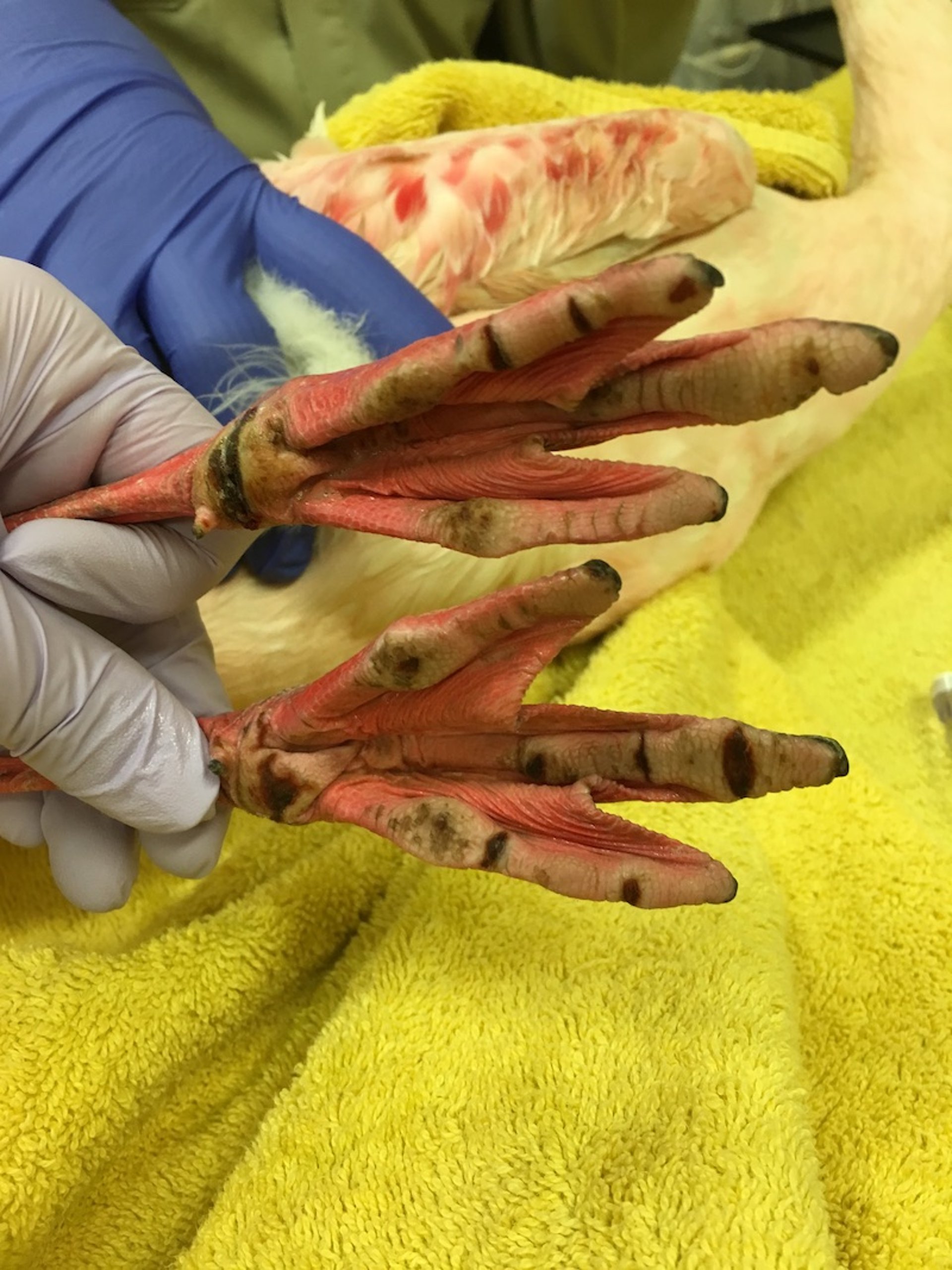
A greater flamingo with pododermatitis. Notice the fissures and bumble lesions that develop when an animal is kept on an inappropriate substrate.
Courtesy of Dr. Meredith M. Clancy.
Toxoplasmosis in Zoo Animals
Many zoo animals are susceptible to toxoplasmosis, including the red panda (Ailurus fulgens), marine carnivores (such as sea otters, sea lions, and other pinnipeds), nonhuman primates (especially squirrel monkeys, Saimiri spp), and marsupials (classically, macropods).
Although cats are definitive hosts of toxoplasmosis that rarely develop disease, the Pallas's cat (Otocolobus manul) is a notable exception, with mortality rates as high as 100% in newborns (20). Excluding stray cats from zoo grounds and placing the enclosures of susceptible species away from those of cats (and on higher ground to avoid environmental contamination from water runoff) can help prevent the spread of Toxoplasma gondii within a collection.
Procedures Performed on Zoo Animals
Dentistry in Zoo Animals
Dentistry in zoo animals presents unique challenges. In particular, dental anatomy differs widely among zoo species and must be researched prior to attempting any dental procedure.
The roots of canine teeth in primates and carnivores are more extensive than the exposed crown. Simple traction and rotation cannot remove such teeth intact, and the use of a dental elevator for extraction is essential. Endodontic therapy (eg, root canals) is indicated when a large canine tooth is fractured and viable pulp exposure occurs. Specialized dental instruments are required to remove the nerve tissue from these elongated canals. Tusks, elongated canines, or incisors, present in many zoo species, require additional specialized instrumentation if extraction is required.
Elephants, macropods and manatees exhibit molar progression (in which molar teeth move sequentially anteriorly along the jaw to replace worn teeth), which presents unique considerations to dental management.
Rodents have elodont, or continuously growing incisor teeth. However, they might have rooted (brachydont) or rootless (elodont) premolars and molars, depending on the species. These animals must be supplied with coarse feed or logs to gnaw on to prevent their incisors from growing excessively and interfering with their ability to feed.
Periodontal disease in zoo animals is treated by routine prophylaxis, including scaling, polishing, and imaging (ie, radiographic evaluation) under general anesthesia.
Geriatric Specimens in Zoos
Because of ongoing improvements in husbandry and health management, many zoo animals live to advanced ages. Care of geriatric specimens is increasingly common, with emphasis on the optimization and routine evaluation of quality of life. Age-related or degenerative disorders, including joint disease, neoplasia, heart disease, and endocrine disorders, require specialized management in zoo species, with therapeutic modalities coming from both the domestic animal and human medicine realms that can be successfully applied to the care of zoo animals.
A holistic approach, including multimodal analgesia along with dietary and husbandry modifications, is recommended when managing chronic degenerative diseases. Quality-of-life assessments, whether qualitative or quantitative, should be performed on geriatric patients or those with chronic diseases or morbidities to ensure that zoo animals are thriving under human care. Once the decision is made that quality of life is compromised, humane euthanasia should be performed following taxon-specific AVMA guidelines.
Hoof and Nail Trims in Zoo Animals
Hoof and nail trims are part of routine care of many zoo species. The skills of an equine farrier can be useful in management of hoof-related issues, and establishment of routine trimming schedules is recommended to avoid excessive overgrowth. Foot care is especially important for megavertebrates such as elephants and giraffes to prevent chronic musculoskeletal problems. Foot and hoof care can usually be accomplished in conscious (nonsedated) elephants and giraffes by training (behavioral restraint) alone. Many other species require chemical immobilization for foot care.
Both artiodactyls and perissodactyls are susceptible to laminitis, inflammation of the laminae that contain the dermoepidermal junctions between the hoof wall and the third phalanx bone. This disease is often caused by impaired blood flow within the laminae and can be secondary to rumen acidosis, endotoxemia from an infection elsewhere in the body, or hoof or limb trauma.
Treatment of laminitis is similar to that for domestic animals and can include antimicrobials, anti-inflammatories, and corrective hoof trimming. Chronic laminitis can affect the animal's quality of life, so humane euthanasia might be indicated for severely affected individuals. Prevention includes routine hoof care and maintenance hoof trimming, good overall health management, using soft substrates such as sand or mats, removing substrates made up of large particles such as rocks or pebbles, and eliminating easily digestible carbohydrates from the diet.
Other problems affecting the hooves of ungulates include interdigital dermatitis and the more severe infectious pododermatitis (foot rot). The latter is commonly associated with anaerobic bacteria (eg, Fusobacterium necrophorum, Dichelobacter nodosus, Trueperella pyogenes). Improvement can be achieved by providing dry pasture, use of corrective hoof trimming, therapeutic foot baths, and systemic antimicrobials and anti-inflammatories. Nondomestic ungulates are also susceptible to dermatophilosis caused by Dermatophilus congolensis and to digital papillomatous dermatitis associated with infection by spirochetes (Treponema spp).
Permanent Prevention of Flight in Zoo Animals
Permanent prevention of flight in birds is accomplished by amputating one wing just distal to the radiocarpal joint (pinion) or, less commonly, by performing a tenectomy and fusing the radiocarpal joint. Pinioning of young birds soon after hatching is easier and more successful, and this method is widely used in the US, although new regulations on deflighting techniques in the EU might decrease its use outside the US (21). Newer technologies of ablating the feather follicle using laser or other methods are gaining acceptance, although techniques and success rates vary (21).
For More Information
AVMA Guidelines for the Euthanasia of Animals: 2020 Edition. American Veterinary Medical Association.
References
Voyles J, Rosenblum EB, Berger L. Interactions between Batrachochytrium dendrobatidis and its amphibian hosts: a review of pathogenesis and immunity. Microbes and Infection. 2011;13(1):25-32. doi:10.1016/j.micinf.2010.09.015
Baitchman EJ, Pessier AP. Pathogenesis, diagnosis, and treatment of amphibian chytridiomycosis. Vet Clin North Am Exot Anim Pract. 2013;16(3):669-685. doi:10.1016/j.cvex.2013.05.009
Papendick RE, Munson L, O'Brien TD, Johnson KH. Systemic AA amyloidosis in captive cheetahs (Acinonyx jubatus). Vet Pathol. 1997;34(6):549-556. doi:10.1177/030098589703400602
Terio KA, O'Brien T, Lamberski N, Famula TR, Munson L. Amyloidosis in black-footed cats (Felis nigripes). Vet Pathol. 2008;45(3):393-400. doi:10.1354/vp.45-3-393
Kim N-H, Park J-H, Hong W-H, Jung J-Y, Kim J-H. Systemic amyloidosis in an African penguin (Spheniscus demersus) with bumblefoot. J Vet Clin. 2022;39(2):81-86. doi:10.17555/jvc.2022.39.2.81
Hyatt MW, Georoff TA, Nollens HH, et al. Voriconazole toxicity in multiple penguin species. J Zoo Wildl Med. 2015;46(4):880-888. doi:10.1638/2015-0128.1
Denner J, Young PR. Koala retroviruses: characterization and impact on the life of koalas. Retrovirology. 2013;10:108. doi:10.1186/1742-4690-10-108
Kayesh MEH, Hashem MA, Tsukiyama-Kohara K. Koala retrovirus epidemiology, transmission mode, pathogenesis, and host immune response in koalas (Phascolarctos cinereus): a review. Arch Virol. 2020;165(11):2409-2417. doi:10.1007/s00705-020-04770-9
Li H, Cunha CW, Taus NS, Knowles DP. Malignant catarrhal fever: inching toward understanding. Annu Rev Anim Biosci. 2014;2:209-233. doi:10.1146/annurev-animal-022513-114156
Schrenzel M, Nicolas M, Witte C, et al. Molecular epidemiology of Mycobacterium avium subsp. avium and Mycobacterium intracellulare in captive birds. Vet Microbiol. 2008;126(1-3):122-131. doi:10.1016/j.vetmic.2007.06.016
Riggs G. Avian mycobacterial disease. In Miller RE, Fowler ME, eds. Fowler's Zoo and Wild Animal Medicine: Current Therapy. Volume 7. Elsevier Saunders; 2012:266-274. doi:10.1016/b978-1-4377-1986-4.00035-4
Kennedy S, Montali RJ, James Jr AE, Bush M. Bone lesions in three tree kangaroos. J Am Vet Med Assoc. 1978;173(9):1094-1098. doi:10.2460/javma.1978.173.09.1094
Philippa J, Dench R. Infectious diseases of orangutans in their home ranges and in zoos. In Miller RE, Lamberski N, Calle P, eds. Fowler's Zoo and Wild Animal Medicine Current Therapy. Volume 9. WB Saunders; 2019:565-573. doi:10.1016/B978-0-323-55228-8.00080-1
Calle PP, Thoen CO, Roskop ML. Tuberculin skin test responses, mycobacteriologic examinations of gatric lavage, and serum enzyme-linked immunosorbent assays in orangutans (Pongo pygmaeus). J Zoo Wildl Med. 1989;20:307-314.
Brock AP, Isaza R, Egelund EF, Hunter RP, Peloquin CA. The pharmacokinetics of a single oral or rectal dose of concurrently administered isoniazid, rifampin, pyrazinamide, and ethambutol in Asian elephants (Elephas maximus). J Vet Pharmacol Therap. 2014;37(5):472-479. doi:10.1111/jvp.12119
Auclair B, Mikota SK, Peloquin CA, Aguilar R, Maslow JN. Population pharmacokinetics of antituberculous drugs and treatment of Mycobacterium bovis infection in bongo antelope (Tragelaphus eurycerus isaaci). J Zoo Wildl Med. 2002;33(3):193-203. doi:10.1638/1042-7260(2002)033[0193:ppoada]2.0.co;2
Backues KA, Wiedner EB. Recommendations for the diagnosis, treatment, and management of tuberculosis, Mycobacterium tuberculosis, in elephants in human care. Int Zoo Yearb. 2019;53(1):116-127. doi:10.1111/izy.12221
Buur J, Saggese MD. Taking a rational approach in the treatment of avian mycobacteriosis. Vet Clin North Am Exot Anim Pract. 2012;15(1):57-70. doi:10.1016/j.cvex.2011.12.001
Manning EJB, Sleeman JM. Johne's disease and free-ranging wildlife. In Miller RE, Fowler ME, eds. Fowler's Zoo and Wild Animal Medicine: Current Therapy. Volume 7. Elsevier Saunders; 2012:628-635. doi10.1016/b978-1-4377-1986-4.00081-0
Riemann HP, Fowler ME, Schulz T, et al. Toxoplasmosis in Pallas's cats. J Wildl Dis. 1974;10(4):471-477. doi:10.7589/0090-3558-10.4.471
Bennett RA, Baumgartner K. Avian deflighting techniques. In Miller RE, Fowler MD, eds. Fowler's Zoo and Wild Animal Medicine. Volume 8. Miller RE, Fowler ME (eds). Saunders; 2015:650-660. doi:10.1016/b978-1-4557-7397-8.00065-7