

The most frequent ophthalmic neoplasms in cattle are the squamous cell carcinoma (SCC) complex and the orbital infiltration associated with lymphosarcoma. The latter, with extensive invasion of the orbital structures, results in progressive bilateral exophthalmos, reduced ocular mobility, exposure keratitis, and corneal ulcerations that can lead to perforation.

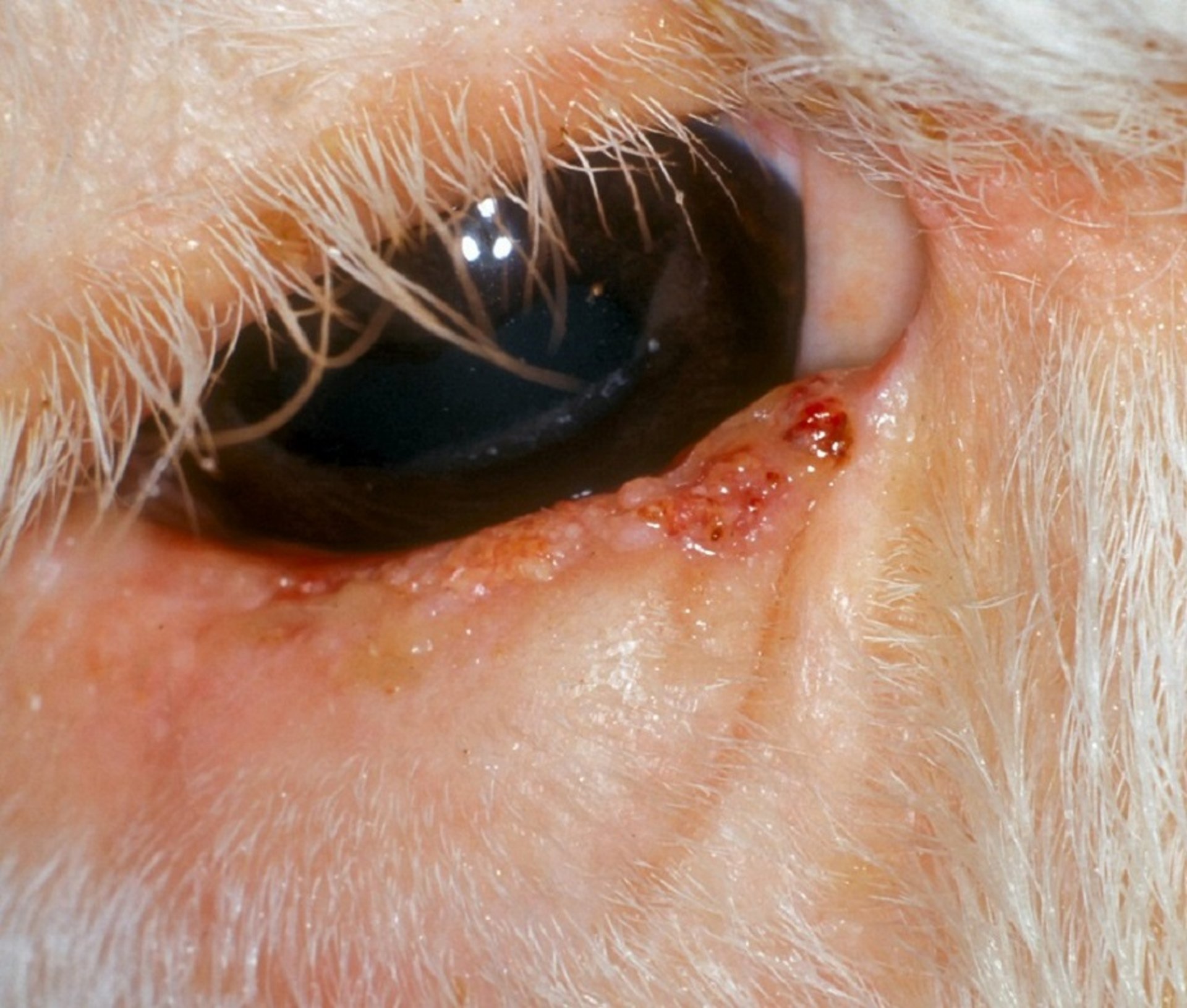
Photograph of the eye of a cow with SCC of the eyelid. The lower eyelid margin has proliferative and erosive lesions along most of the margin.
Courtesy of K. Gelatt.
Ocular SCC is the most common neoplasm of cattle. It results in appreciable economic loss because of condemnation at slaughter and a shortened productive life. It occurs more frequently in Bos taurus breeds than in Bos indicus breeds, and it occurs most often in Herefords, less often in Simmentals and Holstein-Friesians, and rarely in other breeds. The peak age of onset is 8 years; tumors are uncommon in cattle < 5 years old and rare in cattle < 3 years old.1Among herds, the observed incidence varies from 0.8% to 5.0% of animals at risk per year1; however, true incidence may be challenging to determine because of the popularity of Herefords and Hereford crosses that have decreased ocular and periocular pigmentation.
Ocular SCC is caused by multiple factors, including heritability, sunlight, nutrition, eyelid pigmentation, and possibly viral involvement. Both age and lack of pigmentation have been documented as risk factors in the development of ocular SCC. Eyelid and conjunctival pigmentation is highly heritable and may be associated with a reduced frequency of eyelid SCCs; however, pigmentation has limited effect on the development of tumors of the conjunctiva and nictitating membrane. Ultraviolet radiation and a high plane of nutrition are also contributing factors. The medial and lateral limbal regions (corneoscleral junction) are affected most frequently; the eyelids, conjunctivae, and nictitating membrane, however, may also be affected. Bilateral involvement varies but occurs in as many as 35% of cases. The cancerous or precancerous lesions are bilateral or multiple in the same eye in ~28% of cases.1 The viruses of infectious bovine rhinotracheitis (bovine herpesvirus 1) and papilloma (bovine papillomaviruses) have been isolated from the neoplasms; their importance is unknown, however.

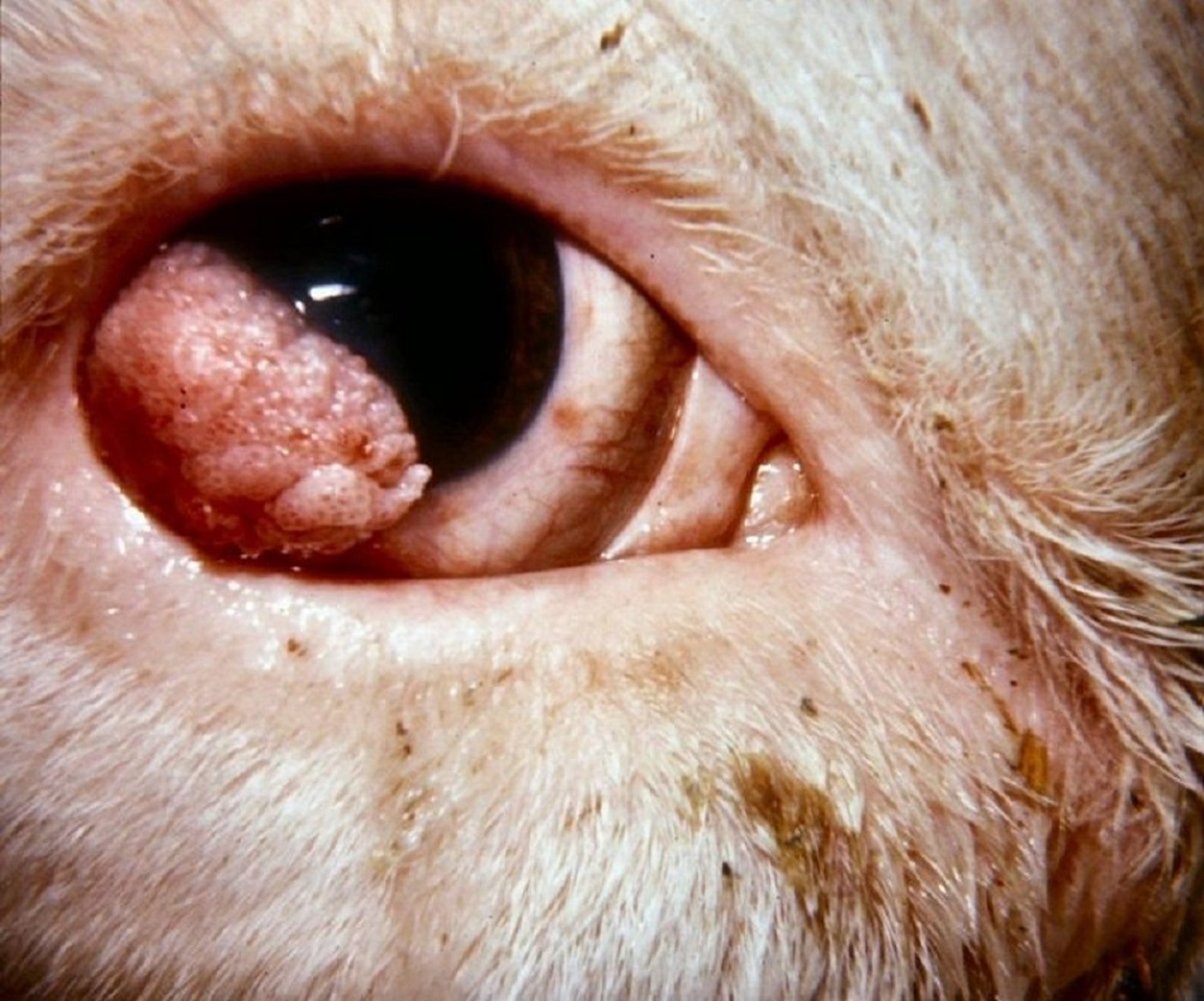
Photograph of the eye of a cow with corneoconjunctival SCC. A firm, pink, proliferative lesion is present in the ventrolateral sclera and cornea.
Courtesy of K. Gelatt.
Corneoconjunctival lesions in cattle usually start at the limbal sclera and grow across the cornea. Typically, they begin as benign, smooth, white plaques on the conjunctival surfaces; they may progress to a papilloma and then to a SCC or go directly to the malignant stage. Lid lesions usually begin as either an ulcerative or a hyperkeratotic lesion (cutaneous horn). While in this benign stage, ~30% may spontaneously regress.1 The tumor may become quite large without invading the globe; however, invasion into the eye and orbit as well as metastasis to parotid and submandibular lymph nodes occur in late stages of the disease. Diagnosis usually is made by the typical clinical appearance; however, it should be confirmed by biopsy and histologic examination. The intraocular tumor invasion must be differentiated from severely damaged and disorganized eyes after trauma or advanced infectious keratoconjunctivitis.
Before treatment, a thorough ocular, periocular and systemic evaluation should be performed in an attempt to determine whether the disease is local, regional, or systemic. Squamous cell carcinomas may respond to wide surgical excision, cryotherapy, hyperthermia, radiation therapy, local chemotherapy, immunotherapy, or often a combination of these therapies. Treatment costs, as well as potential slaughter withdrawal times for any chemotherapy, must be considered. The best success rates are found with treatment of smaller lesions and with a combination of surgical excision and some form of adjunctive treatment. Surgical excision is indicated for small lesions or to debulk larger lesions before cryotherapy, beta-irradiation, hyperthermia, or other adjunctive treatment. Superficial keratectomy can be used to excise limbal plaques, papillomas, and SCCs. After superficial keratectomy and tumor removal, cryotherapy, hyperthermia, or a permanent bulbar conjunctival graft have yielded excellent short-term results; however, the rate of recurrence at the same site is 3%–25%, depending on lesion location and type of adjunctive therapy. Regardless of initial location and treatment, all patients should be monitored carefully after surgery for any signs of recurrence or new lesions in another site.
For advanced lesions confined to the globe, enucleation is recommended. When adjacent tissues are affected, removal of the globe and all orbital contents (exenteration) should be performed. Immunotherapy is still experimental, and the resulting tumor regression may be temporary. Photodynamic therapy and radiation therapy are not readily available, but they are options for valuable animals.
Owners of problem herds should be advised of the heritability factor, with affected animals and their offspring culled to decrease the incidence of tumors. Active breeding bulls with ocular SCC should be culled.
References
Pearce JW, Moore CP. Food Animal Ophthalmology, pp: 1610–1674; In: Veterinary Ophthalmology, 5th Edition; Editors: Gelatt KN, Gilger BC, Kern TJ; 2013; John Wiley & Sons, Inc; Oxford, UK.


