

Ophthalmic manifestations can occur with inherited, infectious, degenerative, parasitic, toxic, nutritional and neoplastic disorders in animals. Often, ophthalmic examinations can assist in timely identification of the systemic disorder. Diseases affecting the vascular and nervous systems may also show ocular manifestations. Animals with bilateral intraocular disease should be carefully evaluated for systemic diseases.

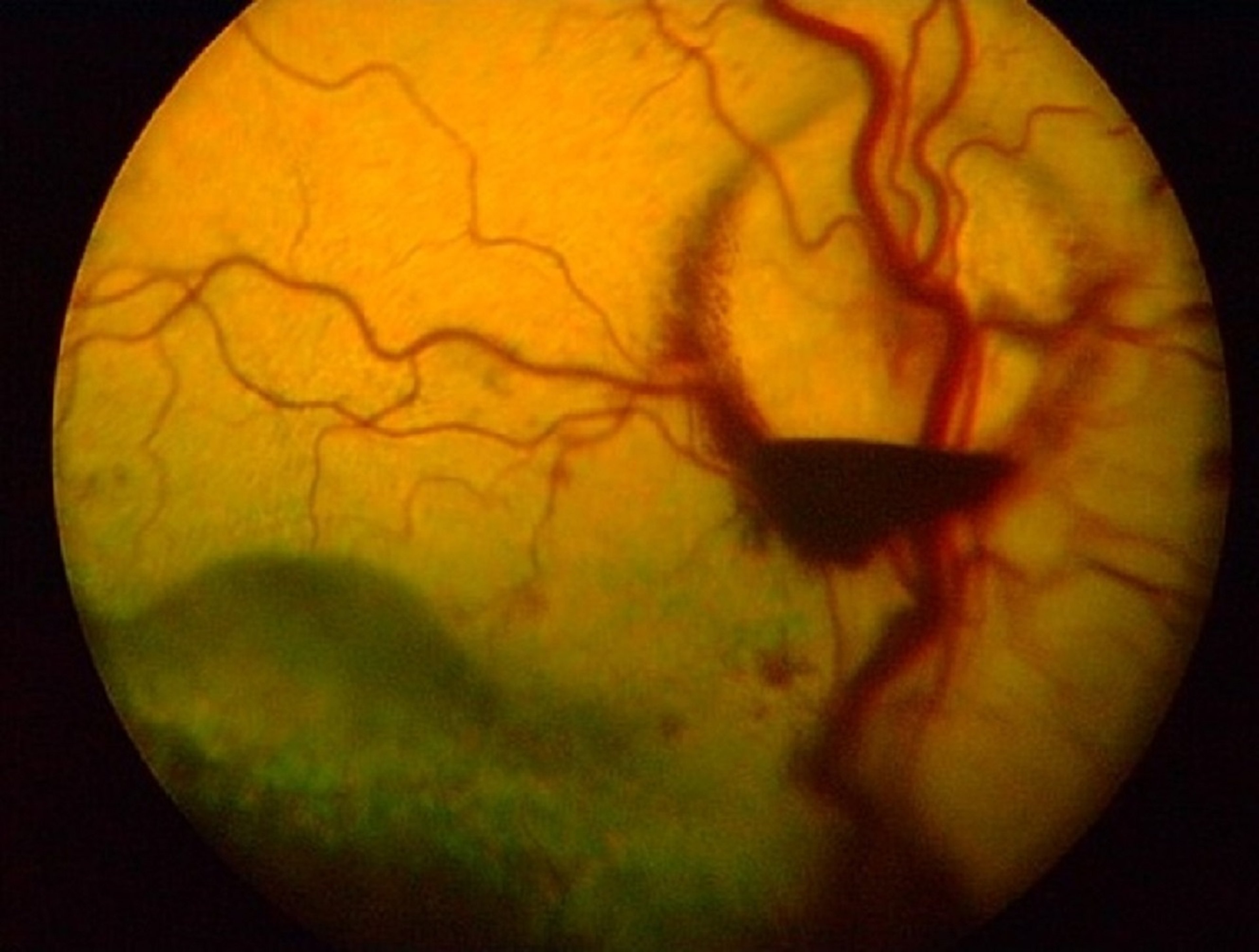
Retinal hemorrhages and partial serous retinal detachment are evident in this photograph showing the eye of a dog that has systemic hypertension.
Courtesy of Dr. Ralph Hamor.
In dogs, ophthalmic diseases, such as retinal dysplasia, microphthalmia, and cataracts, have been associated with dwarfism, albinism, and merle coloration. Infectious diseases often involve the uveal tract and present as iridocyclitis, choroiditis, and panuveitis. They may be caused by viruses (distemper, infectious hepatitis), rickettsial diseases (ehrlichiosis and Rocky Mountain spotted fever), bacteria (Brucella canis and Borrelia burgdorferi), fungi (Blastomyces, Coccidioides, Histoplasma, Cryptococcus, and Aspergillus), protozoa (Toxoplasma, Neospora, Leishmania, and Hepatozoon), algae (Prototheca), or parasites (Dirofilaria, Toxocara, and Diptera species). Metabolic diseases associated with eye diseases in the dog include diabetes mellitus (cataract formation), hypocalcemia (cataracts), hyperadrenocorticism (corneal disease, cataracts, and lipemia retinalis), and hypothyroidism (keratoconjunctivitis sicca [KCS], intraocular hemorrhages from increased systemic blood pressure, and lipemia retinalis [hyperlipidemia]). Blood and vascular disorders may present as intraocular hemorrhage, retinal detachment, secondary glaucoma, and papilledema. Metastatic neoplasms, such as lymphosarcoma, can present as persistent uveitis, overt intraocular masses, intraocular hemorrhage, secondary glaucoma, and/or retinal detachment. Systemic hypertension often results in retinal hemorrhage and/or serous retinal detachments. With appropriate diagnosis and treatment, these retinal lesions can resolve and vision can be retained or returned.

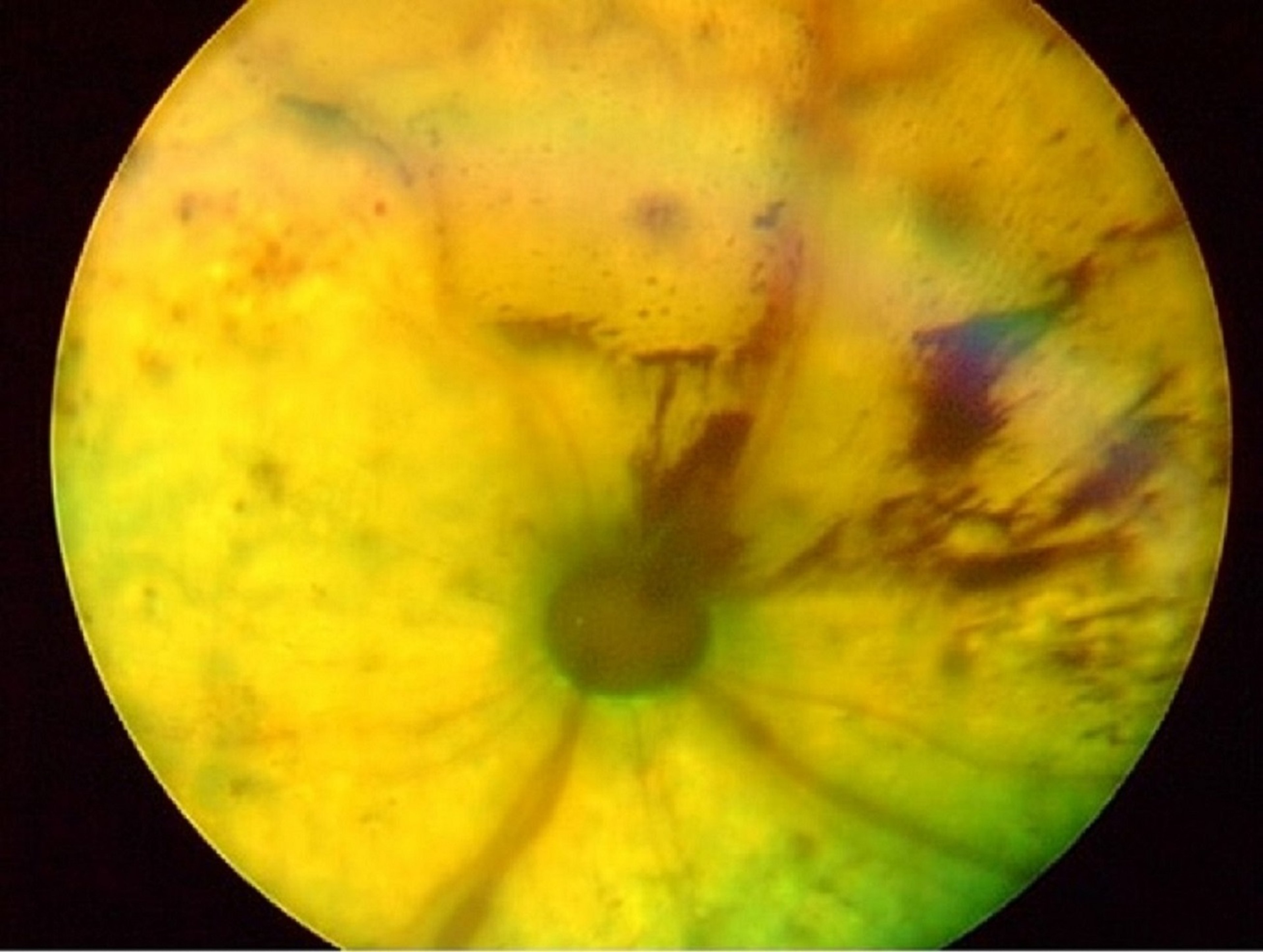
Intraocular hemorrhage and retinal detachment are evident in this photograph showing the eye of a cat that has systemic hypertension.
Courtesy of Dr. Ralph Hamor.
In cats, systemic diseases frequently affect the eye and associated structures. Eyelid inflammation may be associated with systemic Demodex cati and D gatoi, Notoedres cati (scabies), ringworm, and immune-mediated skin diseases. The pathogens that commonly cause infectious diseases of cats—eg, feline herpesvirus 1 (FHV-1), Chlamydia, and Mycoplasma—frequently present as acute and recurrent conjunctivitis. FHV-1 is also associated with ulcerative and stromal keratitis, proliferative keratoconjunctivitis, corneal sequestrum, corneal symblepharon, and KCS. Feline infectious peritonitis (FIP), toxoplasmosis, feline immunodeficiency virus (FIV), and feline leukemia virus often present as anterior and posterior uveitis, chronic uveitis, retinal detachment, and secondary glaucoma. Acute vision loss with intraocular hemorrhage and retinal detachment in older cats may be secondary to systemic hypertension and is often associated with chronic renal failure or hyperthyroidism. Resolution of intraocular hemorrhages, repair of retinal detachment, and possible restoration of vision depend on the successful lowering of blood pressure to normal levels; this is most often achieved by treatment with amlodapine.

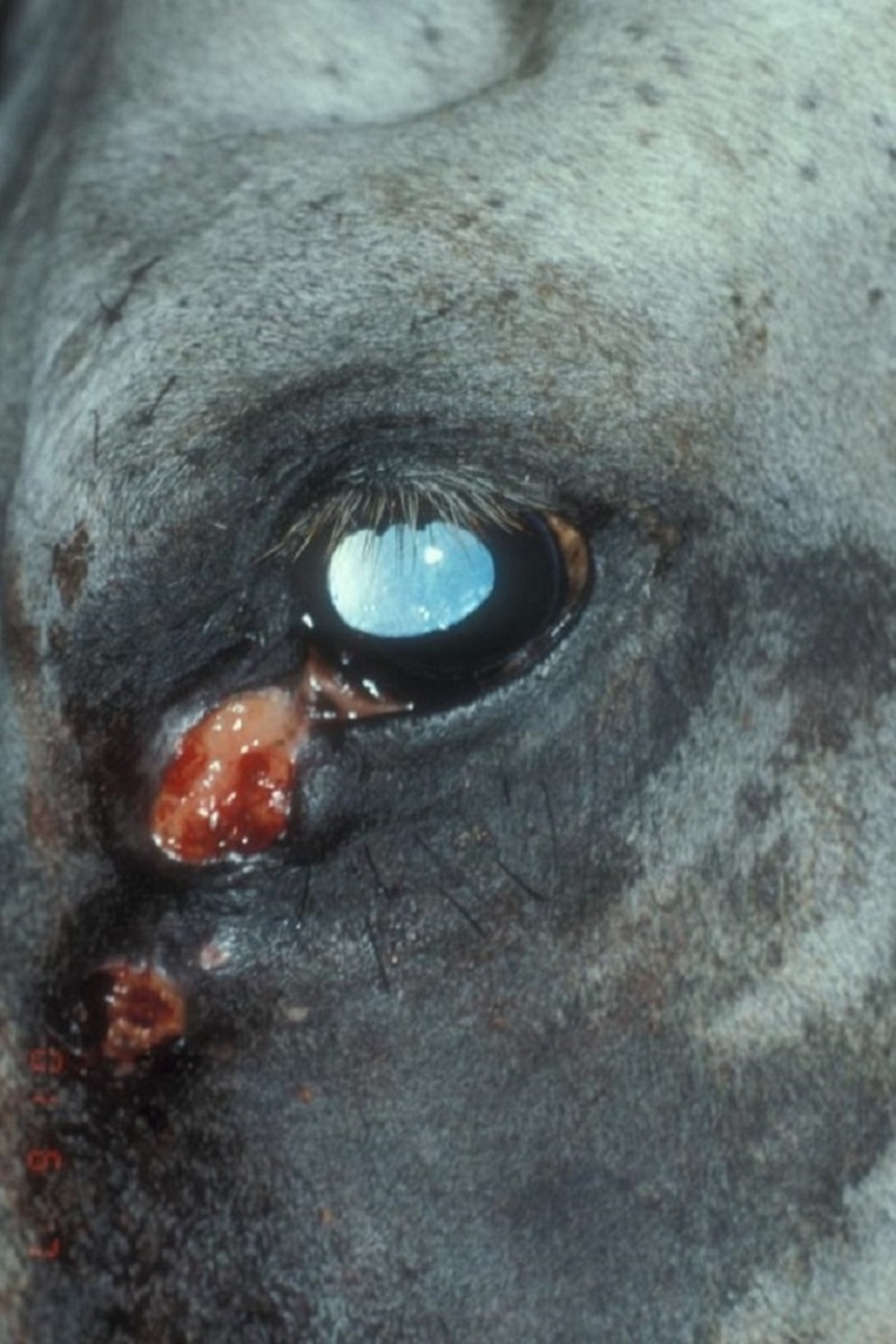
Conjunctival habronemiasis in the eye of a horse. Two proliferative and erosive masses are evident in the medial canthus.
Courtesy of K. Gelatt.
In horses, systemic infectious diseases, such as adenovirus in immunodeficient Arabian foals, equine influenza, strangles (Streptococcus equi), Rhodococcus equi infection, leptospirosis, Lyme disease (Borrelia burgdorferi), and salmonellosis, may present as conjunctivitis, anterior uveitis, or posterior uveitis. Ophthalmic onchocerciasis can present with anterior and posterior uveitis, peripapillary chorioretinitis, keratitis, keratoconjunctivitis, or lateral conjunctival vitiligo; treatment is systemic ivermectin. Habronemiasis ("summer sores") presents with inflammatory conjunctival masses of the periocular area (especially the medial canthus) associated with the aberrant migration of larvae of Habronema muscae, H microstoma, and Draschia megastoma. Treatment is usually to administer systemic ivermectin or moxidectin, debride the lesions, institute topical and environmental fly control, and protect the skin from the moisture of tear drainage with a barrier ointment.
In cattle, microphthalmia, cataracts, retinal dysplasia, and retinal detachments are associated with hydrocephalus and in utero infection of calves with bovine viral diarrhea. The same ophthalmic defects occur in lambs affected in utero with bluetongue virus. Vitamin A deficiency causes microphthalmia in piglets, and blindness and optic nerve hypoplasia in calves. Vitamin A deficiency in adult or growing cattle results in night blindness, mydriasis, and eventually total blindness. Ophthalmoscopic abnormalities include papilledema, retinal degeneration, and optic nerve atrophy. Vitamin A supplementation may restore vision in animals with night blindness only. Lymphosarcoma in cattle may present as bilateral progressive exophthalmos. Many infectious diseases, such as rhinotracheitis, malignant catarrhal fever, thromboembolic meningoencephalitis, and neonatal septicemia, may present with conjunctivitis or anterior or posterior uveitis. Intoxications such as male fern poisoning (Dryopteris filix), bracken fern poisoning (Pteridium aquilinum) in sheep, coumarin poisoning (sweet clover poisoning) in cattle, and phenothiazine toxicosis in cattle present with clinical signs of blindness from retinal degeneration, intraocular hemorrhage, or corneal edema. (Also see Toxicology Introduction.)


