

The same structural anomalies of the brain as described for large animals are found in small animals.
Congenital Cerebral Disorders in Dogs and Cats
Hydranencephaly in Dogs and Cats
In hydranencephaly, there is a marked loss of cerebral cortical tissue (primarily the neocortex) within a cranial vault of normal conformation. The resultant cavity communicates with the ventricular system, has an incomplete ependymal cell lining, and is filled with CSF.
Hydranencephaly develops as a result of the destruction of developing neural tissues and is sometimes accompanied by cerebellar hypoplasia and arthrogryposis.
In small animals, hydranencephaly has been described mainly in kittens after in utero exposure to feline panleukopenia virus (ie, feline parvovirus); however, it has been reported sporadically in dogs without an identifiable viral infection. Brainstem malformations and cerebellar hypoplasia can occur concomitantly.
Clinical signs of hydranencephaly include lethargy, propulsive circling, head pressing, and blindness.
Hydrocephalus in Dogs
Hydrocephalus, an increase in volume of the CSF, can appear similar to hydranencephaly; in hydrocephalus, however, the ventricles retain a complete ependymal lining.
When born alive, affected animals often have a characteristic dome-shaped head (see ); they might also have decreased appetite, seizures, lethargy, and altered mental status. There can be extensive expansion of the lateral ventricles in the frontal lobes.


A puppy with hydrocephalus. Note the domed head and ventrolateral strabismus (“setting sun”) appearance of the eyes.
Courtesy of Dr. Rebecca Packer.
In small animals, hydrocephalus is the most common in dogs, particularly in toy and brachycephalic breeds. It can be classified as communicating (nonobstructive), in which CSF can flow freely into the subarachnoid space; or noncommunicating (obstructive). Known causes of noncommunicating hydrocephalus include atresia of the mesencephalic aqueduct, perinatal encephalitis, or adhesions caused by intraventricular hemorrhage at birth.
Clinical signs of hydrocephalus usually indicate cerebral dysfunction, and they often progress. However, some animals remain subclinically affected. The fontanelles are often patent, and affected animals might have ventrolateral strabismus.
Hydrocephalus has been observed in Saint Bernard puppies in association with aphakia (absence of the lens) and multiple ocular defects.
Imaging by ultrasonography (through the fontanelle), CT, or MRI can confirm a diagnosis of hydrocephalus (see ), and CSF analysis should show encephalitis.


T2-weighted transverse MRI of a dog with hydrocephalus. Note the dramatic enlargement of the lateral ventricles. The fluid-filled ventricles appear white in this image.
Courtesy of Dr. Rebecca Packer.
In experimentally induced models of hydrocephalus, aquaporin-4 and IL-6 were increased in dogs with idiopathic communicating internal hydrocephalus (1).
Treatment of hydrocephalus in dogs relies on omeprazole(1–10 mg/kg, PO, every 24 hours, indefinitely) (2, 3) with or without diuretics such as acetazolamide (10 mg/kg, PO, every 6–8 hours, indefinitely) to decrease CSF production (3). If necessary, prednisone can be administered (0.5–1 mg/kg, PO, every 24 hours) (3), or surgery can be done to shunt CSF into the peritoneum.
Lissencephaly in Dogs
Lissencephaly, an absence of or decrease in cerebral gyri, is a rare disorder that occurs in Lhasa Apsos, Pekingese, and Australian Kelpies. It also occurs in association with cerebellar hypoplasia in Irish Setters, Wire Fox Terriers, and Samoyeds; and in Korat cats with microencephaly.
Clinical signs of lissencephaly consist of mild behavioral abnormalities and seizures.
Pendular Nystagmus in Cats
Pendular nystagmus occurs in various Asian breeds of cats, such as Siamese, Himalayan, and Persian cats. Compared with pathological forms of nystagmus, in pendular nystagmus there is no fast or slow phase, and the nystagmus arcs are similar to the pendulum movement of a clock.
Pendular nystagmus is caused by a congenital abnormality in the visual pathway. No other neurological signs are present, and the condition is nonprogressive and of no clinical importance.
Congenital Deafness in Dogs
In dogs, congenital deafness is associated primarily with Dalmatians, but it has also been recorded in other dog breeds, including Doberman Pinscher, Puli, the "Blue Heeler" Australian Cattle Dog variety, Australian Shepherd, English Setter, Boston Terrier, and Old English Sheepdog.
Congenital deafness is pigment associated in Dalmatians and not pigment associated in Doberman Pinschers and Pulis. It is linked to blue eye color in white cats.
The brainstem auditory evoked response (BAER) is a useful diagnostic test that is used primarily to identify carriers in a litter of affected animals.
Also see Deafness in Animals.
Inherited Cerebral Disorders in Small Animals
Necrotizing Meningoencephalitis
Necrotizing meningoencephalitis (also known as pug encephalitis) is an ultimately fatal disease that might have a familial basis. The condition has some association with mutations on dog leukocyte antigen II loci in Pugs, Maltese, and Chihuahuas, as well as ILR7 and FBXW7 mutations. Affected Pugs show behavioral changes, seizures, and CSF pleocytosis.
A similar nonsuppurative, necrotizing encephalitis has been reported in several other toy-breed dogs, including Yorkshire Terriers, Chihuahuas, and Malteses.
Neonatal Encephalopathy in Standard Poodles
Neonatal encephalopathy is an inherited genetic disorder that is caused by a mutation in the ATF2 gene and has been described in Standard Poodles. Affected dogs appear stunted and weak from birth and begin having seizures at the age of 4–5 weeks.
Neonatal encephalopathy is fatal. A genetic test is available for diagnosis of this disease in Standard Poodles.
Polymicrogyria in Standard Poodles
Polymicrogyria is an inherited disease, identified in Standard Poodles, that results in focal areas of the brain having smaller and more numerous gyri than normal, resulting in disruption of function.
The most common clinical signs are visual disturbances; however, ataxia, behavior problems, and asymmetrical lateral ventricle dilatation (ventriculomegaly) or hydrocephalus can also occur.
Signs are attributable to the portion of the brain affected by the abnormal gyri.
Idiopathic (Familial) Epilepsy in Dogs
Idiopathic, or familial, epilepsy can be inherited in many dog breeds, including Beagles, Keeshonden, Irish Setters, Belgian Tervurens, Siberian Huskies, English Springer Spaniels, Labrador Retrievers, Golden Retrievers, and German Shepherd Dogs. A mutation in the CFA37 chromosome is suspected in Belgian Shepherd dogs. Focal epilepsy of Lagotto Ramagnolo dogs is thought to be due to a mutation in the Lgi2 gene.
Diagnosis of idiopathic epilepsy depends on eliminating other causes of seizures, particularly structural brain abnormalities (such as hydrocephalus or juvenile tumors), encephalitis, or metabolic causes (eg, hepatic encephalopathy).
Paroxysmal Dyskinesia in Dogs
Paroxysmal dyskinesia is a type of movement disorder identified in several dog breeds, including the Soft Coated Wheaten Terrier (for which a mutation in the PIGN gene has been identified), the Cavalier King Charles Spaniel (episodic falling; for which a mutation in the BCAN gene has been identified), the Border Terrier (Spike's disease), and the Chinook, among other pure and mixed breeds.
Paroxysmal dyskinesia is characterized by episodes of involuntary movement, including hyper- or hypotonicity or dysmetria, and it can resemble partial or generalized seizures, except that conscious awareness is maintained and no autonomic signs occur (see ).

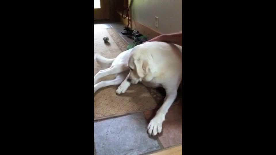
One or more limbs can be affected at once or in sequence, and voluntary movements and ambulation are typically (but not always) inhibited by involuntary movements in the limbs. Episodes can last minutes to hours and occur as seldom as once over several months or as often as multiple times daily.
Hepatic Encephalopathy in Dogs and Cats
Hepatic encephalopathy in dogs and cats is usually caused by a congenital portosystemic shunt. The shunt can be a single large vessel, or there might be microscopic shunting of blood within the liver. Dog breeds often affected include Havanese, Yorkshire Terriers, Maltese, Miniature Schnauzers, Cairn Terriers, and Old English Sheepdogs (4).
The clinical signs of hepatic encephalopathy are usually noticed before the age of 6 months, and they reflect primarily cerebral dysfunction, including staring into space, inappropriate vocalizing, aggression, and agitation. Advanced neurological alterations can cause depression, blindness, myoclonus, stupor, coma, or seizures. In cats, these signs are often accompanied by excessive salivation.
A rare cause of hepatic encephalopathy is a deficiency of hepatic urea cycle enzymes.
Pre- and postprandial bile acid testscan support the diagnosis of hepatic encephalopathy, indicating abnormal liver clearance of bile acids from the blood when the values are elevated. Definitive diagnosis might be facilitated by radiographic imaging techniques such as positive-contrast portography, CT, transcolonic portal scintigraphy, or diagnostic gray-scale ultrasonography.
Blood tests such as the ammonia tolerance test should be used with caution in cases of suspected hepatic encephalopathy because of the risk of causing an encephalopathic crisis. Resting ammonia concentrations can be measured and are supportive of hepatic encephalopathy when elevated above 46 mcmol/L, indicating abnormal clearance by the liver (5); however, ammonia concentrations have been poorly correlated with both diagnosis and the presence of clinical signs.
Pearls & Pitfalls
|
Lysosomal Storage Disorders in Small Animals
Lysosomal storage disorders that commonly cause cerebral signs in small animals include ceroid lipofuscinosis and fucosidosis; however, there are many other forms of lysosomal storage disorders, as well as other inborn errors of metabolism.
Genetic and enzymatic testing is available for many lysosomal storage disorders. When specific tests are not available, organic acid screens might support the general diagnosis of metabolic error.
Puppy Hypoglycemia
Puppy hypoglycemia is an idiopathic syndrome in toy breeds of dogs that occurs in the first 6 months of life. It seems to relate to a relative immaturity of the liver, which affects glycogenolysis and can usually be managed by providing frequent meals of a commercial puppy diet.
The problem usually resolves as the puppy matures.
Narcolepsy and Cataplexy in Dogs and Cats
Narcolepsy, a disorder of sleep-wake control (typically characterized by excessive sleepiness or sudden paroxysmal attacks of flaccid paralysis with conservation of consciousness), is an autosomal-recessive inherited disorder in Doberman Pinschers, Labrador Retrievers, and Dachshunds, and it has been described in other dog breeds as well. It is rare in cats.
A mutation in the hypocretin (orexin) receptor 2 (Hcrtr2) gene has been identified in Doberman Pinschers.
Attacks of narcolepsy are often stimulated by excitement and sometimes are accompanied by cataplexy (sudden loss of muscle tone, with collapse). The disorder must be differentiated from various types of syncope.
Physostigmine (0.025–0.1 mg/kg, IV) potentiates the frequency and severity of cataleptic attacks and can be used as a onetime medication in a hospital setting to stimulate an episode to help diagnose the condition (6). Imipramine (0.5–2 mg/kg, PO, every 8–12 hours as needed, indefinitely) can be used to control the severity of cataplexy (7, 8, 9).
Key Points
Congenital malformations of the cerebrum commonly involve either lack of development of all or a portion of the cerebrum or protrusion of meninges and possibly cerebral tissue outside the cranial vault.
Physical appearance of the animal might provide presumptive or definitive diagnosis of a cerebral disorder; however normal appearance does not rule out a congenital or inherited condition.
Congenital cerebral malformations in small animals can be due to genetic causes or environmental causes, or they can occur sporadically.
Inherited disorders such as idiopathic epilepsy, paroxysmal dyskinesia, and narcolepsy cause sporadic abnormal episodes, and they can be differentiated by effects on consciousness, presence of autonomic signs, and effects on muscles (hypertonicity versus hypotonicity).
Congenital metabolic dysfunction can lead to forebrain clinical signs in small animals, highlighting the need for a systemic workup, including blood glucose monitoring and liver function testing.
For More Information
Mandigers PJJ, Santifort KM, Lowrie M, Garosi L. Canine paroxysmal dyskinesia—a review. Front Vet Sci. 2024;11:1441332
Hülsmeyer V-I, Fischer A, Mandigers PJJ, et al. International Veterinary Epilepsy Task Force's current understanding of idiopathic epilepsy of genetic or suspected genetic origin in purebred dogs. BMC Vet Res. 2015;11:175.
Estey CM. Congenital hydrocephalus. Vet Clin North Am Small Anim Pract. 2016;46(2):217-229.
Also see pet owner content regarding brain, spinal cord, and nerve disorders of dogs and cats.
References
Schmidt MJ, Rummel C, Hauer J, et al. Increased CSF aquaporin-4, and interleukin-6 levels in dogs with idiopathic communicating internal hydrocephalus and a decrease after ventriculo-peritoneal shunting. Fluids Barriers CNS. 2016;13:12. doi:10.1186/s12987-016-0034-1
Pelegrini LF, Silva NF, Campos OPS, et al. Medical therapy using omeprazole in 12 hydrocephalic dogs: clinical, diagnostic, and therapeutic findings. Pesqui Veterinária Bras. 2019;39(10):823–829. doi:10.1590/1678-5150-PVB-6332
Estey CM. Congenital hydrocephalus. Vet Clin North Am Small Anim Pract. 2016;46(2):217–229. doi:10.1016/j.cvsm.2015.10.003
Tobias KM, Rohrbach BW. Association of breed with the diagnosis of congenital portosystemic shunts in dogs: 2,400 cases (1980–2002). J Am Vet Med Assoc. 2003;223(11):1636-1639. doi:10.2460/javma.2003.223.1636
Gerritzen-Bruning MJ, van den Ingh TSGAM, Rothuizen J. Diagnostic value of fasting plasma ammonia and bile acid concentrations in the identification of portosystemic shunting in dogs. J Vet Intern Med. 2006;20(1):13-19. doi:10.1111/j.1939-1676.2006.tb02818.x
Sturges BK, Knipe MF. Sleep disorders. In: Ettinger SJ, Feldman EC, eds. Textbook of Veterinary Medicine: Diseases of the Dog and Cat. 7th ed. Vol 1. Elsevier; 2010:234-237.
Dantas LMS, Ogata N. Veterinary psychopharmacology. Vet Clin North Am Small Anim Pract. 2024;54(1):195-205. doi:10.1016/j.cvsm.2023.07.003
Fadel C, Łebkowska-Wieruszewskac B, Lisowski A, et al. Imipramine in dogs: a pharmacokinetic study following oral administration under fasted and fed conditions. Vet J. 2024;308:106250. doi:10.1016/j.tvjl.2024.106250
Santifort KM, Ives EJ, Fenn J, et al. Suspected acquired narcolepsy in 8 dogs. J Vet Intern Med. 2021;35(3):1448-1454. doi:10.1111/jvim.16116


