


"Pyoderma" generally refers to bacterial dermatitis. Clinical signs of pyoderma vary depending on the pathogenic organism involved and the depth of infection. Diagnosis involves cytological evaluation, culture, and other dermatological diagnostic testing, such as to rule out dermatophytosis or identify an underlying trigger. Treatment involves topical therapy, appropriately chosen systemic antimicrobial therapy, and controlling underlying predisposing conditions, such as allergic dermatitis or ectoparasitism.
"Pyoderma" is an umbrella term for any purulent skin disease; it means, literally, “pus in the skin.” Any condition (whether infectious, inflammatory, immune- mediated, or neoplastic) that results in the accumulation of neutrophilic exudate can be termed "pyoderma"; however, the term usually refers to bacterial infections of the skin (ie, bacterial folliculitis and dermatitis).
Dogs and cats can have a simple pyoderma infection triggered by a onetime or simple event—eg, flea infestation. Complex pyoderma infections are recurrent and are associated with underlying diseases such as the following:
allergies (flea allergy, atopic dermatitis, food allergy)
internal diseases (particularly endocrinopathies such as hypothyroidism or hyperadrenocorticism)
seborrheic conditions (including follicular or sebaceous gland diseases)
parasitic diseases (eg, demodicosis)
anatomical predispositions (eg, skin folds)
immune-mediated conditions (eg, pemphigus foliaceus)
Bacterial pyodermas limited to the epidermis and hair follicles are referred to as superficial pyoderma. The term "bacterial impetigo" is used especially if infection is confined to nonfollicular (glabrous) areas of skin and the predominant lesions are pustules; in young dogs, this is often called "puppy pyoderma."
For superficial bacterial infections that are confined to the superficial portion of the hair follicle, the term "superficial bacterial folliculitis" can be used. "Mucocutaneous pyoderma" generally refers to a superficial bacterial infection in dogs that is present around the lips and perioral skin; it can also be present in periocular skin and other areas.
Bacterial pyodermas that involve the dermis (especially deep dermis) or that cause furunculosis are referred to as deep pyoderma.
In dogs, pyodermas are common, and superficial bacterial pyoderma is the most frequent reason for antimicrobial use in small animal practice. Pyodermas are uncommon in cats.
Etiology of Pyoderma in Dogs and Cats
Pyoderma can be classified by the depth of infection:
Surface pyoderma includes acute moist dermatitis (hot spots), fold pyoderma (intertrigo), and bacterial overgrowth syndrome (erythema with large numbers of bacteria but no other clinical signs).
Superficial pyoderma (especially bacterial folliculitis) extends into the follicular ostium and epidermis.
Deep pyoderma is less common but more serious because it expands into the dermis, with a higher risk of bacteremia. Deep pyoderma can develop from any underlying trigger or acquired immunodeficiency, and it is commonly associated with demodicosis.
Interdigital furunculosis is a disorder of ruptured hair follicles in the dermis, secondary to numerous primary causes (eg, pressure from conformation or licking, demodicosis, and dermatophytosis), and it is commonly secondarily infected.
Etiological classification of pyoderma is based on the pathogenic organism involved in the infection (staphylococci, streptococci, etc).
Bacterial pyoderma usually develops after a triggering event from organisms that are part of the healthy animal's cutaneous and mucosal microbiota. The dog's stratum corneum and hair follicle can be particularly susceptible to overcolonization by bacteria, which is why pyoderma is more common in this species.
The primary pathogen associated with pyoderma in dogs is Staphylococcus pseudintermedius.
Superficial pyoderma in cats is usually caused by Staphylococcus spp. In cats, Staphylococcus aureus and S pseudintermedius are common coagulase-positive staphylococci isolated from pyoderma lesions.
Normal resident bacteria in canine skin include S pseudintermedius, coagulase-negative staphylococci, streptococci, Micrococcus spp, and Acinetobacter spp. Transient bacteria in canine skin include Bacillus spp, Corynebacterium spp, Escherichia coli, Proteus mirabilis, and Pseudomonas spp.
Normal resident bacteria in feline skin include Acinetobacter spp, Micrococcus spp, coagulase-negative staphylococci, and alpha-hemolytic streptococci. Transient bacteria in feline skin include Alcaligenes spp, Bacillus spp, E coli, P mirabilis, Pseudomonas spp, coagulase-positive and coagulase-negative staphylococci, and alpha-hemolytic streptococci.
Microbiota analysis results can differ, depending on the techniques used (culture versus molecular).
Warm, moist areas on the skin, such as lip folds, facial folds, neck folds, axillary areas, dorsal or plantar interdigital areas, vulvar folds, and tail folds, often have higher bacterial counts than other areas of skin and are at an increased risk of infection. Pressure points, such as elbows and hocks, are also prone to infections, possibly because of follicular irritation and rupture due to chronic repeated trauma and pressure.
Any condition that changes the normally dry environment of the skin to a more moist one can predispose the host to overcolonization of the skin by resident and transient bacteria.
Skin infections are usually secondary to ectoparasite infestations, allergies, local trauma, keratinization disorders, endocrinopathies, or immune-mediated conditions.
The most common underlying triggers of superficial pyoderma in dogs include the following:
ectoparasites
allergies (atopic dermatitis, food allergy and flea bite allergy)
endocrinopathies (hypothyroidism, hyperadrenocorticism)
keratinization disorders
immune-mediated conditions
follicular dysplasias
poor grooming
Feline pyoderma is most common in allergic skin diseases, parasitic diseases, and feline chin acne. Chin acne itself is a disorder of keratinization with suspected infectious, environmental, and stress triggers; secondary infections are common.
Recurrent, nonhealing, deep pyoderma in cats can be associated with systemic disease, such as infection by feline immunodeficiency virus, feline leukemia virus, or atypical Mycobacterium spp. Infected feline chin acne can also lead to furunculosis and deep pyoderma.
Clinical Findings of Pyoderma in Dogs and Cats
In dogs, superficial pyoderma commonly has the following clinical signs:
multifocal areas of alopecia
follicular papules or pustules
epidermal collarettes
crusts and scales
The trunk and ventrum are the most commonly affected regions in cases of pyoderma (see ).

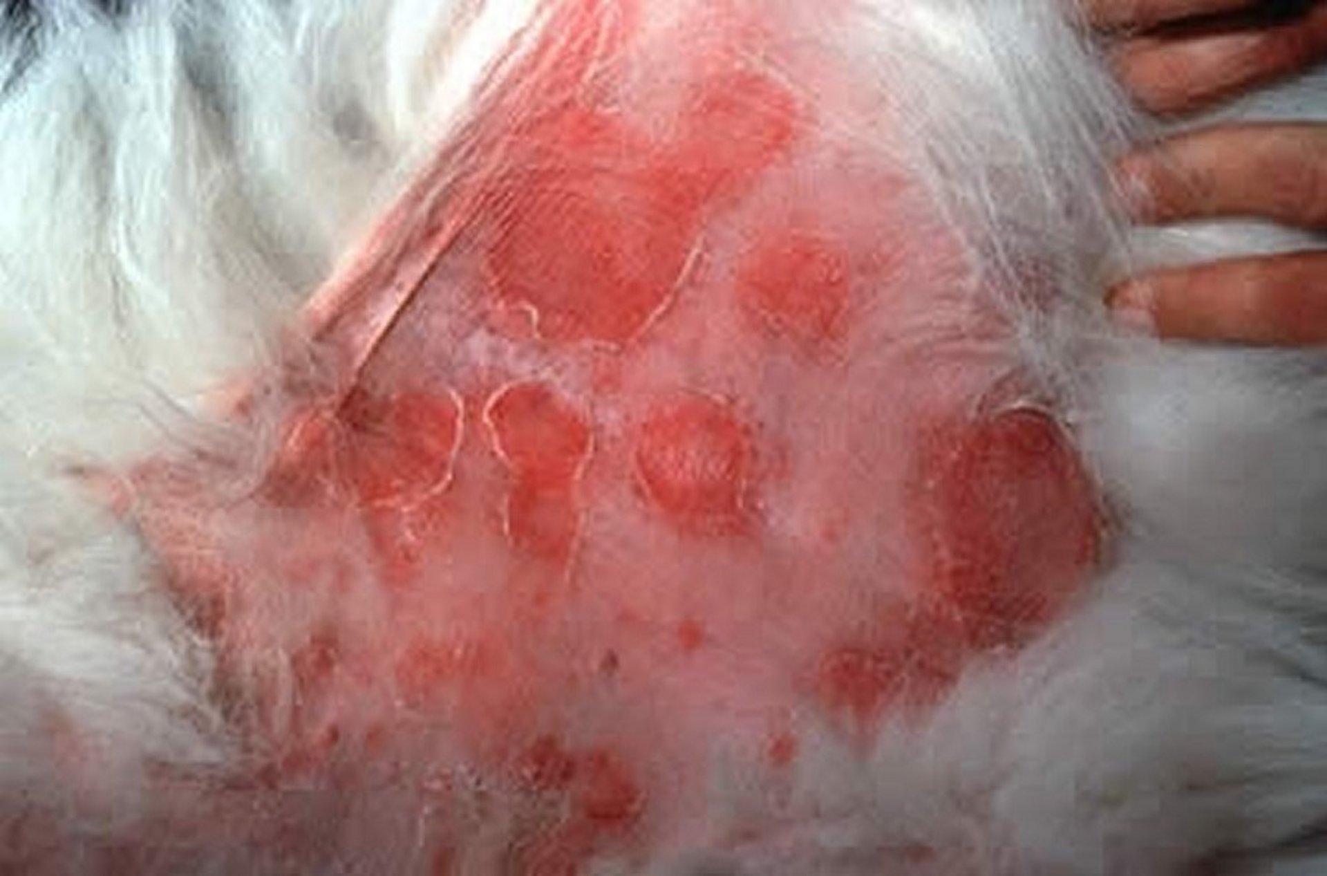
Multiple epidermal collarettes and erythematous papules in a dog with superficial pyoderma.
Courtesy of Dr. Stephen White.
Less common lesions include large, fluctuant pustules (bullous impetigo, occurring often in older animals with concurrent endocrinopathies), large areas of alopecia with an erythematous, scaly edge (superficial spreading pyoderma), and erosions or ulcerations on the mucocutaneous junctions (mucocutaneous pyoderma).
The hallmarks of deep pyoderma in dogs are pain, crusting, odor, and exudation of blood and pus. Erythema, swelling, ulcerations, hemorrhagic crusts and bullae, hair loss, and draining tracts with serohemorrhagic or purulent exudate might also be present. The bridge of the muzzle, chin, elbows, hocks, interdigital areas, and lateral aspects of the stifle joints are more prone to deep infections; however, any area can be involved.
Deep pyoderma can be present in areas of acral lick dermatitis, draining tracts on the dorsum (postgrooming furunculosis), and papules and nodules on the dorsal muzzle (nasal furunculosis).
In cats, superficial pyoderma commonly has the following clinical signs:
crusted papules (miliary dermatitis)
eosinophilic plaques
Superficial pyoderma in cats is often overlooked and underdiagnosed.
Cats with deep pyoderma often present with alopecia, ulcerations, hemorrhagic crusts, and draining tracts.
Diagnosis of Pyoderma in Dogs and Cats
Clinical evaluation
Cytological evaluation
Bacterial culture
Ruling out other causes
Diagnosis of pyoderma is based on the presence of characteristic lesions, confirmation of the presence of bacteria, and ruling out other common causes of folliculitis, such as demodicosis and dermatophytosis.
Use of a Wood's lamp, direct examination of hairs for the presence of hyphae or spores, fungal PCR assay, or fungal culture can be performed to rule out dermatophytosis; negative deep skin scrapings rule out follicular demodicosis in most cases.
Microscopic evaluation of cytological preparations from skin lesions is one of the most valuable tools for diagnosing pyoderma, enabling the identification of inflammatory cells and bacteria (see ). Skin cytological evaluation can also help identify Malassezia spp, which cause a common coinfection.


Photomicrographs of impression smears from a skin lesion in a dog with pyoderma. (A) Note the many neutrophils with few extracellular and intracellular cocci. (B) Note the degenerative neutrophils, with purple nuclear material streaming from ruptured cells, overlaid with numerous clusters of extracellular cocci. For both images: commercial Romanowsky stain variant; scale bar = 20 mcm.
Courtesy of Dr. Mitzi Clark.
Bacterial culture and susceptibility testing are particularly important in cases of recurrent pyoderma. Because of antimicrobial resistance, they are essential in judicious selection of systemic antimicrobial therapy.
Cytological evaluation should be performed from the area of culture to confirm the presence of bacteria. The technique used to collect samples varies, depending on the type and location of the lesion:
Impression smears are used to collect samples after opening a pustule or papule or after removing a crust.
Clear adhesive tape can be used to collect samples from lesions that are dry, oily, or difficult to reach with a microscope slide (eg, interdigital spaces).
Cotton swabs are used to collect samples from ear canals, draining tracts, nail folds, and moist skin lesions.
A microspatula or No. 10 scalpel blade can be used to collect samples from places difficult to reach, such as nail folds, and to expose the skin from underneath a crust or scale before performing an impression smear.
Fine-needle aspiration can be used to collect samples from nodules, plaques, and hemorrhagic bullae.
Because pyodermas are triggered by underlying problems, appropriate diagnostic testing to identify underlying triggers is necessary.
Treatment of Pyoderma in Dogs and Cats
Topical medications
Systemic antimicrobial therapy
Immunomodulators
Treatment of pyoderma can involve topical therapy, systemic antimicrobial therapy, and immunomodulatory therapy. The most common causes of recurrent bacterial pyoderma include failure to identify an underlying trigger, antimicrobial undertreatment (dosage too low or duration of therapy too short), inappropriate use of glucocorticoids or other immunosuppressive therapy, wrong antimicrobial choice, incorrect dosage, and failure to recheck the patient.
Pearls & Pitfalls
|
Topical Therapy for Pyoderma
In most cases, topical therapy should be the sole treatment for surface or superficial pyoderma, including cases with methicillin resistance.
The best formulation to use depends on the extent of the lesions, type of coat, and ease of application. Shampoos, creams, gels, ointments, sprays, and mousses are available. Shampoos and sprays might be better for more widespread lesions.
Medicated bathing should be performed 2–3 times weekly with a contact time of 10–15 minutes each time. Other topical antimicrobials should be used 1–2 times daily during active infection. The duration of therapy should extend to clinical cure, as determined by recheck examination.
Active ingredients in antimicrobial shampoos include the following:
Chlorhexidine (2–4%), which kills bacteria by coagulating bacterial cytoplasmic proteins and deteriorating bacterial cell membranes. At 3-4%, it is an antifungal.
Benzoyl peroxide (2.5–3%), an oxidizing agent that disrupts the bacterial cell wall membrane by increasing permeability or causing the membrane's rupture. It is also degreasing, so it could be drying. Benzoyl peroxide can be used for surface infections, but there are better choices for superficial pyoderma.
Chlorhexidine (2%) in combination with miconazole (2%), both of which are antistaphylococcal, with antifungal properties as well.
Sodium hypochlorite (< 0.05%), a well-tolerated antiseptic that also exhibits anti-inflammatory properties via superoxide radical generation. It has concentration-dependent activity against gram-negative and gram-positive bacteria.
For focal lesions, a topical wipe or cream might be more appropriate. It is imperative to manage the underlying condition that predisposes the skin to infection. In cases of interdigital furunculosis, for instance, anti-inflammatory glucocorticoids or immunomodulators such as modified cyclosporine are often needed in conjunction with infection control.
For feline chin acne, a spot-on product containing phytosphingosine applied twice weekly can be useful after the infection has been managed with topical or systemic antimicrobials.
In allergic animals, antipruritic therapy is often necessary.
Topical therapy can also be used as an adjunctive treatment for deep pyodermas, to hasten healing.
Systemic Antimicrobial Therapy for Pyoderma
Systemic antimicrobial therapy should be reserved for cases of deep pyoderma and for the rare cases of superficial pyoderma that cannot be treated with topical therapy alone.
With the spread of methicillin-resistant and multidrug-resistant Staphylococcus spp, the treatment of pyoderma has become more challenging. Empirical selection of systemic antimicrobials is increasingly difficult. Treatment should be based on the results of bacterial culture and susceptibility testing.
Because S pseudintermedius, the most common pathogen associated with pyoderma, produces beta-lactamase, empirical use of penicillin, ampicillin, and amoxicillin should be avoided. Most strains of S pseudintermedius are also resistant to tetracycline and streptomycin.
Pearls & Pitfalls
|
As a consideration for systemic therapy, antimicrobial agents can be classified into first-tier and second-tier drugs, depending on the likelihood that they will be effective against staphylococci and on their spectrum of activity against gram-negative pathogens.
First-tier drugs can be used empirically in animals with no history of methicillin resistance. First-tier drugs include the following:
clindamycin (11 mg/kg, PO, every 24 hours for up to 21 days) (1, 2)
first-generation cephalosporins, such as cephalexin (25–30 mg/kg, PO, every 12 hours for 21 to 42 days) (3, 4)
amoxicillin-clavulanate (12.5–25 mg/kg, PO, every 12 hours for up to 30 days) (5, 6)
The duration of therapy is important for successful management. Antimicrobial therapy should be continued until complete resolution of clinical lesions and no evidence of cytological infection as determined on recheck exam (7). However, if there is no response after 3–4 days of treatment, the diagnosis should be reconsidered.
Second-tier agents, such as fluoroquinolones, should be chosen according to results from bacterial culture and antimicrobial susceptibility testing.
Of note, inducible clindamycin resistance can be present in S aureus and, rarely, S pseudintermedius canine isolates. Therefore, these strains are often erythromycin resistant but clindamycin susceptible on culture and susceptibility testing reports. However, without checking for inducible clindamycin resistance in vitro via a D test (disk diffusion test), clindamycin should not be selected for these patients.
Immunomodulators of Pyoderma
Bacterins are defined as suspensions, typically of lysed or attenuated bacteria, that are used as vaccines to increase immunity to particular pathogens or to a particular disease. They have been used sporadically in dogs for recurrent pyoderma.
The mechanism of action of bacterins is poorly understood. Improvement is determined by a decrease in the frequency or severity of signs. When using bacterins, it is important to control the infection first by giving concurrent antimicrobial therapy for the first 4–6 weeks.The usual bacterin dosage is 0.5 mL, SC, twice weekly for 10–12 weeks.
If bacterin treatment is efficacious, the frequency can be decreased to once a week or less; in most cases, however, lifelong therapy is needed.
Key Points
In dogs, superficial bacterial pyoderma is common. It is the most common reason for antimicrobial use in small animal practice.
With the identification of methicillin-resistant and multidrug-resistant Staphylococcus spp, the treatment of pyoderma has become more challenging.
Because Staphylococcus pseudintermedius, the most common pathogen associated with canine pyoderma, produces beta-lactamase, empirical use of penicillin, amoxicillin, and ampicillin should be avoided.
Topical therapy should be the sole therapy used for surface and superficial pyoderma.
For More Information
Loeffler A, Cain C, Ferrer L, et al. Antimicrobial use guidelines for canine pyoderma by the International Society for Companion Animal Infectious Diseases (ISCAID). Vet Dermatol. 2025;36(3):234-282.
Loeffler A, Lloyd DH. What has changed in canine pyoderma? A narrative review. Vet J. 2018;235:73–82.
Summers JF, Brodbelt DC, Forsythe PJ, Loeffler A, Hendricks A. The effectiveness of systemic antimicrobial treatment in canine superficial and deep pyoderma: a systematic review. Vet Dermatol. 2012;23(4):305-e61.
Also see pet owner content regarding pyoderma in dogs and cats.
References
Saridomichelakis MN, Athanasiou LV, Salame M, Chatzis MK, Katsoudas V, Pappas IS. Serum pharmacokinetics of clindamycin hydrochloride in normal dogs when administered at two dosage regimens. Vet Dermatol. 2011;22(5):429-435. doi:10.1111/j.1365-3164.2011.00969.x
Batzias GC, Delis GA, Athanasiou LV. Clindamycin bioavailability and pharmacokinetics following oral administration of clindamycin hydrochloride capsules in dogs. Vet J. 2005;170(3):339-345. doi:10.1016/j.tvjl.2004.06.007
Guaguere E, Salomon C, Maynard L. Using cephalexin in the treatment of canine pyoderma: comparing the efficacy of different dosages. Pratique Medical & Chirurgicale de l'Animale de Compagnie. 1998;33:237-246.
Frank LA, Kunkle GA. Comparison of the efficacy of cefadroxil and generic and proprietary cephalexin in the treatment of pyoderma in dogs. J Am Vet Med Assoc. 1993;203(4):530-533. doi:10.2460/javma.1993.203.04.0530
Stegemann MR, Coati N, Passmore CA, Sherington J. Clinical efficacy and safety of cefovecin in the treatment of canine pyoderma and wound infections. J Small Anim Pract. 2007;48(7):378-386. doi:10.1111/j.1748-5827.2007.00363.x
Lloyd DH, Carlotti DN, Koch HJ, Van den Broek AH. Treatment of canine pyoderma with co-amoxyclav: a comparison of two dose rates. Vet Rec. 1997;141(17):439-441. doi:10.1136/vr.141.17.439
Loeffler A, Cain C, Ferrer L, et al. Antimicrobial use guidelines for canine pyoderma by the International Society for Companion Animal Infectious Diseases (ISCAID). Vet Dermatol. 2025;36(3):234-282. doi:10.1111/vde.13342


