


Hepatic lobule
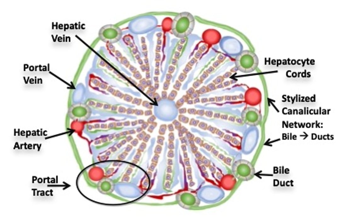
Schema of a classical hepatic lobule in cross-section. Linear cords of hepatocytes are oriented radially surround the hepatic vein and extend to portal tracts. Portal tracts display profiles of hepatic artery, portal vein, and bile duct. Most perfusion is venous (blue shading), with less arterial perfusion (red shading). These intermix in sinusoids. Tiny arterial branches extend to the bile ducts (peribiliary arterial plexus). The hepatic vein is most vulnerable to hypoxic or ischemic events receiving the last sinusoidal distribution of blood. Canalicular conduits (not shown) are located at the intercellular juxtaposition of hepatocytes (see Hepatic lobule, zones). Green shading in areas other than well-defined bile ducts reflects bile flow in canalicular and cholangiolar conduits.
Courtesy of Dr. Sharon Center.