


Courtesy of Dr. Ronald Green.
Patellar luxation, a hereditary disorder in dogs and cats, is characterized by ectopic development of the patella medial or lateral to the trochlear groove of the femur. Patellar luxation can be associated with multiple deformities of the hindlimb, involving the hip joint, femur, and tibia. Medial patellar luxations can be involved with a reduced coxofemoral angle (coxa vara), lateral bowing of the femur, internal rotation of the tibia, shallow trochlear groove, and hypoplasia of the medial femoral condyle; lateral luxations cause the reverse changes.
Clinical signs are variable and based on the severity of luxation. Animals of any age may be affected. In general, cats and small and miniature breeds of dogs have a medial luxation, and large dogs have a lateral luxation. Affected animals are lame or ambulate with a skipping gait. Palpation of the stifle joint reveals displacement of the patella.
Patellar luxation can be classified according to the level of severity:
Grade I — Clinical signs are mild and infrequent, and the patella can be manually luxated but easily returns to the trochlear groove.
Grade II — The patella luxates during flexion of the joint and is repositioned during extension, causing animals to have a resolvable skipping lameness.
Grade III — the dislocated patella is more frequently out of, instead of in, the trochlear groove, and lameness is consistent. Bone deformities are evident in these animals.
Grade IV — Lameness and limb deformations are most severe.
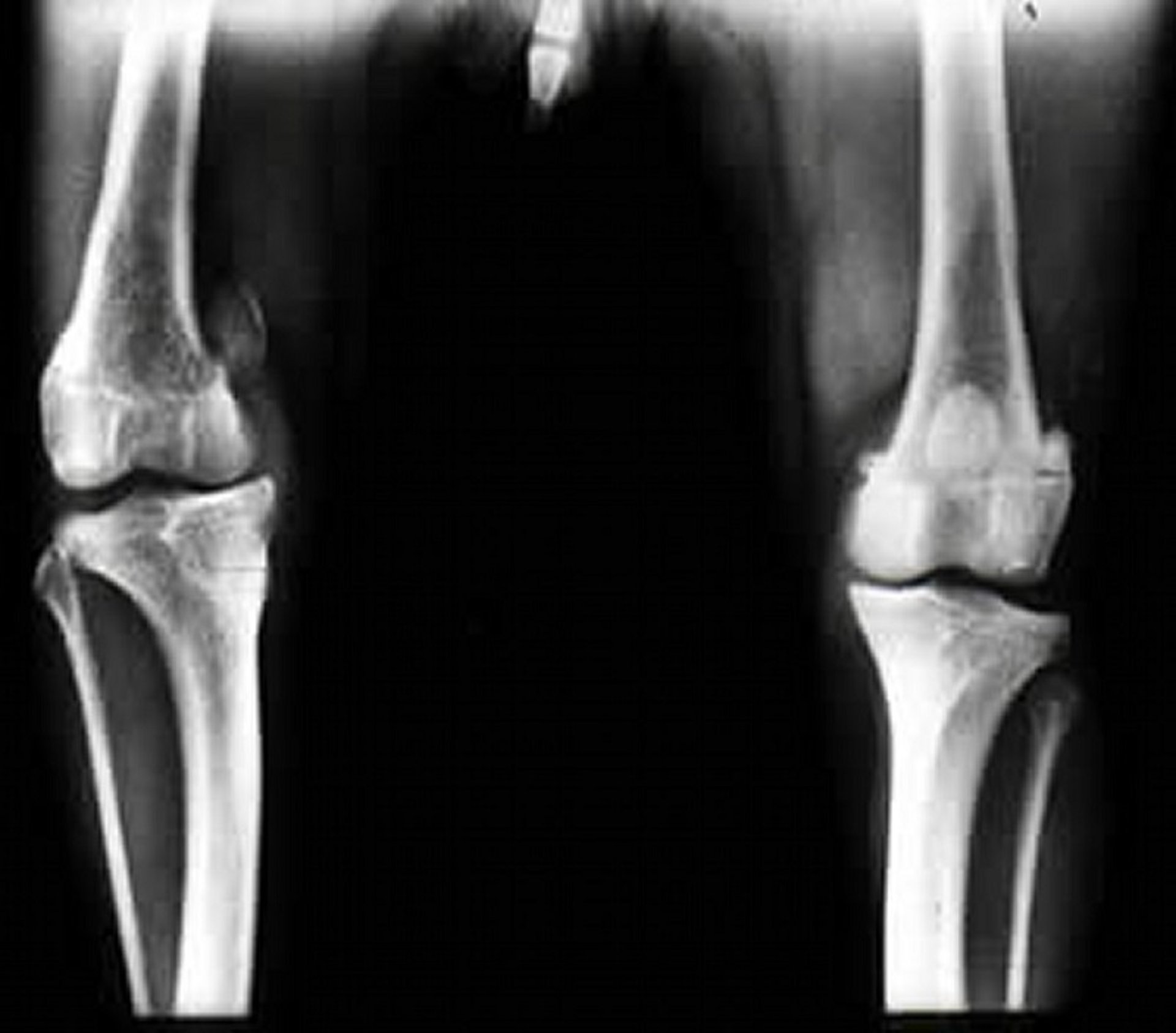
Radiography of affected animals reveals various degrees of limb changes based on the grade of the luxation.
Surgical correction is the usual treatment. The type of surgery is based on the severity of the luxation and can include both orthopedic and soft-tissue procedures. Useful procedures involve fascial releasing incisions (on the side of the luxation), joint capsule and retinaculum imbrications (on the side opposite the luxation), deepening of the trochlear groove, tibial crest transposition, and fabella to tibial tuberosity derotation sutures. Severe deformations may require femoral or tibial osteotomies, stifle joint arthrodesis, or limb amputation.
Prognosis for recovery is good in mild or moderately affected animals. Concurrent cranial cruciate ligament and medial meniscal injuries should be identified and treated. Cats are less severely affected than dogs and have an excellent prognosis.
For More Information


