

The functional unit of skeletal muscle is the motor unit, which consists of an individual motor neuron and all the muscle fibers it innervates. The motor neuron's cell body is located in the central horn of the spinal cord; the cell body is the start of the pathway to the motor neuron's peripheral axon, the neuromuscular junction, and the muscle fibers innervated by the neuron.
Each component of the motor unit must be functionally intact for the muscle to contract appropriately, resulting in movement of the skeleton. The motor unit is the final common pathway conducting neural impulses from the CNS to the muscle.
Transmission of a nerve impulse at the neuromuscular junction involves the release of acetylcholine from small synaptic vesicles in the presynaptic terminal of neurons. The released acetylcholine fills the synaptic cleft between the nerve terminal and the muscle fiber membrane before binding to the postsynaptic receptor. The binding of acetylcholine to the postsynaptic receptor excites the postsynaptic muscle fiber membrane, markedly increasing membrane permeability to sodium ions and enabling a rapid influx of extracellular sodium into the muscle membrane. The sodium influx changes the membrane potential, and if that change is strong enough, an action potential is propagated over the surface of the skeletal muscle membrane. Muscle contraction results when this occurs in all the muscle fibers innervated by each motor neuron.
Normal muscle, comprising many motor units, is dynamic, and many diseases can influence its function and structure. Primary muscular dysfunctions of infectious, toxic, or congenital origin can cause complete paralysis, paresis, or ataxia. However, more commonly (eg, in diseases such as tetanus, equine herpesvirus infection, canine distemper, or protozoal myelitis), the primary disorder is not muscular in origin but can be attributed to the nervous system, with the muscular system representing the effector organ.
Disorders that affect the neuromuscular junction (eg, myasthenia gravis, hypocalcemia, hypermagnesemia) can result in muscle fatigue, weakness, and paralysis. The neuromuscular junction can also be affected by muscle-relaxing drugs (eg, curare, succinylcholine), certain antimicrobials, and toxins (eg, botulism, tetanus, venoms).
Primary disorders of the muscle membrane and, to some extent, of muscle fibers are called myopathies. Myopathies can be hereditary (eg, myotonia congenita in goats) or acquired (eg, vitamin E and selenium deficiencies, hypothyroidism, and hypokalemia). Myopathies involving the muscle fiber components include muscular dystrophy, polymyositis, eosinophilic myositis, white muscle disease, and exertional rhabdomyolysis. Various laboratory tests—eg, histological examination, determination of serum enzyme levels, electromyographic studies and determinations of conduction velocity—are instrumental in confirming a specific diagnosis.
Muscles are affected by several mechanisms: contusions, strains and ruptures, contractures and fibrosis, and skeletal tumors.
Contusions of muscle are caused by blunt trauma, secondary to a fracture, or they can occur iatrogenically during surgery. When a contusion occurs, the cutaneous, subcutaneous, and intramuscular vasculatures of the affected area are damaged, resulting in local hemorrhage. Pain, discoloration (bruising of skin), and potential limitations in muscle contractions can result from compression and partial tearing of muscle fibers. Injuries due to contusions can lead to inflammation and localized hematomas within the muscle tissue. Most contusions heal without long-term problems; however, more severe contusions involving muscle strains and ruptures can result in residual deficits.
Muscle strains and ruptures occur when muscles stretch too far and partially or completely tear. Strains are likely the most common muscle injury in small animals and horses; when mild, however, many of these cases are not reported to veterinarians. Strains typically occur at the musculotendinous junction at the origin or insertion of the muscle; they can also occur within the muscle belly. A strain that is severe enough to cause a muscle tear might result in hemorrhage and subsequent inflammation (see ). As a strained or torn muscle heals, the subsequent hematoma, edema, inflammatory reaction, and fibrosis (scar tissue) can limit the muscle's ability to contract normally, thus limiting the animal's normal function.

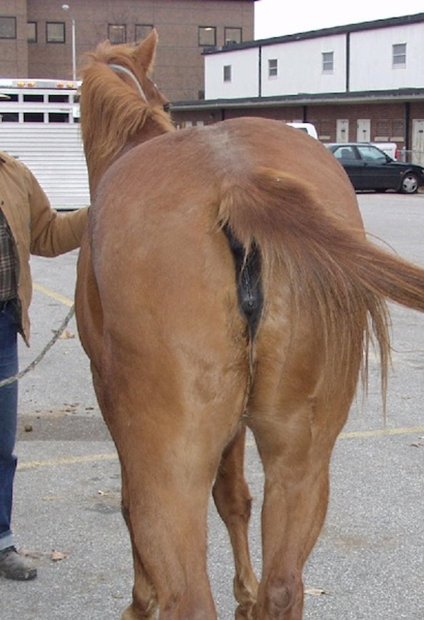
Rupture of the gastrocnemius muscle has caused lameness and marked swelling of the left thigh in this horse.
Courtesy of Dr. Stephen Adams.
Muscle contractures and fibrosis can be devastating injuries resulting in shortening of functional muscle length and impairment of the muscle's function (see ). The development of excessive scar tissue between muscle fibers inhibits the contractility of the muscle, limiting its ability to flex or extend to its greatest capacity. Injuries such as muscle lacerations, muscle tearing (as can occur during a fracture), iatrogenic damage due to surgical trauma, and formation of excessive scar tissue after an injury or surgery can result in abnormal muscle and joint function. In severe cases, muscle contracture and fibrosis can ultimately inhibit the ability to flex a major joint, as can occur in the stifle of young dogs with quadriceps tie-down after femoral fracture.


Contracture of the quadriceps muscle in a dog. This dog had a previous chronic femoral fracture that healed with contracture and fibrosis of the quadriceps muscles. Note the straight alignment of the right hindlimb. This dog was unable to flex its stifle and coxofemoral joint because of fibrous adhesions in the muscle.
Courtesy of Dr. Michael Jaffe.
Primary striated (skeletal) muscle tumors are relatively uncommon in animals and can be benign (rhabdomyoma) or malignant (rhabdomyosarcoma). These tumors can occur in a variety of tissue types, including heart, larynx, and other skeletal muscle tissues. Rhabdomyosarcomas can be locally invasive and can metastasize, most commonly to the lungs and less commonly to other organs (liver, spleen, kidneys, or adrenal glands). Clinical signs depend on the location of the tumor and can include localized swelling, lameness, and a palpable mass. Provisional diagnosis, based on physical examination and imaging, is confirmed by biopsy and histological examination. Surgical excision is the primary treatment recommended, when possible. Limb amputation might be necessary when primary mass excision cannot be performed. Chemotherapy and radiation therapy might be necessary for incompletely excised tumors or when surgery is not possible. The prognosis for patients with rhabdomyosarcomas is guarded to poor, depending on the invasiveness and metastasis of the tumor.
Also see Muscular Trauma in Dogs and Cats.
For More Information
Steiss JE. Muscle disorders and rehabilitation in canine athletes. Vet Clin North Am Small Anim Pract. 2002;32(1):267-285.
Aleman M. A review of equine muscle disorders. Neuromuscul Disord. 2008;18(4):277-287.
Karthika K, Ramkumar PK. Diseases and disorders of the musculoskeletal system in dogs and cats. In: Rana T, ed. Introduction to Diseases, Diagnosis, and Management of Dogs and Cats. Elsevier; 2024:321-337.
Also see pet owner content regarding muscle disorders in dogs, cats, and horses.


