

Hemorrhagic enteritis virus belongs to the family Adenoviridae, genus Siadenovirus, species turkey adenovirus 3 (TAdV-3). It is serologically and antigenically indistinct from marble spleen disease virus. Clinical signs include splenomegaly in both turkeys and chickens, bloody diarrhea in turkeys, and acute respiratory disease in chickens. Clinical signs and lesions can allow a provisional diagnosis, which is normally confirmed by molecular diagnostic tests, mainly PCR assay. There is no direct treatment available. However, live virus vaccines can be used for prevention and to control outbreaks. Hygiene measures should be implemented to decrease the risk of secondary infection as a consequence of virus-induced immunosuppression.
Hemorrhagic enteritis (HE) is an acute disorder affecting young turkeys ≥ 4 weeks old. In its most severe form, it is characterized by depression and hemorrhagic droppings. Mortality is now relatively low in most operations because of the extensive use of vaccines.
Marble spleen disease (MSD) is an acute respiratory disease of pheasants characterized by depression, enlarged and mottled spleens, pulmonary congestion, and death.
HE and MSD are caused by genetically similar viruses. Species-specific differences in the clinical response are thought to be related to differences in the target organs for anaphylaxis and variation in viral pathotype. Infection with less virulent pathotypes in either host can often go undetected until secondary bacterial infections begin to develop as a result of viral-induced immunosuppression.
Diseases similar to HE and MSD have been observed sporadically in other species of birds, such as chickens (avian adenovirus splenomegaly [AAS]), guinea fowl, peafowl, and chukar partridges. Other siadenoviruses have been reported to cause disease in pigeons, finches, penguins, and various psittacine species.
Etiology and Epidemiology of Hemorrhagic Enteritis and Marble Spleen Disease
Hemorrhagic enteritis in turkeys and marble spleen disease in pheasants were reported for the first time in 1937 and 1972, respectively. The etiological agent of HE and related infections such as MSD and avian adenovirus splenomegaly is a nonenveloped, icosahedral DNA virus, 70–90 nm in diameter. It is a member of the family Adenoviridae and the genus Siadenovirus, species turkey adenovirus 3 (TAdV-3). Differences in presentation within host species suggest that numerous viral pathotypes exist. These differ slightly at the DNA level but are indistinguishable serologically.
HE, MSD, and AAS are geographically widespread and considered endemic in areas where turkeys, pheasants, and chickens are raised commercially. Because of the widespread use of HE vaccines in turkey-producing countries, clinical HE is less frequently observed; however, hemorrhagic enteritis virus (HEV)–associated immunosuppression can still be prevalent, with subsequent losses due to secondary infections.
The usual route of HEV infection is fecal-oral/cloacal ("cloacal drinking"), and the virus is often introduced onto previously uninfected premises via personnel or equipment contaminated with infectious feces. In the case of MSD, aerosol-based transmission cannot be ruled out. As infection begins to cycle through a flock, large quantities of the virus are shed in the feces, facilitating rapid transmission to susceptible birds. Persistent infections with latent shedding can be detected in clinically recovered birds.
Turkey poults and pheasants younger than 3–4 weeks old with HEV normally do not develop clinical signs, because of age-related resistance or, more commonly, the presence of maternally derived antibodies. The virus can survive under moist conditions (ie, in litter) well beyond the refractory period.
Clinical Findings of Hemorrhagic Enteritis and Marble Spleen Disease
Morbidity usually approaches 100% for both hemorrhagic enteritis and marble spleen disease. In commercial operations, HE typically affects turkeys 6–12 weeks old. However, it is most common between 7 and 9 weeks old, when maternally derived HE antibodies have waned.
In HE and MSD outbreaks involving virulent pathotypes, clinical signs can include depression, pallor, and bloody droppings, which are observed within 5–6 days after oral infection and progress quickly. Acute mortality can range from 1% to 60%, with an average of 10–15% throughout a 2-week period (1, 2).
Birds that survive the acute phase of HE and MSD experience transient immunosuppression related to the lymphotrophic, lymphocytopathic nature of the virus. This immunosuppression often manifests in the form of secondary bacterial infections, (eg, colibacillosis) approximately 10–14 days after exposure to the virus. Thus, a second peak in mortality, potentially overlapping the first, can occur and, in less virulent outbreaks, can actually dominate the clinical picture.
The second wave of HE and MSD mortality often lasts 2–4 weeks and is characterized by lesions commonly associated with bacterial respiratory disease or septicemia (eg, fibrinopurulent pneumonia, airsacculitis, pericarditis, peritonitis, perihepatitis, hepatomegaly, and splenomegaly). Concomitant or prior exposure to necrotic enteritis, coccidiosis, Newcastle disease virus, Bordetella avium, or Mycoplasma gallisepticum and Mycoplasma synoviae can exacerbate the problem. Similar multiple-agent interactions have been implicated in mortality associated with the use of live virus vaccines for hemorrhagic enteritis.
MSD typically affects pheasants kept in captivity when 3–8 months old. Onset is acute, with dyspnea, asphyxiation, and sudden death occurring as a result of pulmonary congestion and edema. Mortality is commonly 2–3%; however, it can reach 15% (3). Secondary bacterial infections as a result of immunosuppression have also been noted.
Lesions
Necropsy of birds infected with hemorrhagic enteritis virus reveals gross congestion and occasional intraluminal hemorrhage in the proximal small intestine (see ). The spleen is usually enlarged, friable, and mottled white (see ), except in birds that have hemorrhaged extensively. Hemorrhage in the intestine is not common in naturally occurring cases observed in production settings or wild populations.

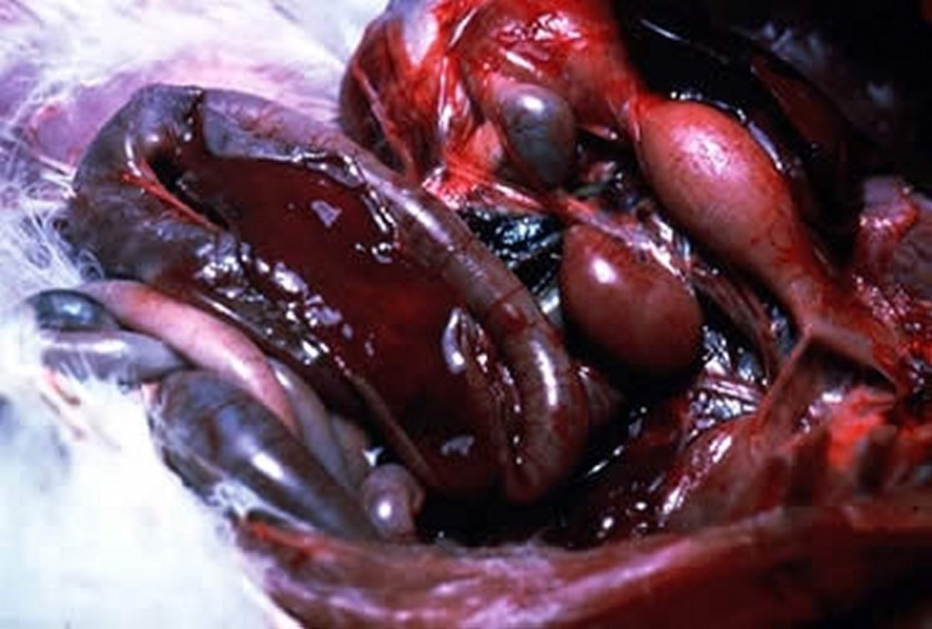
Frank hemorrhage into the intestinal lumen of a turkey with hemorrhagic enteritis.
Courtesy of Dr. Jean Sander.

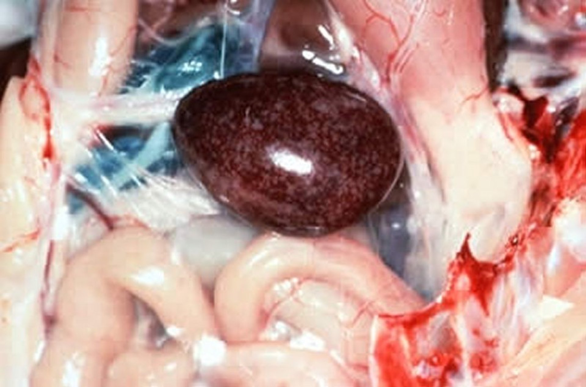
Enlarged, mottled spleen in a turkey with hemorrhagic enteritis.
Courtesy of Dr. Jean Sander.
Histopathological changes of HE and MSD include duodenal congestion, hemorrhage, and epithelial necrosis. These lesions, in particular, are thought to result from a virally induced, cytokine-mediated anaphylactic reaction, with the GI tract considered the target shock organ in turkeys.
Basophilic intranuclear inclusions caused by HE and MSD can be found in lymphocytes and macrophages in a variety of tissues (eg, intestine, liver, and lungs). However, they are predominantly observed in the spleen, where lymphoreticular hyperplasia and lymphoid necrosis are noted. Intranuclear inclusions in the renal tubular epithelial cells of the kidneys can be present in turkeys that have recovered from hemorrhagic enteritis.
Histopathological evaluation of pheasants with MSD often reveals atrial and tertiary bronchial flooding with fibrin and RBCs along with generalized pulmonary vascular congestion and focal necrosis. As with HE, this response can be anaphylactic in nature, with the lung considered the target shock organ in the pheasant.
Splenomegaly with lymphoreticular hyperplasia and lymphoid necrosis also occur and are the characteristic lesions for which MSD is named. Basophilic or magenta-colored intranuclear inclusions can be found in a variety of tissues outside of the GI tract, with the highest concentration of virus found in the spleen.
Pearls & Pitfalls
|
Diagnosis of Hemorrhagic Enteritis and Marble Spleen Disease
PCR assay
ELISA
Diagnosis of virulent outbreaks of hemorrhagic enteritis or marble spleen disease can often be made on the basis of clinical signs and gross lesions. Confirmation is by histopathology and the presence of seroprecipitating virus in the spleen as determined by agar gel immunodiffusion. Hemorrhagic enteritis virus isolation and propagation in the cell line MDTC-RP19, spleen or B cell suspension cultures, or naive birds (turkeys and chickens) is possible but time-consuming.
Various PCR assay techniques to detect HEV viral DNA in tissues have also been described and are in regular use. Sequencing approaches can be used to differentiate vaccine and virulent field strains. However, differences at the nucleic acid level are minor, so sequencing must go beyond a single viral gene; most likely, multiple genes will need to be sequenced (4).
To determine whether HEV or MSD virus is a predisposing factor in cases of bacterial respiratory disease or septicemia, or to verify a primary diagnosis, acute and convalescent sera (3 weeks apart) can be tested using either agar gel immunodiffusion or ELISA. ELISA is also commercially available.
In turkeys, differential diagnoses include colibacillosis, pasteurellosis, paratyphoid, and erysipelas. Reticuloendotheliosis or lymphoproliferative disease should be considered when lymphoreticular hyperplasia is the predominant lesion. GI lesions without splenic involvement should evoke consideration of other viral, bacterial, parasitic, and toxic enteritides of turkeys.
In pheasants with acute respiratory disease, differential diagnoses include Newcastle disease, avian influenza, Syngamus trachea, and, in the case of birds reared in confinement, gaseous toxins.
Treatment, Control, and Prevention of Hemorrhagic Enteritis and Marble Spleen Disease
Proper vaccination with live attenuated vaccine or avirulent strains
Improved hygiene measures
Prevention of hemorrhagic enteritis or marble spleen disease normally relies on the use of live virus vaccines administered in the drinking water to turkeys at approximately 4–5 weeks of age, when maternally derived antibodies are below the breakthrough concentration of the vaccine. Tissue culture products and crude splenic preparations containing avirulent isolates produce lifelong protection, possibly because of virus persistence, and are licensed in different countries. If there is high variation in maternally derived antibody concentrations within a flock, a second vaccination may be necessary to achieve sufficient protection.
A subunit vaccine and a full-antigen inactivated vaccine to prevent HE in turkeys have been described in Europe. Antibodies have been suggested to be the main determinant of protection. However, cell-mediated immunity has also been shown to be stimulated after infection. Virus-neutralizing epitopes have been identified within the hexon protein, the major structural capsid protein of the virus, and protection is not strain-specific. It is possible that vaccination can contribute to antigenic drift in field strains (ie, genetic changes in viral surface proteins that could allow the virus to evade immune recognition).
Vaccines intended for use in turkeys should not be used in pheasants, and vice versa, because the avirulent isolates used for vaccinating one species are typically virulent in the other. Because of the potential for interaction with other agents, including live virus vaccines, regular disease monitoring and careful integration of HE and MSD vaccines into flock vaccination protocols are encouraged. Vaccines should not be administered to birds exhibiting clinical signs or within 2 weeks after any other vaccination.
Pearls & Pitfalls
|
Secondary bacterial infections can be treated with antimicrobials according to the resistance profile of the bacterial isolates. Vaccination must be accompanied by biosecurity and hygiene measures. Chlorine, iodine, and quaternary ammonium-based disinfectants have varying effectiveness against the nonenveloped viruses; chlorine is highly effective against adenoviruses. All-in, all-out management practices can help decrease the risk of transmission between flocks. However, elimination of the virus on farms with multiple flocks of different ages is difficult.
HE outbreaks have also been successfully treated and controlled by injection of exposed birds with 0.5–1 mL of antiserum obtained from recovered flocks. It is presumed that a similar approach can be effective for pheasants. Because of the amount of labor involved, this approach is rarely used.
Key Points
Vaccination, in combination with hygiene measures, is important for the control of HE and MSD.
Maternal antibodies can interfere with the vaccine response, so vaccine administration should be timed for birds approximately 4–5 weeks old.
In the case of high variation in maternally derived antibody concentrations within a flock, a second vaccination could be necessary to achieve sufficient protection.
For More Information
Fitzgerald SD, Rautenschlein S, Mahsoub HM, Pierson FW, Reed WM, Jack SW. Adenovirus infections. In: Swayne DE, ed. Boulianne M, Logue CM, McDougald LR, Nair V, Suarez DL, associate eds. Diseases of Poultry. 14th ed. Wiley Blackwell; 2020:321-363.
Dhama K, Gowthaman V, Karthik K, et al. Hemorrhagic enteritis of turkeys - current knowledge. Vet Q. 2017;37(1):31-42.
Mahsoub HM. Real Time PCR-Based Infectivity Assay and Characterization of Cell Surface Receptors for Turkey Hemorrhagic Enteritis Virus. Dissertation. Virginia Polytechnic Institute and State University; 2015.
Jakowski RM, Wyand DS. Marble spleen disease in ring-necked pheasants: demonstration of agar gel precipitin antibody in pheasants from an infected flock. J Wildl Dis 1972;8(3):261-263.
Carlson HC, Pettit JR, Hemsley RV, Mitchell WR. 1973. Marble Spleen Disease of Pheasants in Ontario. Can J Comp Med 1973;37(3):281-286.
Domermuth CH, Gross WB, Schwartz LD, Mallinson ET, Britt R. Vaccination of Ring-Necked Pheasant for Marble Spleen Disease. Avian Dis 1979;23(1):30-38.
References
Gross WB, Moore WE. Hemorrhagic enteritis of turkeys. Avian Dis 1967;11(2):296-307.
Pomeroy BS, Fenstermacher R. Hemorrhagic enteritis in turkeys. Poult Sci 1937;16(6):378-382. doi:10.3382/ps.0160378
Fitzgerald SD, Rautenschlein S, Mahsoub HM, Pierson FW, Reed WM, Jack SW. Adenovirus infections. In: Swayne DE, ed. Boulianne M, Logue CM, McDougald LR, Nair V, Suarez DL, associate eds. Diseases of Poultry. 14th ed. Wiley Blackwell; 2020:321-363. doi:10.1002/9781119371199.ch9
Beach NM, Duncan RB, Larsen CT, Meng X-J, Sriranganathan N, Pierson FW. Comparison of 12 turkey hemorrhagic enteritis virus isolates allows prediction of genetic factors affecting virulence. J Gen Virol 2009;90:1978-1985. doi:10.1099/vir.0.010090-0


