Fasciolopsiasis is a foodborne parasitic disease that is prevalent in Asian and Indian regions. It is transmitted through consumption of parasitized plants, the second intermediate host. The parasite embeds itself in the duodenum and can cause intestinal signs in affected humans and animals.
Etiology and Pathogenesis of Fasciolopsiasis
Fasciolopsiasis is a parasitic zoonosis caused by the trematode Fasciolopsis buski, known as the giant intestinal fluke because it measures approximately 20–75 × 8–20 mm. F buski is found in many parts of Asia and the Indian subcontinent. Fasciolopsiasis is an important foodborne infection; an estimated 10 million people are infected worldwide.
F buski causes serious illness and substantial economic losses in agriculture. Although global estimates of animal fasciolopsiasis are unavailable, individual reports from several Asian countries indicate that its prevalence in pigs can exceed 40% (1, 2, 3).
F buski has three hosts in its life cycle (1, 3):
First intermediate host: freshwater snails
Second intermediate host: aquatic plants such as bamboo shoots, watercress, water chestnuts, or lotus tubers
Definitive host or reservoir: humans and aquatic plant-eating animals
F buski infects snails in the Planorbidae family, including species in the genera Segmentina, Hippeutis, Giraulus, Lymnaea, and Indoplanorbis. In addition to humans and pigs, some aquatic, plant-eating animals, such as swamp buffalo, ducks, and chickens, are also suspected reservoir hosts (2, 3).
The life cycle of F buski (see ) begins with adult flukes attached to the duodenal wall of definitive hosts (most commonly humans and pigs). These parasites release eggs in the feces, which can contaminate water and be ingested by freshwater snails. Miracidia (the larval stage) first pass through several larval stages to become cercariae (free-swimming larvae). Snails then release cercariae, which encyst as metacercariae, the resting or mature stage, on aquatic plants.

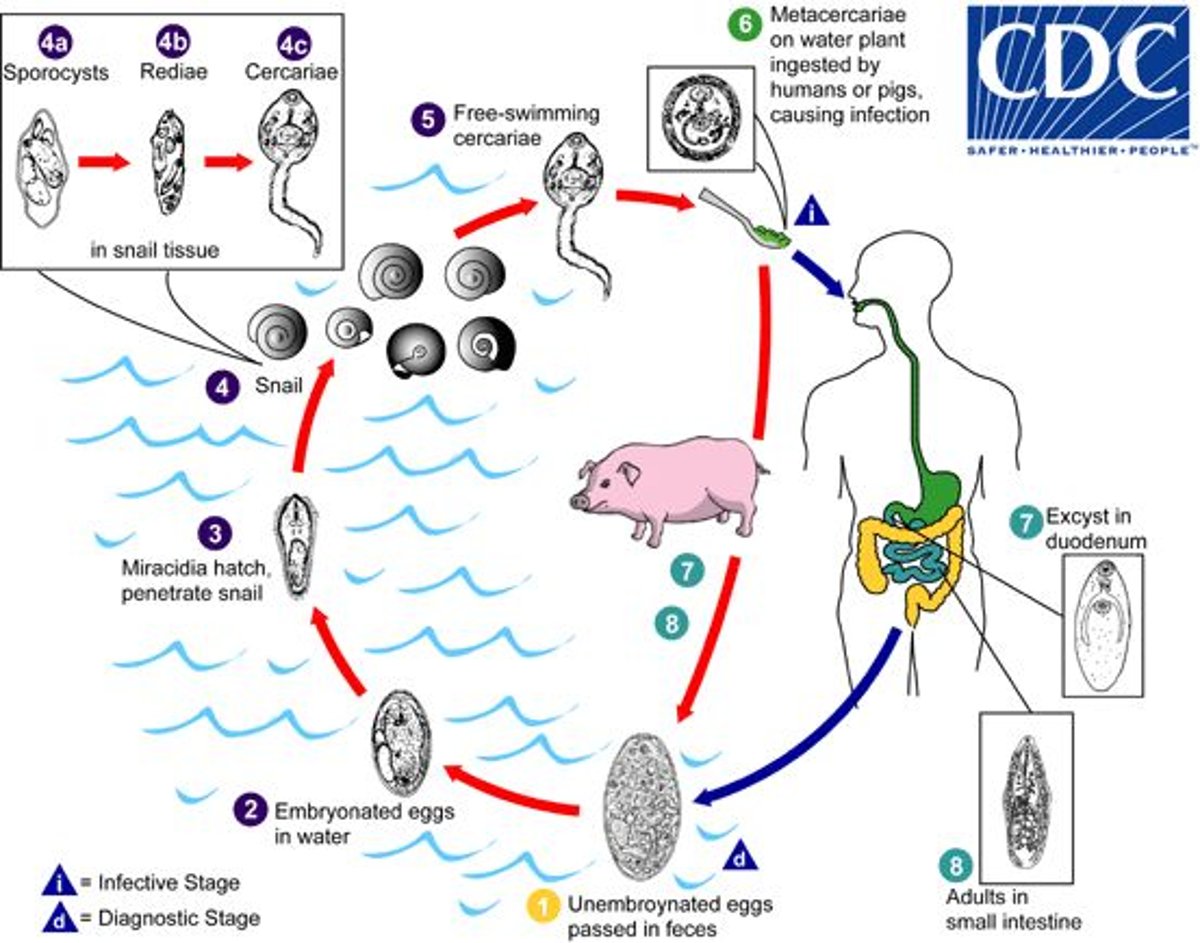
The life cycle of Fasciolopsis buski, the fluke that causes fasciolopsiasis. Immature eggs are discharged into the intestine and feces of mammalian hosts (humans and pigs). Eggs embryonate in water and release miracidia, which invade a snail intermediate host. In the snail, miracidia undergo several developmental stages (sporocyst, redia, and cercaria). Cercariae are released from the snail and encyst as metacercariae on aquatic plants. Mammalian hosts become infected by ingesting these metacercariae along with aquatic plants. Metacercariae then excyst in the mammal's duodenum, attach to the intestinal wall, and develop in approximately 3 months into adult flukes (20–75 × 8–20 mm) that live approximately 1 year.
Developed by the CDC. This diagram is available for no charge at https://www.cdc.gov/dpdx/fasciolopsiasis. (Reference to specific commercial products, manufacturers, companies, or trademarks does not constitute their endorsement or recommendation by the US Government, Department of Health and Human Services, or Centers for Disease Control and Prevention.)
Humans and other mammals can become infected with F buski by consuming infected plants. Metacercariae are released in the duodenum, where they attach to the intestinal wall. There they develop into adults in approximately 3 months and live approximately 1 year (1).
Practices that contribute to F buski transmission include poor sanitation and open defecation, the use of contaminated water, and the consumption of undercooked or raw aquatic plants. Animals that defecate widely can accelerate egg transfer into ponds or rivers and worsen epidemics (2, 4).
Clinical Findings of Fasciolopsiasis
Diagnosis of Fasciolopsiasis
Detection of eggs via microscopy
Detection of adult flukes in feces or vomit
Microscopic detection of eggs is the most common method of identifying F buski. Less commonly, adult flukes can be found in feces or vomitus. F buski eggs are ellipsoidal; have a thin shell and a small, indistinct operculum; and measure 130–150 × 60–90 mcm. The eggs cannot be easily distinguished from those of Fasciola sp (1).
Control of Fasciolopsiasis
Routine targeted deworming of animals
Thorough cooking of aquatic plants before consumption
Sanitation to eliminate intermediate hosts
To decrease the risk of fasciolopsiasis and decrease endemicity, the following veterinary public health measures can be helpful:
Feeding raw aquatic plants to animals in endemic areas should be avoided.
Domestic animals should be treated with praziquantel.
Snail control is important in endemic areas.
Sanitation should be improved by promoting latrines, decreasing fecal contamination of fresh water, and providing a safe drinking-water supply.
Adequate cooking of aquatic plants and safe food-handling practices should be promoted.
Contaminated ponds and distribution of infected plants and snails in endemic areas should be monitored.
Currently, no commercial vaccine against F buski is available (2, 3, 4).
Key Points
Fasciolopsiasis is caused by the trematode Fasciolopsis buski, which is prevalent in parts of Asia and the Indian subcontinent.
F buski causes serious illness and substantial economic losses in agriculture.
F buski requires three hosts in its life cycle: freshwater snails; aquatic plants; and humans and aquatic plant-eating animals.
Poor sanitation, open defecation, and the consumption of undercooked or raw aquatic plants contribute to the transmission of F buski.
For More Information
Fasciolopsiasis. CDC.
References
CDC. Fasciolopsiasis. June 5, 2024. https://www.cdc.gov/dpdx/fasciolopsiasis/index.html
Siles-Lucas M, Becerro-Recio D, Serrat J, González-Miguel J. Fascioliasis and fasciolopsiasis: current knowledge and future trends. Res Vet Sci. 2021;134:27-35. doi:10.1016/j.rvsc.2020.10.011
Ridha MR, Indriyati L, Andiarsa D, Wardhana AH. A review of Fasciolopsis buski distribution and control in Indonesia. Vet World. 2021;14(10):2757-2763. doi:10.14202/vetworld.2021.2757-2763
CDC. About Fasciolopsis. April 25, 2024. Accessed October 12, 2026. https://www.cdc.gov/fasciolopsis/about