Ovarian Abnormalities
The most common congenital abnormality of the ovary is ovarian dysgenesis, also known as ovarian hypoplasia (see ). Ovarian dysgenesis has been described in several domestic animal species and associated with various chromosomal abnormalities (monosomy X or Turner syndrome, trisomy XXX, or Klinefelter syndrome XXY). The ovaries in affected animals are very small and lack follicular activity.


Laparoscopic diagnosis of ovarian hypoplasia in an alpaca. The tip of the uterine horn is visible in the center of the image (blue arrow). The adjacent ovarian tissue is rudimentary in size and appearance (yellow arrow).
Courtesy of Dr. Ahmed Tibary.
Segmental Aplasia of the Paramesonephric Ducts
The paramesonephric ducts are paired embryonic ducts that develop into the anterior vagina, cervix, uterus, and uterine tube. Segmental aplasia of the paramesonephric ducts results in anomalies of those organs. The aplasia (obstruction) can be located in a segment of the uterine tube, uterine horn, cervix, or vagina. Ovarian development is normal. Accumulation of secretions proximal to the obstruction occurs secondarily (hydrosalpinx, hydrometra, mucometra, hematocolpometra).
See images of ; ; ; and .

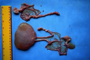
Hydrosalpinx due to segmental aplasia in a female dromedary.
Hydrosalpinx due to segmental aplasia in a female dromedary.
Courtesy of Dr. Ahmed Tibary.


Ultrasonographic diagnosis of mucometra due to vaginal aplasia in an alpaca. The uterine horn (arrow) is distended with anechoic fluid due to the obstruction of the vaginal opening.
Ultrasonographic diagnosis of mucometra due to vaginal aplasia in an alpaca. The uterine horn (arrow) is distended with
Courtesy of Dr. Ahmed Tibary.

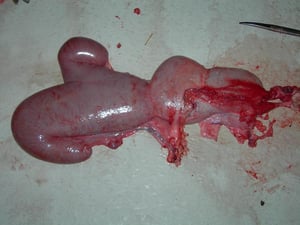
Mucometra and mucocolpos due to vaginal aplasia in an alpaca.
Mucometra and mucocolpos due to vaginal aplasia in an alpaca.
Courtesy of Dr. Ahmed Tibary.


Uterus unicornis in an alpaca. The left horn (arrow A), left ovary (arrow B), and the right ovary (arrow C) are present. The right horn is absent.
Uterus unicornis in an alpaca. The left horn (arrow A), left ovary (arrow B), and the right ovary (arrow C) are present
Courtesy of Dr. Ahmed Tibary.
Hydrosalpinx due to segmental aplasia in a female dromedary.
Hydrosalpinx due to segmental aplasia in a female dromedary.
Courtesy of Dr. Ahmed Tibary.
Ultrasonographic diagnosis of mucometra due to vaginal aplasia in an alpaca. The uterine horn (arrow) is distended with anechoic fluid due to the obstruction of the vaginal opening.
Ultrasonographic diagnosis of mucometra due to vaginal aplasia in an alpaca. The uterine horn (arrow) is distended with
Courtesy of Dr. Ahmed Tibary.
Mucometra and mucocolpos due to vaginal aplasia in an alpaca.
Mucometra and mucocolpos due to vaginal aplasia in an alpaca.
Courtesy of Dr. Ahmed Tibary.
Uterus unicornis in an alpaca. The left horn (arrow A), left ovary (arrow B), and the right ovary (arrow C) are present. The right horn is absent.
Uterus unicornis in an alpaca. The left horn (arrow A), left ovary (arrow B), and the right ovary (arrow C) are present
Courtesy of Dr. Ahmed Tibary.
Segmental aplasia of the uterus can involve one horn (resulting in a condition called uterus unicornis), both horns, or only part of one horn (which can result in cystic dilatation of the uterine horn anterior to the area of dilatation). Uterus unicornis has been described in several domestic animal species. The condition is among the most commonly diagnosed congenital abnormalities of the reproductive tract in female camelids (1). Affected females can become pregnant and carry the pregnancy to term in the existing uterine horn. Cervical aplasia has been described in a few cases but is not as common, in the author's experience (2).
True persistence of the hymen or imperforate hymen is the most commonly reported paramesonephric duct anomaly in domestic animals. Fluid accumulates in the vagina and uterus, resulting in protrusion of the hymen at the vulva when the animal is lying down or straining. Hymenal defects are most common in white Shorthorn cattle (white heifer disease).
Cervical Abnormalities
Double external os of the cervix is due to a failure of the paramesonephric ducts to fuse. It might present as a band of tissue caudal to, or in, the external os of the cervix. In other cases, there is a true double external os opening into a single caudal part of the cervical canal. Affected cows usually conceive normally.
Rarely, a true double cervix, with a complete septum between the two cervical canals, each opening into its respective uterine horn (uterus didelphys), occurs.
Vaginal and Vulvar Abnormalities
Vaginal stricture or vestibulovaginal hypoplasia has been described in mares, dogs, and camelids (1, 3, 4).
Gartner ducts, located beneath the mucosa of the floor of the vagina, can develop multiple cysts, which are generally of no clinical importance.
Vulvar atresia or hypoplasia (atresia vulvi) has been described primarily in camelids (1, 5). In extreme cases, the labia are completely fused. The condition is believed to be due to an autosomal recessive gene.
Rectovaginal constriction is a connective tissue disorder of Jersey cattle characterized by stenosis of the anus and/or vestibulovaginal sphincter. Rectovaginal constriction is inherited as a simple autosomal recessive defect. Anal stenosis renders transrectal palpation difficult. Affected females experience severe dystocia. The prevalence of the disease has been substantially decreased because of identification of carrier bulls.
Bilateral stenosis of the milk veins has also been described and results in udder edema and ischemic necrosis.
See images of ; ; ; ; and .

Cystic dilation of Gartner ducts in a heifer (yellow arrows).
Cystic dilation of Gartner ducts in a heifer (yellow arrows).
Courtesy of Dr. Ahmed Tibary.
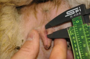
Vulvar atresia/hypoplasia in an alpaca.
Vulvar atresia/hypoplasia in an alpaca.
Courtesy of Dr. Ahmed Tibary.


Reproductive tract from a 1-day-old alpaca with atresia vulvi and fused labia (site of fused labia: yellow arrow).
Reproductive tract from a 1-day-old alpaca with atresia vulvi and fused labia (site of fused labia: yellow arrow).
Courtesy of Dr. Ahmed Tibary.
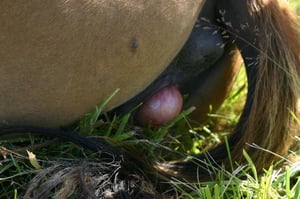
Persistent hymen (complete imperforate hymen) in a mare. Note the protrusion of the hymen due to presence of fluid in the vagina.
Persistent hymen (complete imperforate hymen) in a mare. Note the protrusion of the hymen due to presence of fluid in t
Courtesy of Dr. Ahmed Tibary.
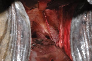
Persistent hymen and incomplete double vagina in a mare. Note the septum separating the vagina into two cavities.
Persistent hymen and incomplete double vagina in a mare. Note the septum separating the vagina into two cavities.
Courtesy of Dr. Ahmed Tibary.
Cystic dilation of Gartner ducts in a heifer (yellow arrows).
Cystic dilation of Gartner ducts in a heifer (yellow arrows).
Courtesy of Dr. Ahmed Tibary.
Vulvar atresia/hypoplasia in an alpaca.
Vulvar atresia/hypoplasia in an alpaca.
Courtesy of Dr. Ahmed Tibary.
Reproductive tract from a 1-day-old alpaca with atresia vulvi and fused labia (site of fused labia: yellow arrow).
Reproductive tract from a 1-day-old alpaca with atresia vulvi and fused labia (site of fused labia: yellow arrow).
Courtesy of Dr. Ahmed Tibary.
Persistent hymen (complete imperforate hymen) in a mare. Note the protrusion of the hymen due to presence of fluid in the vagina.
Persistent hymen (complete imperforate hymen) in a mare. Note the protrusion of the hymen due to presence of fluid in t
Courtesy of Dr. Ahmed Tibary.
Persistent hymen and incomplete double vagina in a mare. Note the septum separating the vagina into two cavities.
Persistent hymen and incomplete double vagina in a mare. Note the septum separating the vagina into two cavities.
Courtesy of Dr. Ahmed Tibary.
For More Information
Parkinson TJ. Infertility in the cow due to functional and management deficiencies. In: Noakes DE, Parkinson TJ, England GCW, eds. Veterinary Reproduction and Obstetrics. 10th ed. Saunders Ltd; 2019:361-407.
Raudsepp T. Genetics of equine reproductive diseases. Vet Clin North Am Equine Pract. 2020;36(2):395-409.
Griffin B, White S, Root Kustritz MV. Disorders of sexual development and common reproductive pathologies. In: White S, ed. High‐Quality, High‐Volume Spay and Neuter and Other Shelter Surgeries. Wiley-Blackwell; 2020:27-51.
References
Sghiri A, Ciccarelli M, Waqas MS, Anouassi A, Tibary A. Advances in the diagnosis of reproductive disorders in female camelids. Animals. 2025;15(19):2902. doi:10.3390/ani15192902
Payan-Carreira R, Quaresma M. Cervical hypoplasia in jennets: a description of two cases. Reprod Domest Anim. 2010;45:540-544. doi:10.1111/j.1439-0531.2008.01231.x
Guimarães T, Oliveira C, Crespo J, Porto B, Cunha R, Rocha A. Congenital stricture of the vestibulo-vaginal fold in a mare with normal karyotype. Equine Vet Educ. 2021;33(11}:e400-e402. doi:10.1111/eve.13425
Palm CA, Segev G, Shipov A, Balsa IM, Phillips KL, Culp WTN. Treatment of a congenital imperforate vestibulovaginal junction and secondary hydrocolpos with endoscopic laser ablation in two dogs. Top Companion Anim Med. 2021;45:100576. doi:10.1016/j.tcam.2021.100576
Wilkins PA, Southwood LL, Bedenice D. Congenital vulvar deformity in 6 alpacas. J Am Vet Med Assoc. 2006;229(2):263-265. doi:10.2460/javma.229.2.263