


Posthitis and vulvitis are inflammatory conditions of the external genitalia in small ruminants, particularly sheep and goats. These diseases are of clinical and reproductive importance, affecting animal welfare, mating behavior, and herd productivity. Two major forms are recognized: enzootic posthitis and vulvitis, which are primarily associated with dietary and environmental factors, and ulcerative or necrotic balanoposthitis and vulvitis, which have a more complex and multifactorial etiology.
Enzootic Posthitis and Vulvitis in Sheep and Goats
Enzootic posthitis, also referred to as pizzle rot or sheath rot, is a chronic inflammatory condition of the prepuce and penis, most frequently observed in castrated males (see images of and ). The disease is caused by an overgrowth of Corynebacterium renale, a gram-positive, urease-producing bacterium that normally inhabits the preputial mucosa.

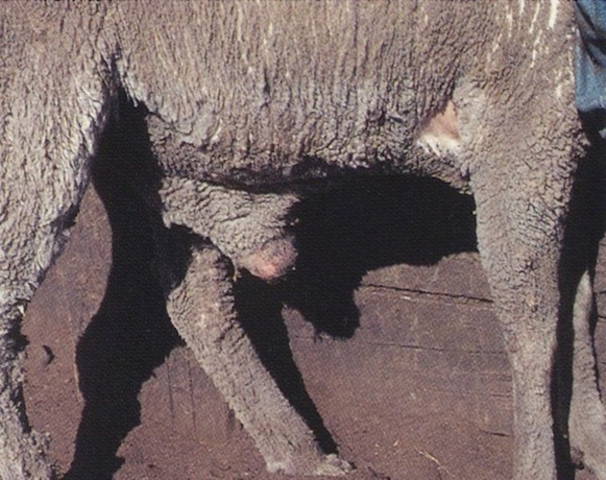
Preputial swelling in an early case of ovine posthitis ("pizzle rot"), ram.
Courtesy of Dr. John Larsen.

Preputial stain and swelling in a ram with ovine posthitis.
Courtesy of Dr. John Larsen.
The pathogenesis of enzootic posthitis is closely linked to high-protein diets, typically those exceeding 16% crude protein. Elevated dietary protein leads to increased urinary urea concentrations, which are hydrolyzed by urease-producing bacteria to release ammonia. This ammonia causes substantial irritation and ulceration of the preputial tissues, creating an environment conducive to bacterial proliferation.
The condition is exacerbated in animals with poor preputial drainage, such as those with hypoplastic penises or incomplete penile-preputial separation, which allows urine to pool and further irritate the tissues. Environmental factors such as caked mud or organic debris around the preputial orifice can also impair drainage and contribute to lesion development.
Breed predispositions have been noted, particularly in Merino and Angora wethers, where long wool retains moisture and promotes bacterial growth.
Enzootic vulvitis is considered the female analog of pizzle rot and follows a similar pathogenesis, with high-protein diets and C renale colonization leading to mucosal irritation and ulceration
Ulcerative Balanoposthitis and Vulvitis in Sheep and Goats
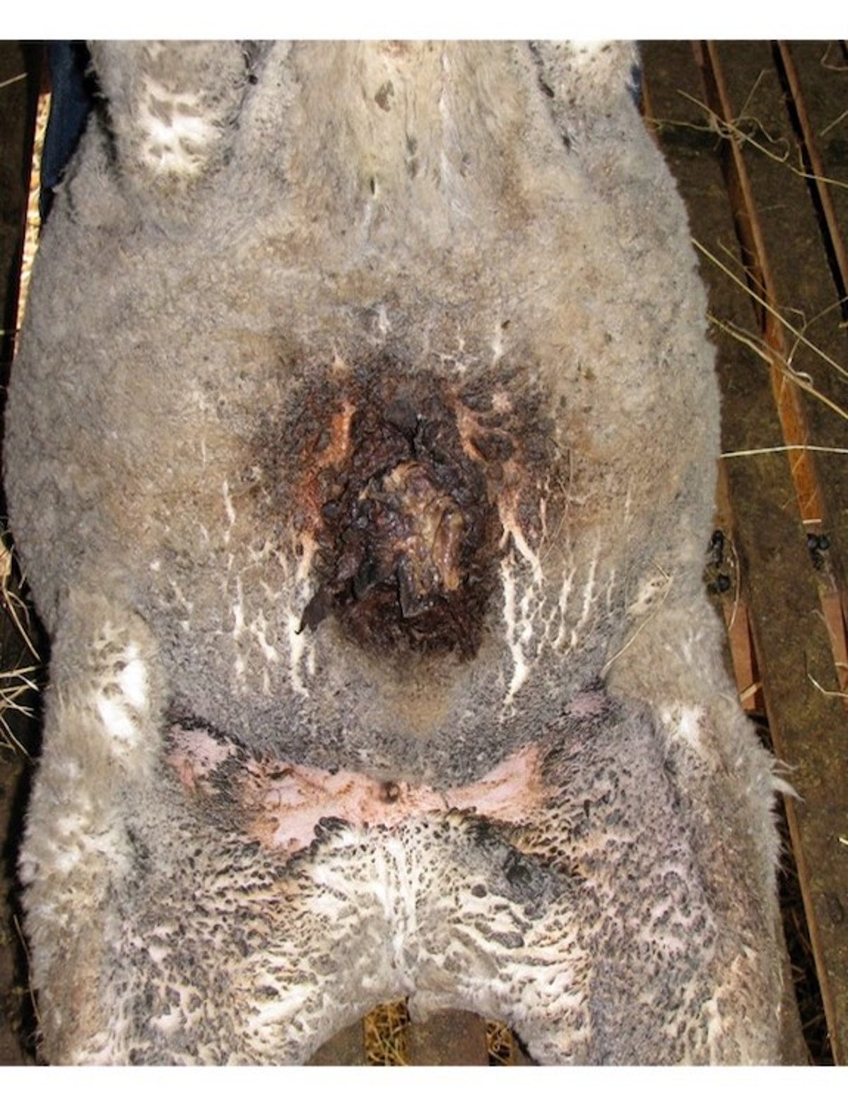
Ulcerative balanoposthitis and vulvitis are characterized by ulceration and inflammation of the glans penis and prepuce in males and the vulva in affected ewes.
Early clinical signs include swelling and bleeding of the prepuce or vulva; on examination, ulcers are typically present and bleed readily when manipulated. In ewes, the ventral aspect of the tail can also be affected due to contact with the inflamed vulva. The condition can spread within a breeding flock, potentially affecting a sizable proportion of animals. Affected rams may show reluctance to mate due to pain, which can lead to decreased pregnancy rates in the flock.
The etiology of ulcerative balanoposthitis and vulvitis remains unclear, though Mycoplasma mycoides mycoides has been isolated from affected sheep, and the bacteria Trueperella pyogenes and Histophilus ovis are frequently present. Viral agents such as ovine herpesvirus 2 and caprine herpesvirus 1 (CapHV1) might also play a role. In goats, CapHV-1 has been associated with both abortion and ulcerative posthitis, highlighting the potential for systemic reproductive consequences.
Ulcerative vulvitis in females is marked by inflammation, swelling, and ulceration of the vulva, often extending to the vestibule and caudal vagina. The glans clitoridis can also be affected, appearing swollen, red, and ulcerated. The condition can be sexually transmitted, particularly during breeding with infected males. Poor vulvar conformation and trauma during parturition or coitus are contributing factors.
Granular vulvitis, a distinct form of vulvar inflammation, is attributed to Ureaplasma spp infection and is characterized by hyperemia and lymphoid proliferation. It is considered a venereal disease and has been linked to infertility and abortion in sheep. Transmission occurs during mating, and clinical signs may include blood around the vulva after coitus. The disease can remain subclinical for several months postexposure.
Diagnosis of posthitis and vulvitis is based on clinical signs, dietary history, and physical examination. Swelling, pain on palpation, and visible ulceration are common findings. Definitive diagnosis requires culture of preputial or vulvar swabs to confirm the presence of causative organisms. Removal of scabs in ulcerative cases typically results in minimal hemorrhage, which helps distinguish these lesions from other conditions. Differential diagnoses include contagious ecthyma (orf), herpes viral infections, neoplasia, trauma, and parasitic infestations.
Treatment strategies depend on the severity of the lesions. Mild cases may respond to local care, including clipping of preputial or vulvar hair, cleaning with nonirritating antiseptic solutions, and application of topical antimicrobials. In more severe cases, systemic antimicrobial therapy with penicillin or cephalosporins might be necessary, particularly if there is concern for ascending infection. Tulathromycin has also been used successfully in some cases.
Surgical intervention may be required to restore preputial patency in cases of stricture, though care must be taken to avoid further adhesions or fibrosis. Acidifying agents such as ammonium chloride may be added to the diet to decrease urine alkalinity and mitigate mucosal irritation. Irrigation of the prepuce and use of mild antiseptic creams may help prevent preputial adhesions. Rams with severe adhesions that prevent full penile extrusion should be culled, as they are unlikely to regain breeding soundness.
Prevention of ulcerative balanoposthitis and vulvitis is considered more effective than treatment. Dietary management is critical, with protein levels ideally maintained below 16% crude protein. Diets should emphasize hay intake and may include urinary acidifiers. Affected animals should be isolated to prevent transmission, and biosecurity measures such as quarantine of new stock, minimizing visitor traffic, and avoiding reuse of needles should be implemented. Hygiene practices, including proper cleaning of clippers and equipment with bactericidal agents, are essential to control spread.
Breeding management, including careful sire selection and monitoring of libido, also plays a role in disease prevention. Recovered animals generally return to normal fertility; however, surveillance is not routinely warranted due to the sporadic nature of outbreaks. However, when the disease goes undetected, its impact on mating behavior and pregnancy rates can be substantial.
In summary, ulcerative and enzootic posthitis and vulvitis are multifactorial diseases involving dietary, infectious, and environmental components. Early diagnosis, appropriate treatment, and preventive management are key to minimizing their impact and maintaining the health and productivity of small ruminant herds.
Key Points
Posthitis and vulvitis are inflammatory conditions of the external genitalia in small ruminants that can impair urination, mating, and overall comfort.
Enzootic forms are linked to high-protein diets and ammonia irritation caused by Corynebacterium renale, especially in castrated males.
Ulcerative forms may involve bacteria such as Mycoplasma and Trueperella and can spread within breeding flocks, reducing fertility.
Diagnosis is based on clinical signs and culture, while treatment includes topical care, antimicrobials, and dietary adjustments.
Prevention focuses on managing protein intake, isolating affected animals, and maintaining good hygiene and biosecurity.
For More Information
Abbott KA. Diseases of the urinary system. In: Abbott KA, ed. Sheep Veterinary Practice. CRC Press; 2024:453-471.
Choudhary S, Davis KJ, Kumar C, Sharma P. In: Rana T, ed. Clinical findings of diseases of goats. Principles of Goat Disease and Prevention. Wiley Blackwell; 2024:33-48.
Falchi L, Pau S, Ledda M, Melosu V, Zedda MT. Lesions of the prepuce and penis in rams: a retrospective study. Vet Res Commun. 2023;47(4):2259-2264.
Scully CM. Management of urologic conditions in small ruminants. Vet Clin North Am Food Anim Pract. 2021;37(1):93-104.
Byers SR. Ulcerative posthitis and vulvitis. In: Smith BP, ed. Large Animal Internal Medicine. 5th ed. Mosby; 2015:895-896.
Jones, M, Miesner MD, Baird AN, Pugh DG. Diseases of the urinary system. In: Pugh DG, Baird AN, eds. Sheep and Goat Medicine. 2nd ed. Elsevier; 2012:325-360.
Greig A. Ulcerative balanitis and vulvitis. In: Aitken ID, ed. Diseases of Sheep. 4th ed. Blackwell; 2007:143-149.


