

When investigating reproductive disorders in the bitch, the male should be assessed first. If the male is a proven sire and the bitch is cycling normally, the probability is as high as 94% that two apparently normal dogs would produce a litter after one or two consecutive estrous cycles with adequate breeding management. The most common cause of infertility in dogs is poor timing.
Prior to an infertility exam, the brood bitch should be examined for both general and reproductive health.
History in Clinical Investigation of Canine Reproduction Disorders
An in-depth history should be taken, regardless of the reason for the infertility. General health history should be collected, including neuter status, prior illnesses, vaccines, deworming, certifications, and diet. This detailed medical history can help to reveal factors that might contribute to infertility or suitability for breeding.
The reproductive history of the patient’s dam, sire, and littermates can provide clues to familial fertility problems (eg, if the patient’s dam had difficulty whelping or was prone to dystocia).
Information about the patient’s past estrous cycles should be collected (eg, age at first estrous cycle, date and duration of estrous cycles, length of interestrous intervals).
It should be noted how the prior breedings were managed and what the outcomes of those breedings were if available. If the client can also provide the progesterone and vaginal cytologic analysis results from previous estrous cycles, it could prove useful to evaluate the trends that may exist. Whether or not those previous estrous cycles resulted in pregnancy and information on the sire used may reveal an area for improvement in the breeding management.
Reproductive Examination in Canine Reproduction Disorders
After a general physical examination, a reproductive examination is performed.
The mammary glands should be carefully palpated for any masses, asymmetry, or any other irregularities.
The external genitalia should be examined. Visual inspection of the vulva’s conformation and digital examination of the vagina may reveal anomalies such as abnormal vulvar conformation, vaginal strictures, or a persistent median septum.
Diagnostic Testing in Canine Reproduction Disorders
Vaginoscopy can aid in identifying the abnormality felt on digital palpation. Visualizing the vaginal mucosa may indicate what stage of the estrous cycle the bitch is currently in based on the amount of edema or crenulation (ie, wrinkling of vaginal mucosa after edema) or evidence of an infectious or inflammatory process.
Special equipment (endoscope) can be purchased for this technique (entire vagina), but a modified otoscope with a long cone is also effective (caudal vagina).

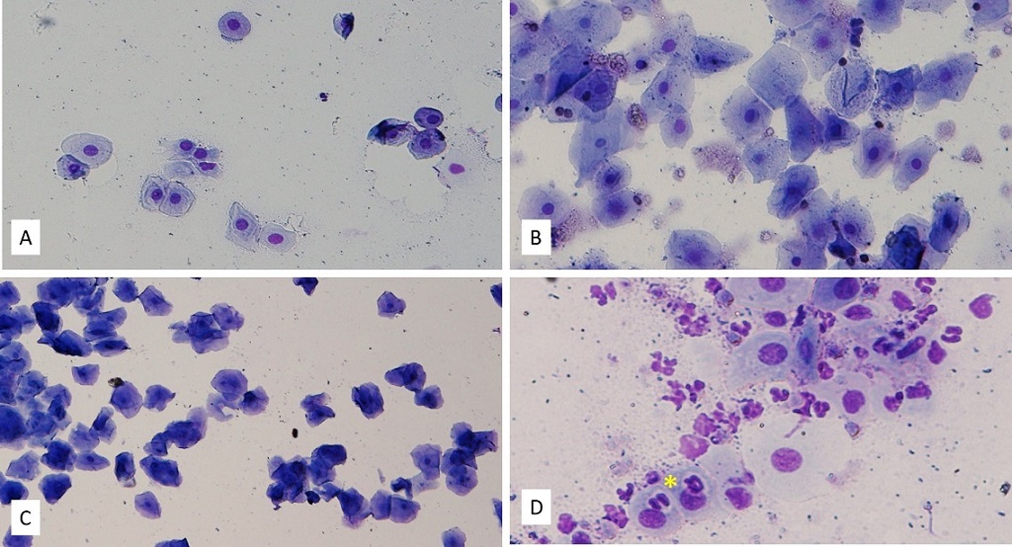
Photomicrograph of exfoliative cytologic slides from dogs in different stages of the estrous cycle.
A. Anestrus. There is low cellularity, and the vaginal epithelial cells are mostly parabasal. Original magnification, 200×; Romanowsky variant stain.
B. Late proestrus. The vaginal epithelial cells are mainly large intermediate cells. Few superficial cells, RBCs, and bacteria are also noted. Original magnification, 200×; Romanowsky variant stain.
C. Estrus. There is a predominance of superficial vaginal epithelial cells (full cornification) and clear background. Few bacteria are noted. Original magnification, 100×; Romanowsky variant stain.
D. Early diestrus. There are small intermediate and parabasal vaginal epithelial cells, bacteria, numerous neutrophils, and moderate debris. Two metestrum cells (asterisk) are evident. Original magnification, 200×; Romanowsky variant stain.
Courtesy of Dr. Viviane Gomes, Louisiana State University.

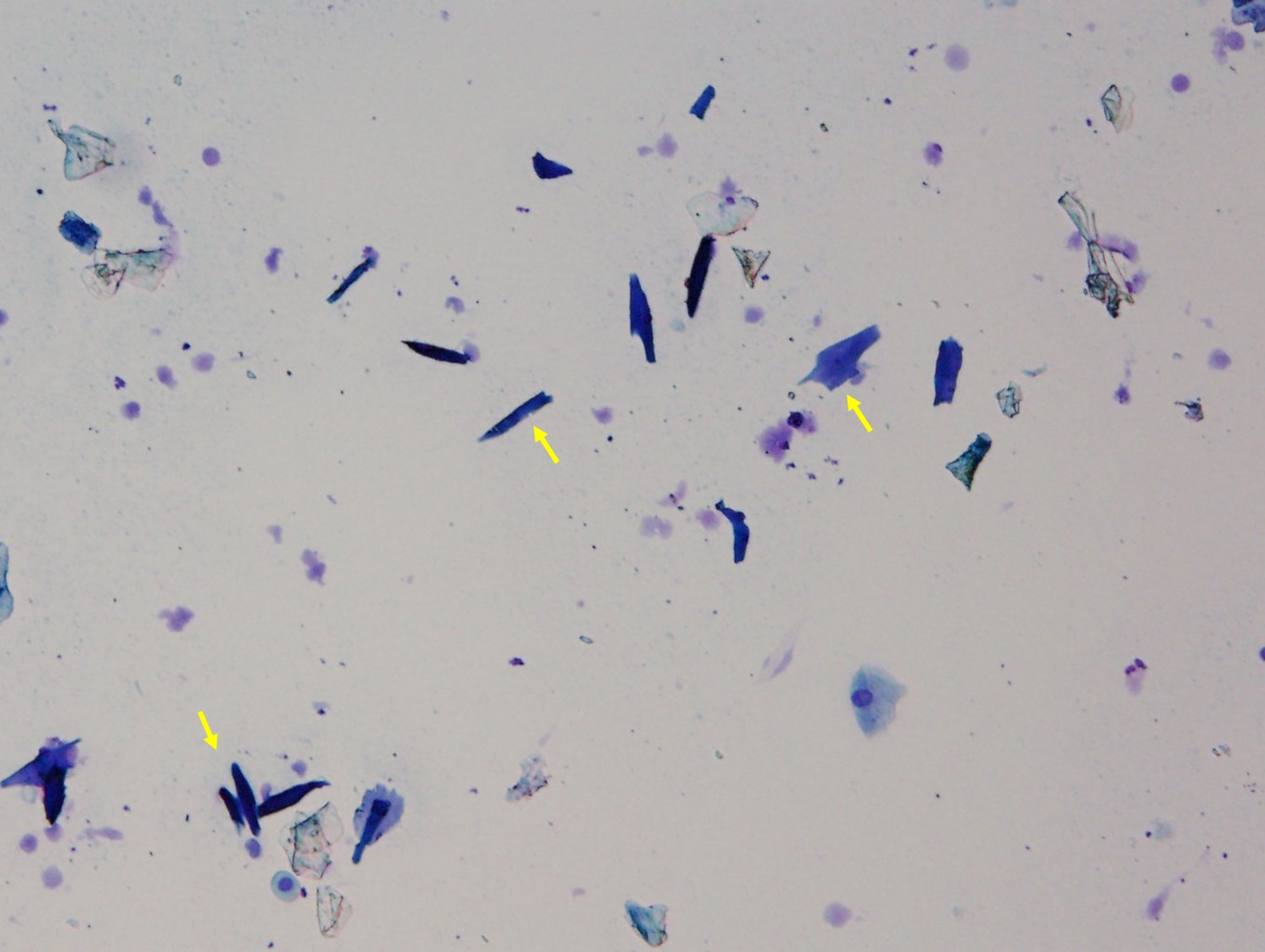
Photomicrograph of exfoliative cytologic slides from the caudal aspect of a dog's vaginal canal. Low cellularity and multiple vestibular cells (arrows) are evident. Original magnification, 100×; Romanowsky variant stain.
Courtesy of Dr. Viviane Gomes, Louisiana State University.

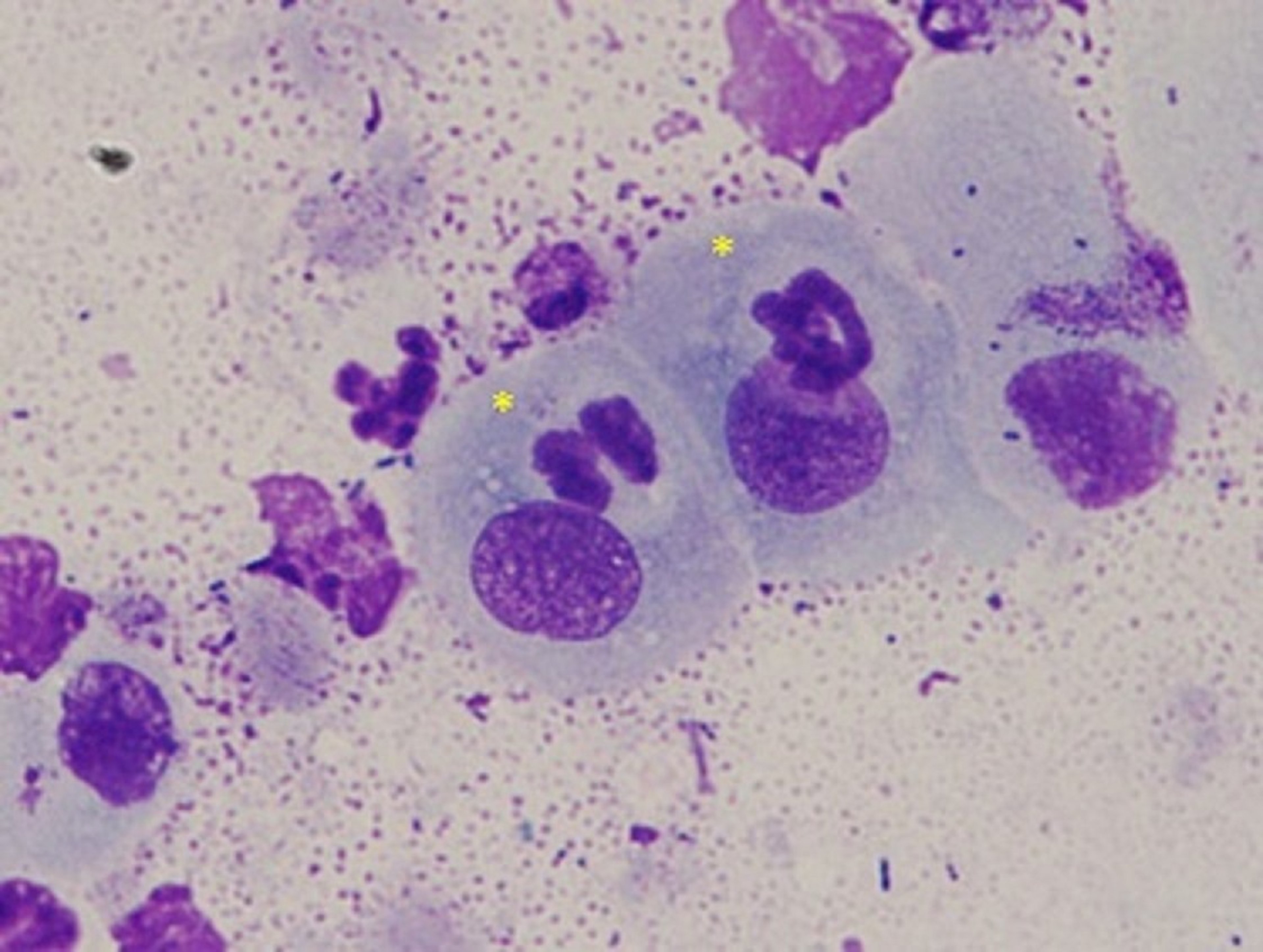
Photomicrograph of exfoliative vaginal cytologic slide of a bitch during early diestrus, showing two metestrum cells (asterisks), a large intermediate cell, neutrophils, and bacteria. Original magnification, 1000×; Romanowsky variant stain.
Courtesy of Dr. Viviane Gomes, Louisiana State University.
Vaginoscopy is a useful method for monitoring the estrous cycle. Permanent longitudinal mucosal folds (plicae) are present in the caudal region of the vagina and change depending on hormonal influences. In proestrus, the plicae become thickened, rounded, and moist. During early estrus, crenulation (wrinkling) of the vaginal folds occurs. In late estrus, the folds are sharp, peaked, and angulated. Throughout diestrus and anestrus, the folds are low and rounded.
Vaginal swab samples for microbial culture should be obtained. The use of a double-guarded swab can help prevent contamination of the sample. Culture results should be interpreted in light of the fact that the vagina contains numerous bacteria that are part of the normal microbiota. Overgrowth of one particular bacterial species over the others would be an abnormal finding.
Using a moistened cotton-tipped applicator, vaginal swab samples should be collected from the cranial vagina for cytologic evaluation to identify the stage of the estrous cycle. The swab is then rolled onto a slide in several nonoverlapping rows, air-dried, and stained with a Romanowsky variant stain. Under a light microscope, the epithelial cells are identifiable and categorized in four main groups.
The parabasal cells are small, round, and have a large heterochromatic nuclei; they are described as resembling a fried egg.
The intermediate cells are larger and the cytoplasm to nucleus ratio has increased. They can be further categorized into large and small intermediate cells.
The superficial cells have pyknotic nuclei with angular-shaped cytoplasm.
The anuclear squamous cells stain deeply basophilic and have no nucleus present; they are described as resembling cornflakes.
Other cells that can be found on vaginal cytologic evaluation include RBCs, WBC, metestrum cells, and bacteria. A metestrum cell is a parabasal cell that has one or more neutrophils in its cytoplasm.
During early proestrus, the vaginal cytologic evaluation findings are characterized by the presence of RBCs, parabasal and small intermediate cells. As proestrus advances, the cornified epithelial cells (superficial and anuclear) start to appear. This could last from 2 to 22 days but average duration is 9 days.
Vaginal cytologic findings in estrus include ≥ 90% cornified vaginal epithelial cells, with or without RBCs and no WBCs. Estrus can range from 4–21 days but on average lasts 9 days.
Vaginal cytologic findings in diestrus include the influx of neutrophils, a decreased population of cornified cells, and an increased population of parabasal cells. Documentation of diestrus can help predict a whelping date (57 days after diestrus).
Measurement of P4 concentration predicts when a bitch will ovulate, but it is not P4 that is responsible for ovulation. Unlike most species, P4 concentration rises before ovulation in the bitch. Therefore, serial P4 concentration assays are useful to predict the LH surge, given that the initial increase in serum P4 concentration is closely associated with the LH peak.
Commercial P4 concentration assay kits are available but tend to be semiquantitative; in-house P4 concentration analyzers may be cost-effective depending on their frequency of use. Reference laboratories offer timely, cost-effective P4 concentration analysis.
When monitoring P4 concentration, trends are more important than individual test results. It is recommended to check every other day, generally excluding weekends. A well-established concentration-time curve can be used to make estimations regarding the P4 concentration for days when samples were not obtained.
When the LH surge is identified, ovulation can be predicted to occur 2 days later. Because it can be challenging to detect LH surge unless measuring LH concentration daily, P4 monitoring is preferable.
Once the LH surge and ovulation are documented, the fertile period is identified as 2–4 days after ovulation. The ova released during ovulation in the bitch are at the primary oocyte stage, and it takes ~2 days for the ovum to become a fertilizable secondary oocyte. The fertile period lasts for 48–72 hours.


