Pharyngitis is an inflammatory condition of the oro- or nasopharynx. It develops secondary to viral or bacterial infections of the upper respiratory tract. Clinical signs of pharyngitis include upper respiratory tract noise, nasal discharge, coughing, and occasionally dysphagia. The diagnosis of pharyngitis is confirmed with upper airway endoscopy. Treatment of pharyngitis includes administration of antimicrobials and anti-inflammatories and restriction of exercise.
Pharyngitis is inflammation of the walls of the oro- or nasopharynx. Pharyngitis can develop secondary to viral or bacterial infections of the upper respiratory tract (eg, strangles in horses and distemper in dogs) (see ).

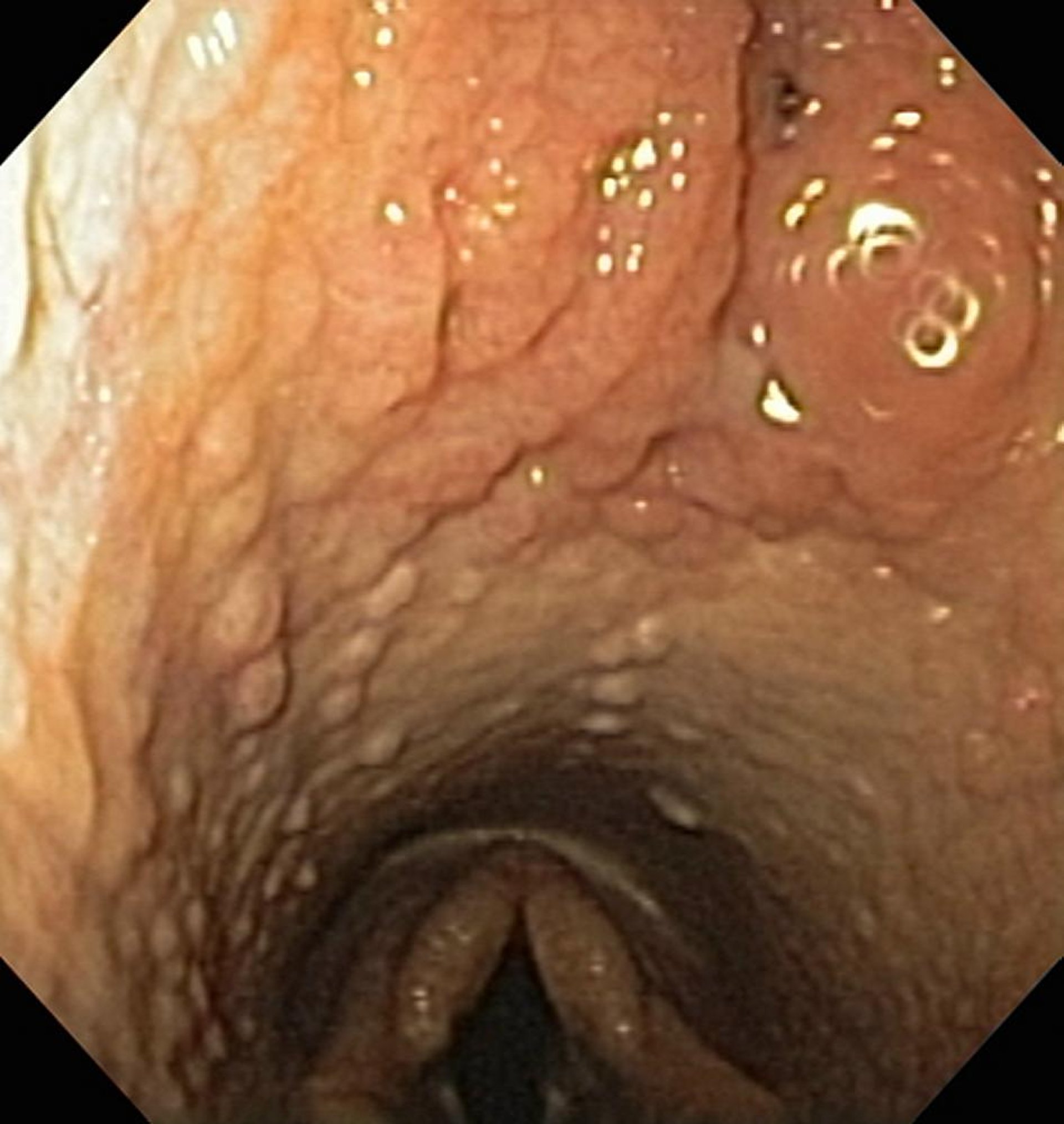
Endoscopic photograph of grade 3.5/4 pharyngeal lymphoid hyperplasia in a horse.
Courtesy of Dr. Jan Hawkins, Purdue University.
In most species, a common pharynx (shared by the respiratory and digestive tracts) is present at times other than deglutition. The unique caudal pharyngeal-laryngeal anatomy of horses shows complete separation of the pharynx into two components, the nasopharynx and the oropharynx. (Also see Pharyngeal Lymphoid Hyperplasia in Horses.)
Clinical Findings of Pharyngitis
Animals affected with pharyngitis have a normal desire to eat and drink; however, they can cough and be dysphagic. Animals with secondary peripharyngeal cellulitis and abscessation can be acutely dyspneic secondary to pharyngeal obstruction. For example, foals affected with suppurative pharyngitis secondary to abscessation of the retropharyngeal lymph nodes can become acutely dyspneic and require an emergency tracheotomy.
Diagnosis of Pharyngitis
Physical examination
Endoscopy
Diagnostic imaging
A diagnosis of pharyngitis can be made with a complete physical examination, radiographic or CT evaluation of the skull, endoscopic evaluation of the pharynx, microbial cultures of draining abscesses, or viral isolation testing of nasopharyngeal swabs. In small animals, oral pain and resistance to having the mouth opened can indicate retropharyngeal abscessation and the presence of a penetrating foreign body or oral or tonsillar neoplasia.
Histopathological analysis of biopsies of abnormal pharyngeal tissue should be performed to exclude pharyngeal neoplasia. In small animals, oral and/or endoscopic examination is the best diagnostic tool for pharyngitis. In large animals, the diagnosis of pharyngitis is easily made by endoscopic examination of the upper respiratory tract.
Treatment of Pharyngitis
Systemic antimicrobials and anti-inflammatories
Topical application of anti-inflammatories
Supportive care with intravenous fluids or extraoral alimentation
Bacterial pharyngitis should be treated with systemic antimicrobials on the basis of results of microbial culture and sensitivity testing. Abscesses should be drained and lavaged when appropriate. Viral-induced pharyngitis should be managed with antimicrobials to prevent secondary bacterial infections and anti-inflammatory medication to address edema and pain associated with the pharyngitis.
Animals affected with either bacterial or viral pharyngitis should be treated with NSAIDs. In some cases, corticosteroid administration can be considered to manage edema and inflammation. Pharyngitis secondary to foreign bodies should be treated with surgical removal of the offending object, drainage, and excision of necrotic tissue.
Racehorses affected by pharyngeal lymphoid hyperplasia can be treated with topical and systemic anti-inflammatory agents such as flunixin meglumine, phenylbutazone, or dexamethasone. A commonly used topical anti-inflammatory treatment includes prednisolone, dimethyl sulfoxide, glycerin, and nitrofurazone.
Large pharyngeal masses can be treated with contact diode laser photoablation. Some veterinarians have anecdotally suggested that hyperimmunization (repeated vaccination of horses with specific antigens to stimulate the production of high levels of circulating antibodies) is helpful in managing pharyngeal lymphoid hyperplasia.
Calicivirus infections in cats can cause mild, moderate, or severe ulceration of the oropharyngeal mucosa. Although specific antiviral therapies are not available, affected cats should be treated with systemic antimicrobials to prevent secondary bacterial infection.
Animals that cannot maintain their own hydration because of severe mucosal ulceration might require nutritional and electrolyte supplementation either intravenously or by extraoral tube feeding (eg, pharyngostomy or esophagostomy tube in small animals, horses, and ruminants or rumenostomy tubes in ruminants).
Key Points
Pharyngitis is an inflammatory condition.
Pharyngitis is managed primarily with administration of antimicrobials and systemic and topical anti-inflammatories.
For More Information