

Mycoplasma hyopneumoniae is a common cause of pneumonia in pigs worldwide. It also frequently leads to subclinical infection that causes lung lesions that can be detected post-mortem. Clinical signs, if present, are often a dry cough and reduced growth. Diagnosis may be based on clinical signs, characteristic lesions, and confirmation by PCR assay. Partial disease control can be achieved via improved management practices, antimicrobial treatment, and vaccination.
Mycoplasmal pneumonia is a chronic, typically clinically mild, infectious pneumonia of pigs. The disease is characterized by a persistent dry cough, impaired growth, occasional flares of overt respiratory distress, and a high incidence of lung lesions in slaughtered pigs. It tends to become endemic in infected herds and occurs worldwide.
Clinical outbreaks of mycoplasmal pneumonia may impair growth rate and feed conversion. The effects of the disease are uneven and unpredictable and place limits on the efficiency and flexibility of large production units. In swine units with good disease control measures, mycoplasmal pneumonia may remain largely subclinical. However, total disease control seems to be achieved only by pathogen eradication.
Etiology and Epidemiology of Mycoplasmal Pneumonia of Pigs
Mycoplasma hyopneumoniae causes mycoplasmal pneumonia in pigs. It is sometimes referred to as enzootic pneumonia, a characteristic disease syndrome caused primarily by M hyopneumoniae. A host-specific pleomorphic organism that lacks a cell wall and is fastidious and smaller than most bacteria. It can be cultured in specially prepared media, but isolation from field cases is difficult. It is rapidly inactivated in the environment and by disinfectants, but it may survive longer in cold weather and depending on the surface of the material. Mycoplasmal pneumonia is also frequently complicated by other mycoplasmas, bacteria, and viruses, which affect the severity of the disease. It is recognized as part of the porcine respiratory disease complex.
In most countries that use modern pig-farming methods, the lungs of 30%–80% of pigs slaughtered show lung lesions of the type associated with M hyopneumoniae infection. Pigs of all ages are susceptible, but within a herd, pigs are colonized in the first few weeks of life either by their dam or by other young pigs after mixing. Transmission to suckling piglets can occur from sows of all parities but is most prevalent in first-parity (gilt) litters.
Prevalence of M hyopneumoniae at weaning age has been suggested as an indicator of disease in finishing pigs. The onset of the disease may be most evident in the finishing stage at ~18–20 weeks of age. The incidence of lung lesions is highest in pigs 3–5 months old. Immunity develops slowly, followed by regression of the lung lesions. Older growing and mature pigs may recover from the disease completely, although persistence of the bacterium in the respiratory tract of infected animals has been confirmed for up to 7 months.
Clinical Findings of Mycoplasmal Pneumonia of Pigs
Nonproductive coughing is the most common sign of mycoplasmal pneumonia and is most obvious when pigs are roused. In endemically infected herds, morbidity is high, but clinical signs may be minimal, and mortality is low. Average daily weight gain and days to market weight are common production parameters negatively affected.
Individual pigs or groups sporadically develop severe pneumonia. Common predisposing factors are season and other stresses such a transient viral infections, parasitic migration, and mixing pigs. The disease is usually more severe when it first enters a naive herd.
Lesions

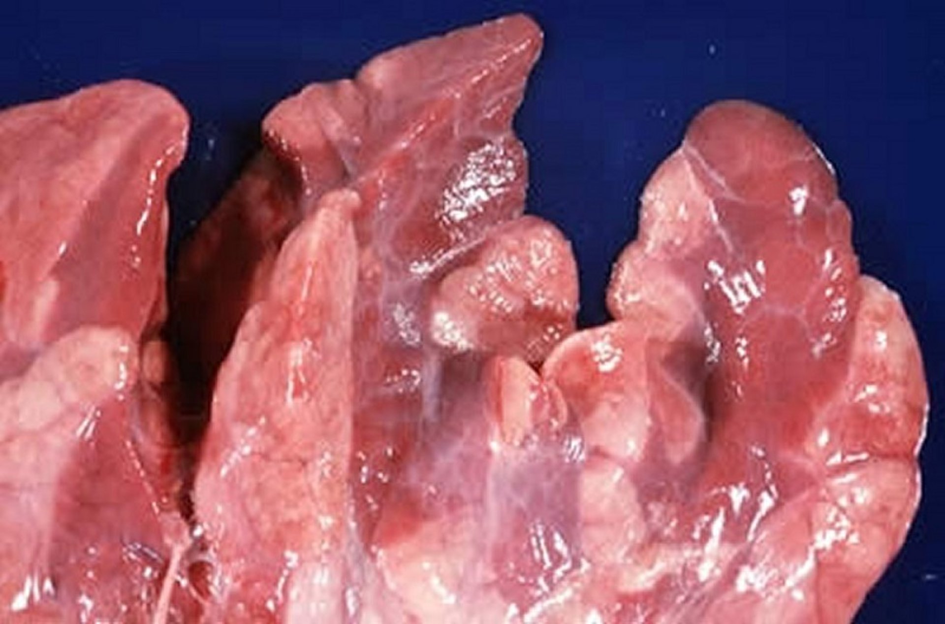
Mycoplasma hyopneumoniae, gross lesions. Note the atelectasis and lobar discoloration.
Courtesy of Dr. Louise Bauck.

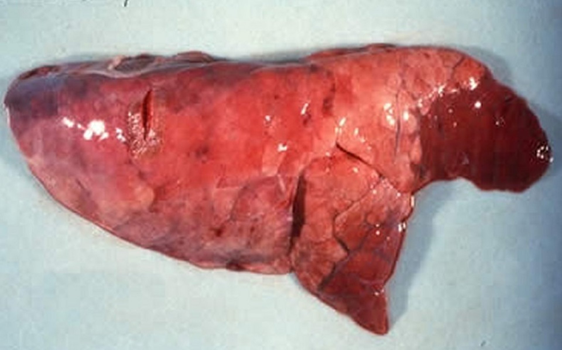
Mycoplasma hyopneumoniae in a pig. Note the cranioventral consolidation characteristic of enzootic pneumonia in pigs.
Courtesy of the Department of Pathobiology, University of Guelph.
On gross examination, areas of affected lungs are gray or purple, most commonly in the apical and cardiac lobes, and consolidated. Old lesions become clearly demarcated and eventually heal, leaving visible scars. The associated lymph nodes may be enlarged. Histologically, inflammatory cells are present in the bronchioles, and there is perivascular and peribronchiolar cuffing and extensive lymphoid hyperplasia.
Diagnosis of Mycoplasmal Pneumonia of Pigs
Based on clinical, histopathologic findings, and epidemiologic indicators in the herd.
Confirmation is by PCR assay testing
Clinical, histopathologic, and herd epidemiologic findings are usually suggestive of mycoplasmal pneumonia. M hyopneumoniae can be demonstrated in impression smears of the cut surface of affected lung, identified by fluorescent antibody technique or by in situ hybridization, and isolated and identified in culture. However, the above-mentioned laboratory tests are not routinely performed by practicing veterinarians.
Diagnosis of M hyopneumoniae infection in live pigs can be challenging. Bacterial isolation is seldom considered for routine diagnostic based on difficulty in growing the organism (low sensitivity). Serologic tests, principally commercially available ELISAs, are used on a herd basis, but results may be difficult to interpret due to slow seroconversion, lack of differentiation between antibodies generated by infection or by vaccination, and the frequent identification of unexpected positive reactors. Recently, PCR tests have become the most commonly used tool for detection of M hyopneumoniae in various samples types. In live pigs, the sensitivity of PCR increases when clinical specimens approach the lower respiratory tract. Thus, samples collected from the trachea, larynx, or nasal or oral cavity show different sensitivity, with tracheal samples exhibiting greater detection rates.
When postmortem sample collection is feasible, bronchial swabs or lung tissue specimens containing airways are preferred, because they are associated with the greatest probability of accurate pathogen detection.
Control of Mycoplasmal Pneumonia of Pigs
Improved management practices, particularly all in/all out management
Antimicrobial treatment
Vaccination may reduce clinical signs but does not prevent infection
The economic effects of mycoplasmal pneumonia in pigs can be reduced by improvements in housing and husbandry, particularly ventilation and space allowance. “All-in/all-out” management of pigs from birth to market is extremely effective at reducing negative effects of disease; following this practice improves growth performance and reduces lung lesions.
Commercial inactivated mycoplasmal bacterins consist of adjuvanted whole-cell preparations. Bacterins induce protection against development of gross lesions and significantly reduce clinical signs (coughing) in growing pigs. However, vaccination does not prevent infection.
When the disease first enters a herd, mass treatment with antibiotics effective against Mycoplasma spp helps to control the severity of signs. When disease increases in endemic herds, treatment of individual pigs with antimicrobials usually results in remission, presumably by controlling secondary bacteria.
Data indicate that prefarrowing management, vaccination with M hyopneumoniae vaccines, or antimicrobial treatment of dams significantly reduces colonization of suckling piglets, leading to fewer respiratory disease issues in downstream flows. In recent years, gilt acclimation practices, aimed at a uniform and controlled exposure of M hyopneumoniae in replacement females at least 8 months prior to first farrowing, have become common in North America. Gilt acclimation has been mainly used to “start the clock” in eradication programs relying on the known end of bacterial persistence. Gilt acclimation is also used to provide disease control in endemically infected farms.
Starting with foundation stock free of mycoplasmal pneumonia and adopting strict precautions against direct and indirect contact with pigs from other herds is advisable. In the USA and parts of Europe, most herds free of mycoplasmal pneumonia were established by repopulation or by partial depopulation. M hyopneumoniae-free herds have been established by segregated early weaning, herd closure, and medication, or by whole-herd medication. The combined success rate of the latter two methods has been reported to be approximately 70%, with reinfections potentially attributable to airborne transmission, breaches in biosecurity, and to a larger extent to lack of eradication with the original program.
Diagnostic monitoring of herds presumably free of mycoplasmal pneumonia can be difficult. It is hypothesized that the organism has not been successfully eliminated but rather coexisted within the population at an undetectable level for extended periods. However, achieving a low M hyopneumoniae prevalence, regardless of lack of pathogen elimination, has proven to be a significant production advantage in large swine units.
Key Points
Mycoplasmal pneumonia is the most important bacterial respiratory disease in pigs.
Clinical outbreaks, subclinical infections, and endemic disease within herds are common.
Partial disease control can be achieved with improved management, vaccination, and medication.
Disease eradication is a growing trend in North America.
For More Information
University of Minnesota College of Veterinary Medicine: How to choose the right sample type for detection of Mycoplasma hyopneumoniae


